

Circular RNA circ_0003028 contributes to tumorigenesis by regulating GOT2 via miR-1298-5p in non-small cell lung cancer
- PMID: 34077306
- PMCID: PMC8806680
- DOI: 10.1080/21655979.2021.1935064
Circular RNA circ_0003028 contributes to tumorigenesis by regulating GOT2 via miR-1298-5p in non-small cell lung cancer
Abstract
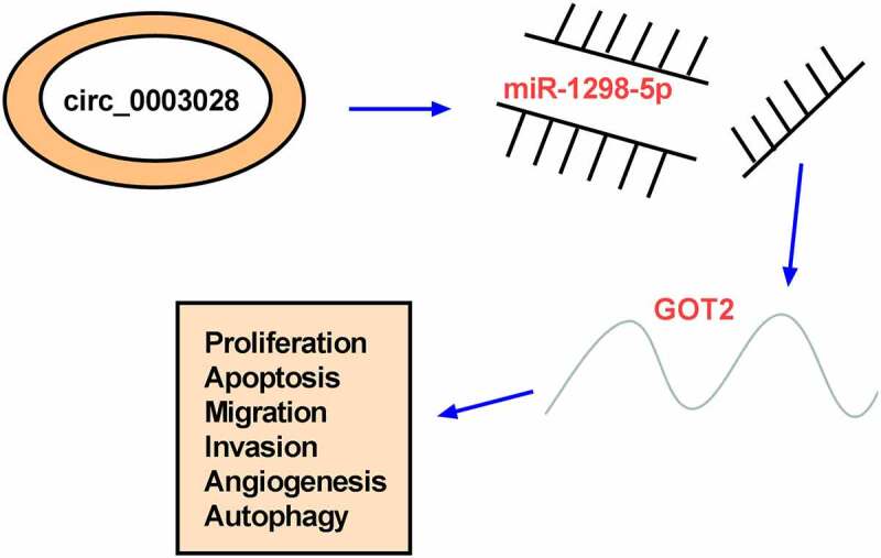
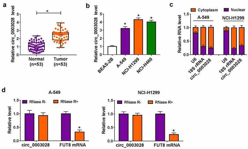
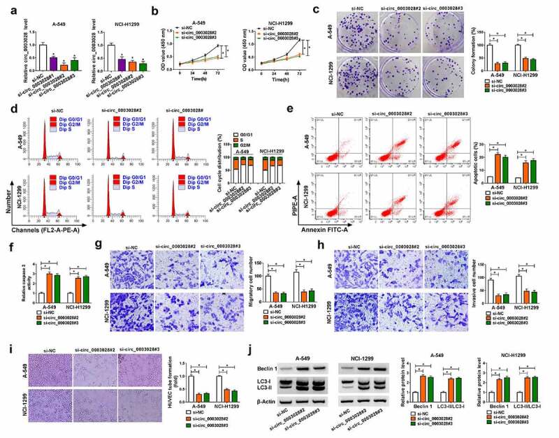
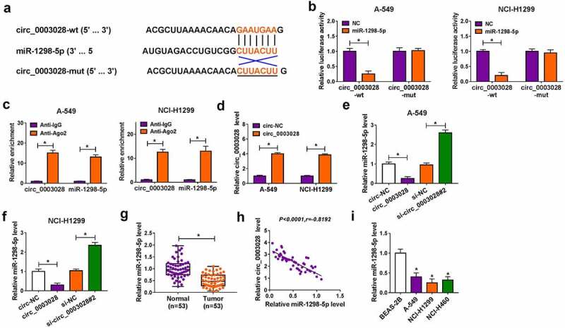
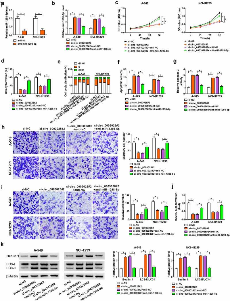
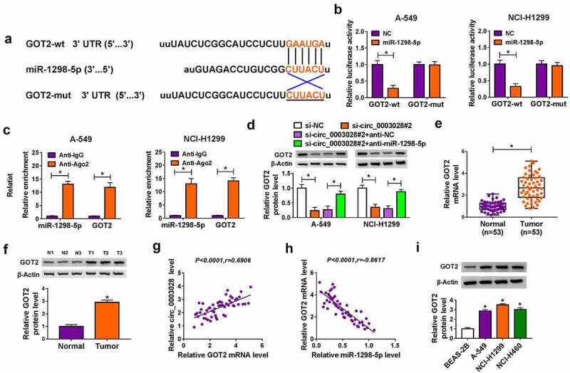
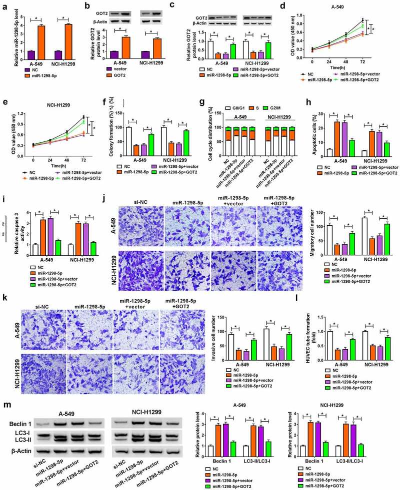
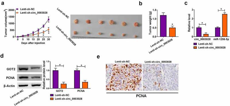
Non-small cell lung cancer (NSCLC) is a common malignant tumor, with high morbidity and mortality. Circular RNA (circRNA) circ_0003028 was reported to be upregulated in NSCLC. This study is designed to explore the role and mechanism of circ_0003028 on NSCLC progression. In this work, circ_0003028, microRNA-1298-5p (miR-1298-5p), and glutamic oxaloacetic transaminase 2 (GOT2) level were detected by real-time quantitative polymerase chain reaction (RT-qPCR). The localization of circ_0003028 was analyzed by subcellular fractionation assay. Cell proliferation, colony number, cell cycle progression, apoptosis, migration, invasion, and angiogenesis were measured by Cell Counting Kit-8 (CCK-8), colony formation, flow cytometry, transwell, and tube formation assays. Protein levels of Beclin1, light chain 3 (LC3)-II/LC3-I, GOT2, proliferating cell nuclear antigen (PCNA) were examined by western blot assay. The binding relationship between miR-1298-5p and circ_0003028 or GOT2 was predicted by circular RNA Interactome or starbase and then verified by dual-luciferase reporter, RNA Immunoprecipitation (RIP), and RNA pull-down assays. The biological role of circ_0003028 on NSCLC tumor growth was examined by the xenograft tumor model in vivo. We reported that circ_0003028 and GOT2 were upregulated, and miR-1298-5p was decreased in NSCLC tissues and cells. Moreover, circ_0003028 knockdown curbed cell proliferative ability, migration, invasion, angiogenesis, and facilitate apoptosis and autophagy in NSCLC cells in vitro. Mechanical analysis discovered that circ_0003028 regulated GOT2 expression by sponging miR-1298-5p. Circ_0003028 silencing hindered the cell growth of NSCLC in vivo. Taken together, circ_0003028 knockdown could suppress NSCLC progression partly by regulating the miR-1298-5p/GOT2 axis, providing an underlying therapeutic target for NSCLC.
Keywords: Circ_0003028; GOT2; miR-1298-5p; non-small cell lung cancer.
Figures
Similar articles
-
Circ_0072088 promotes the development of non-small cell lung cancer via the miR-377-5p/NOVA2 axis.Thorac Cancer. 2020 Aug;11(8):2224-2236. doi: 10.1111/1759-7714.13529. Epub 2020 Jun 11. Thorac Cancer. 2020. PMID: 32524752 Free PMC article.
-
Circ_0006988 promotes the proliferation, metastasis and angiogenesis of non-small cell lung cancer cells by modulating miR-491-5p/MAP3K3 axis.Cell Cycle. 2021 Jul;20(13):1334-1346. doi: 10.1080/15384101.2021.1941612. Epub 2021 Jun 30. Cell Cycle. 2021. PMID: 34189997 Free PMC article.
-
Circ_0082374 Promotes the Tumorigenesis and Suppresses Ferroptosis in Non-small Cell Lung Cancer by Up-Regulating GPX4 Through Sequestering miR-491-5p.Mol Biotechnol. 2025 Feb;67(2):484-495. doi: 10.1007/s12033-024-01059-z. Epub 2024 Mar 4. Mol Biotechnol. 2025. PMID: 38438754
-
Circ-CSPP1 knockdown suppresses hepatocellular carcinoma progression through miR-493-5p releasing-mediated HMGB1 downregulation.Cell Signal. 2021 Oct;86:110065. doi: 10.1016/j.cellsig.2021.110065. Epub 2021 Jun 26. Cell Signal. 2021. PMID: 34182091 Review.
-
Circ_CDR1as: A circular RNA with roles in the carcinogenesis.Pathol Res Pract. 2022 Aug;236:153968. doi: 10.1016/j.prp.2022.153968. Epub 2022 Jun 1. Pathol Res Pract. 2022. PMID: 35667198 Review.
Cited by
-
MicroRNA-1298-5p in granulosa cells facilitates cell autophagy in polycystic ovary syndrome by suppressing glutathione-disulfide reductase.Cell Tissue Res. 2023 Jun;392(3):763-778. doi: 10.1007/s00441-023-03747-9. Epub 2023 Feb 14. Cell Tissue Res. 2023. PMID: 36781484
-
Combining single-cell and transcriptomic analysis revealed the immunomodulatory effect of GOT2 on a glutamine-dependent manner in cutaneous melanoma.Front Pharmacol. 2023 Aug 24;14:1241454. doi: 10.3389/fphar.2023.1241454. eCollection 2023. Front Pharmacol. 2023. PMID: 37693904 Free PMC article.
-
Circ_0003028 enhances the proliferation and glycolytic capacity and suppresses apoptosis in non-small cell lung cancer cells via the miR-1305/miR-1322-SLC5A1 axis.Ann Transl Med. 2023 Mar 15;11(5):215. doi: 10.21037/atm-23-178. Ann Transl Med. 2023. PMID: 37007569 Free PMC article.
-
The potential of targeting autophagy-related non-coding RNAs in the treatment of lung cancer.Front Pharmacol. 2025 May 14;16:1551258. doi: 10.3389/fphar.2025.1551258. eCollection 2025. Front Pharmacol. 2025. PMID: 40438586 Free PMC article. Review.
-
Theoretical perspectives and clinical applications of non-coding RNA in lung cancer metastasis: a systematic review.Discov Oncol. 2025 Feb 12;16(1):169. doi: 10.1007/s12672-025-01919-3. Discov Oncol. 2025. PMID: 39937377 Free PMC article. Review.
References
-
- Siegel RL, Miller KD.. Cancer statistics, 2021. CA Cancer J Clin. 2021;71:7–33. - PubMed
-
- Nasim F, Sabath BF, Eapen GA.. Lung cancer. Med Clin North Am. 2019;103:463–473. - PubMed
-
- Herbst RS, Morgensztern D, Boshoff C. The biology and management of non-small cell lung cancer. Nature. 2018;553:446–454. - PubMed
-
- Sekihara K, Hishida T, Yoshida J, et al. Long-term survival outcome after postoperative recurrence of non-small-cell lung cancer: who is ‘cured’ from postoperative recurrence? Eur J Cardiothorac Surg. 2017;52:522–528. - PubMed
MeSH terms
Substances
LinkOut - more resources
Full Text Sources
Other Literature Sources
Medical
Miscellaneous