Case Reports
. 2021 Jun 2;5(6):e21.00055-7.
doi: 10.5435/JAAOSGlobal-D-21-00055.

Intramedullary Fixation With a Short Nail in a Young Patient Presenting With a Pathological Proximal Femur Fracture

Affiliations
Case Reports

Intramedullary Fixation With a Short Nail in a Young Patient Presenting With a Pathological Proximal Femur Fracture

Marcos Roberto González et al. J Am Acad Orthop Surg Glob Res Rev. .

Abstract

An 18-year-old man presented with a pathological fracture of the right proximal femur. Desmoplastic fibroma was diagnosed through histological studies. Surgical management involved extended intralesional curettage and fracture stabilization by open reduction with intramedullary nailing, using a short Gamma nail. At 42-month follow-up, the patient presented no limitations or recurrence. Internal fixation after prior intralesional curettage is a valid treatment strategy for pathological fractures in young patients. A short nail was chosen to prevent direct tumor cell seeding throughout the femur and future recurrence. Fracture consolidation was achieved because of the healing potential of a young patient.


Figures

Figure 1
Figure 1
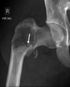
AP radiograph of the right hip at presentation demonstrates a pathological fracture through a sharply marginated lytic lesion in the intertrochanteric region of the right femur. Note small bony fracture fragments (arrow) in the dependent portion of the lytic lesion, simulating the “fallen fragment” sign seen in unicameral bone cysts.
Figure 2
Figure 2
MRI of the right hip with and without contrast: A, Coronal T1-weighted MRI demonstrates the intertrochanteric pathological fracture with varus angulation. The underlying tumor is well-defined by a hypointense rim at both its superior and inferior margins (arrowheads). Intratumoral bone fragments can also be appreciated (dashed arrow). B, Coronal fat-suppressed T2-weighted image demonstrates the T2 hyperintense tumor in the proximal femur, with extensive surrounding bone marrow edema and hemorrhage caused by the pathological fracture. Note the marked thinning and endosteal scalloping along both the medial and lateral cortices (arrows). C, Axial contrast-enhanced fat-suppressed T1-weighted MRI shows somewhat heterogeneous enhancement in the intraosseous tumor, above the level of the fracture. Note the well-defined boundary with normal marrow in the mid-cervical region (arrow).
Figure 3
Figure 3
Axial CT above the pathological fracture demonstrates the lytic lesion with well-defined margins, high-grade endosteal scalloping, and posterior cortical buckling.
Figure 4
Figure 4
Histopathological features of desmoplastic fibroma: A, Desmoplastic fibroma–spindled cells with bland small nuclei, evenly dispersed in the collagenous stroma (low power). B, Slender spindled cells set within abundant eosinophilic collagen matrix (low power). C, Spindled cell proliferation accompanied by collagenous stroma-desmoplastic fibroma infiltrating bone (low power). D, Spindled cells with indistinct cytoplasmic borders and bland ovoid nuclei with smooth contours show finely dispersed chromatin. Cells appear to merge with the intercellular collagenous matrix. No mitoses present (high power). E, Interface of desmoplastic fibroma and bone-spindled cells with elongated nuclei (lower left) enmeshed between wavy collagen fibers abut large polyhedral osteoblasts (upper right) within and surrounding the nascent osteoid (high power).
Figure 5
Figure 5
Postoperative AP radiograph of the right hip after intralesional treatment with curetting and bone grafting, fracture reduction, and internal fixation with a short intramedullary nail.
Figure 6
Figure 6
Three-year follow-up AP radiograph demonstrates complete incorporation of the graft and healing of the fracture, with no evidence of recurrent disease or implant loosening.

References

    1. Ishizaka T, Susa M, Sato C, et al. : Desmoplastic fibroma of bone arising in the cortex of the proximal femur. J Orthop Sci 2018;26:306-310. - PubMed
    1. Cho BH, Tye GW, Fuller CE, Rhodes JL: Desmoplastic fibroma of the pediatric cranium: Case report and review of the literature. Childs Nerv Syst 2013;29:2311-2315. - PubMed
    1. Gong YB, Qu LM, Qi X, Liu JG: Desmoplastic fibroma in the proximal femur: A case report with long-term follow-up. Oncol Lett 2015;10:2465-2467. - PMC - PubMed
    1. Kang DM, Juhng SK, Sohn YJ, Kim HS: Imaging findings of desmoplastic fibroma rarely involving the clavicle: Case report. Korean J Radiol 2014;15:130. - PMC - PubMed
    1. Takazawa K, Tsuchiya H, Yamamoto N, et al. : Osteosarcoma arising from desmoplastic fibroma treated 16 years earlier: A case report. J Orthop Sci 2003;8:864-868. - PubMed

Publication types