

Adult skin acute stress responses to short-term environmental and internal aggression from exposome factors
- PMID: 34077579
- PMCID: PMC8519049
- DOI: 10.1111/jdv.17432
Adult skin acute stress responses to short-term environmental and internal aggression from exposome factors
Abstract
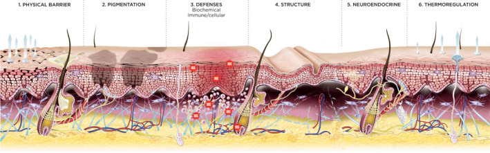
Exposome factors that lead to stressed skin can be defined as any disturbance to homeostasis from environmental (meteorological factors, solar radiation, pollution or tobacco smoke) and/or internal exposure (unhealthy diet, hormonal variations, lack of sleep, psychosocial stress). The clinical and biological impact of chronic exposome effects on skin functions has been extensively reviewed, whereas there is a paucity of information on the impact of short-term acute exposure. Acute stress, which would typically last minutes to hours (and generally no more than a week), provokes a transient but robust neuroendocrine-immune and tissue remodelling response in the skin and can alter the skin barrier. Firstly, we provide an overview of the biological effects of various acute stressors on six key skin functions, namely the skin physical barrier, pigmentation, defences (antioxidant, immune cell-mediated, microbial and microbiome maintenance), structure (extracellular matrix and appendages), neuroendocrine and thermoregulation functions. Secondly, we describe the biological and clinical effects on adult skin from individual exposome factors that elicit an acute stress response and their consequences in skin health maintenance. Clinical manifestations of acutely stressed skin may include dry skin that might accentuate fine lines, oily skin, sensitive skin, pruritus, erythema, pale skin, sweating, oedema and flares of inflammatory skin conditions such as acne, rosacea, atopic dermatitis, pigmentation disorders and skin superinfection such as viral reactivation. Acute stresses can also induce scalp sensitivity, telogen effluvium and worsen alopecia.
© 2021 The Authors. Journal of the European Academy of Dermatology and Venereology published by John Wiley & Sons Ltd on behalf of European Academy of Dermatology and Venereology.
Figures
References
-
- Krutmann J, Bouloc A, Sore G, Bernard BA, Passeron T. The skin aging exposome. J Dermatol Sci 2017; 85(3): 152–161. - PubMed
-
- Passeron T, Krutmann J, Andersen ML, Katta R, Zouboulis CC. Clinical and biological impact of the exposome on the skin. J Eur Acad Dermatol Venereol 2020; 34(Suppl 4): 4–25. - PubMed
-
- Cecchi L, D'Amato G, Annesi‐Maesano I. External exposome and allergic respiratory and skin diseases. J Allergy Clin Immunol 2018; 141(3): 846–857. - PubMed
Publication types
MeSH terms
LinkOut - more resources
Full Text Sources
