. 2021 Jun 24;184(13):3486-3501.e21.
doi: 10.1016/j.cell.2021.05.001. Epub 2021 Jun 1.

Protective neutralizing antibodies from human survivors of Crimean-Congo hemorrhagic fever

Affiliations

Protective neutralizing antibodies from human survivors of Crimean-Congo hemorrhagic fever

J Maximilian Fels et al. Cell. .

Abstract

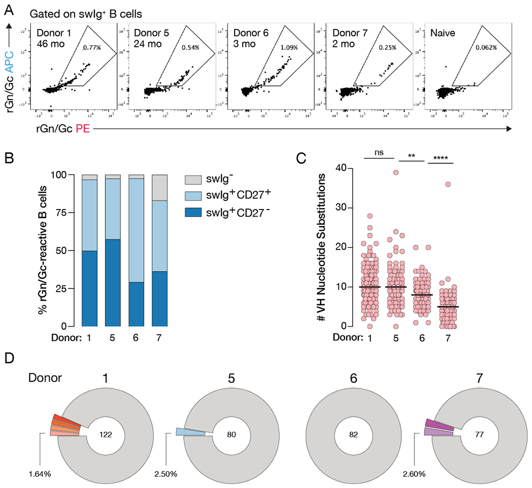
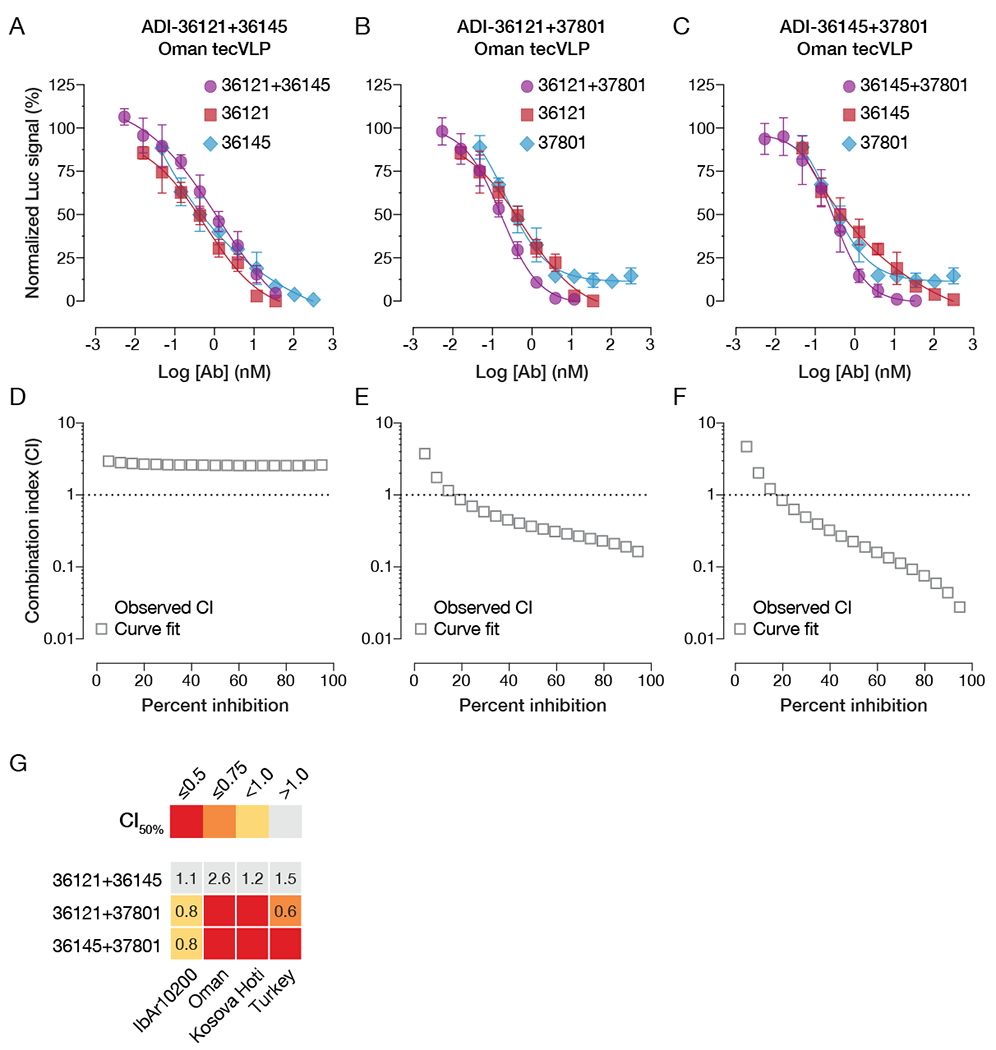
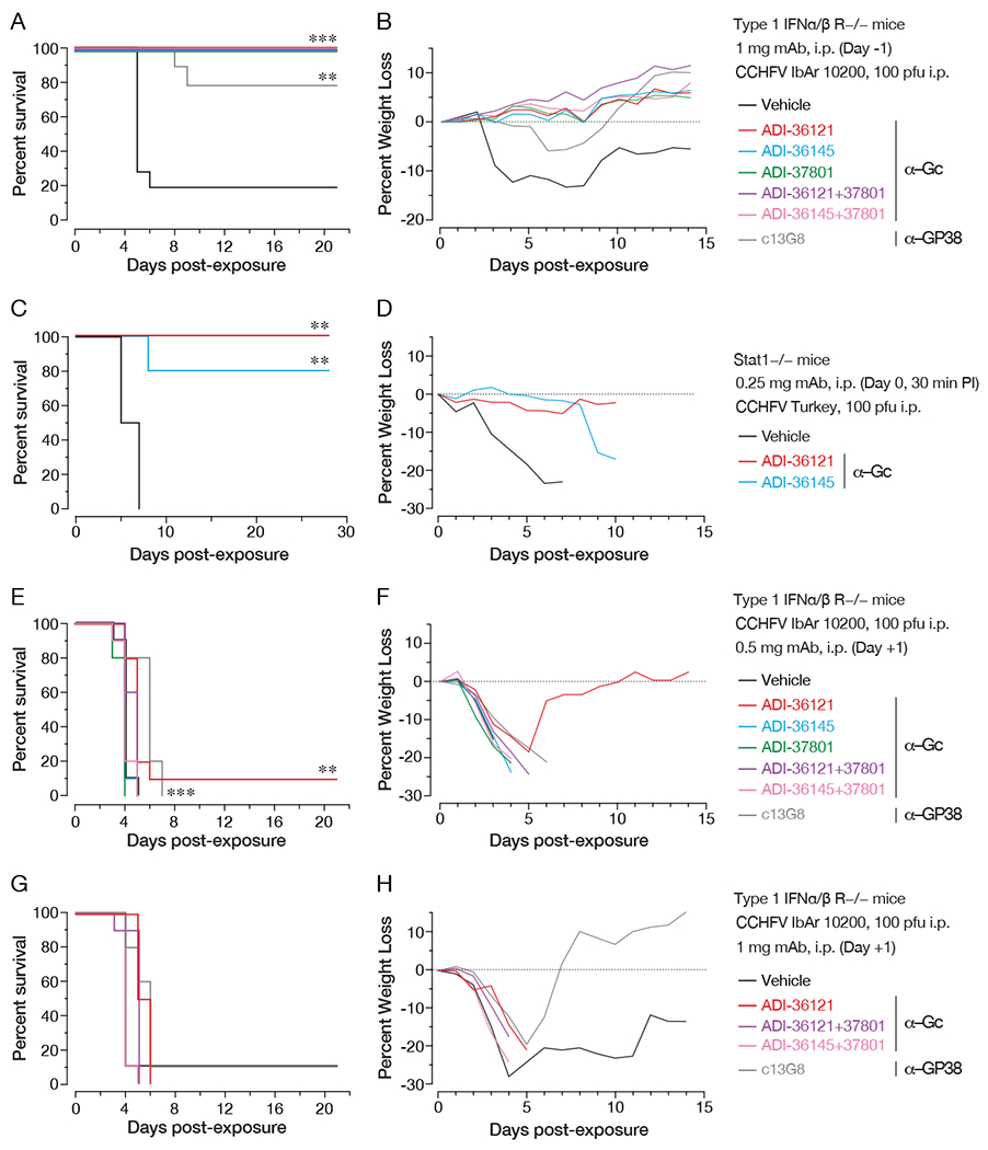
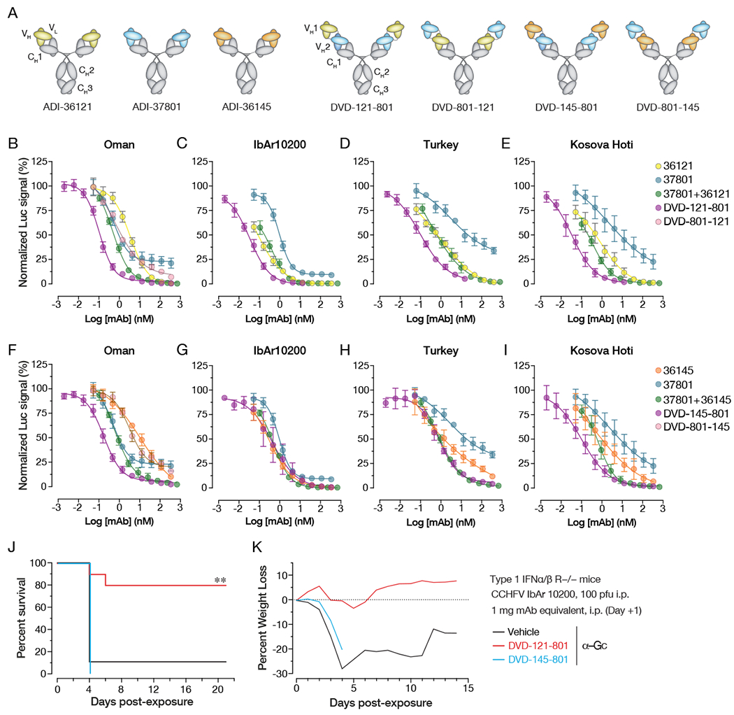
Crimean-Congo hemorrhagic fever virus (CCHFV) is a World Health Organization priority pathogen. CCHFV infections cause a highly lethal hemorrhagic fever for which specific treatments and vaccines are urgently needed. Here, we characterize the human immune response to natural CCHFV infection to identify potent neutralizing monoclonal antibodies (nAbs) targeting the viral glycoprotein. Competition experiments showed that these nAbs bind six distinct antigenic sites in the Gc subunit. These sites were further delineated through mutagenesis and mapped onto a prefusion model of Gc. Pairwise screening identified combinations of non-competing nAbs that afford synergistic neutralization. Further enhancements in neutralization breadth and potency were attained by physically linking variable domains of synergistic nAb pairs through bispecific antibody (bsAb) engineering. Although multiple nAbs protected mice from lethal CCHFV challenge in pre- or post-exposure prophylactic settings, only a single bsAb, DVD-121-801, afforded therapeutic protection. DVD-121-801 is a promising candidate suitable for clinical development as a CCHFV therapeutic.

Keywords: CCHFV; Crimean-Congo hemorrhagic fever virus; antibody therapeutic; bunyavirus; emerging virus; human monoclonal antibody; monoclonal antibody; nairovirus; tickborne; viral glycoprotein.


Conflict of interest statement

Declaration of interests K.C. is a scientific advisory board member of Integrum Scientific and Biovaxys Technology Corporation. K.C., J.S.M., and J.R.L. are scientific advisory board members of the Pandemic Security Initiative of Celdara Medical. N.T.P. and L.M.W. are employees and shareholders of Adimab. D.P.M. is a shareholder of Adimab. Z.A.B., D.M.A., C.L.M., and L.Z. are shareholders and employees of Mapp Biopharmaceutical. Mapp Biopharmaceutical has filed a patent application related to this work.

Figures

Figure 1.
Figure 1.. Isolated CCHFV rGn/Gc-specific mAbs represent diverse lineages.
(A) Flow cytometric analysis of rGnGc-specific B cells in four Ugandan donors. Flow plots are gated on CD14/CD16/CD3/PI/IgM/IgD CD19/CD20+ rGn/Gc+ B cells. Inset denotes donor ID and approximate time between infection and blood draw. (B) Frequency of B cell subsets that encoded the 361 antibody genes described throughout, swIg = class-switched immunoglobulin (IgM/IgD). (C) Load of somatic hypermutation as determined by the number of VH nucleotide substitutions away from the predicted VH germline. Statistical comparison was made by the Mann-Whitney test (**** P < 0.0001, ** P < 0.01, ns = not significant). See also Table S1. (D) Clonal analysis of isolated antibody genes. Clones that encode the same germline genes, CDR3 lengths, and have >80% nucleotide sequence homology within CDRH3 are considered to be within the same lineage. Colored and expanded slices represent lineages for which more than one clone was identified. Unique clones are shown as a single gray segment. Numbers in the center of the pie represent the total number of mAbs with binding verified by biolayer interferometry. See also Figure S1.
Figure 2.
Figure 2.. mAbs mainly target conserved antigenic sites in Gc.
Apparent KD values (KDApp) of mAbs binding IbAr10200 rGn/Gc (x-axis) and IbAr10200 Gc (A), China rGn/Gc (B), or Kosova Hoti rGn/Gc (C) (y-axis), as determined by biolayer interferometry. NB/PF denotes mAbs with no binding or poor fits. Each circle represents a single mAb and is color coded as in D and E. (D) Frequency of mAbs from each donor that bind to rGn/Gc broken down by subunit. (E) Cross-reactivity profiles of antibodies isolated from each donor showing the number of strains (IbAr10200, China, and Kosova Hoti) bound by mAbs. Binding was determined by > 0.1 nm response in the BLI assay. See also Figure S2.
Figure 3.
Figure 3.. Distribution of antigenic sites targeted and mapping of epitopes.
(A) Summary of mAb binding data together with targeted antigenic sites as determined by binding of IgG to rGn/Gc with pre-complexed competitor or to rGP38 by BLI. Each column denotes the antigen reactivity of an individual mAb, colored by KDApp (see legend in panel C). Each of the top five rows indicates the test antigen. The last row denotes the antigenic site, color coded according to the legend in panel C. (B) Distribution of targeted antigenic sites by Gc-specific antibodies for which a targeted site was determined in panel A. Named mAbs (e.g. ADI-36193) shown with each group represent the Fab used in the competition assay. mAbs for which the targeted site could not be readily defined (“Unknown”) or that bound GP38 are not shown. (C) Distribution of targeted antigenic sites by Gc-specific mAbs across all donors. mAbs with “Unknown” antigenic sites include those with weak affinities and/or weak competition (<10-fold reduction in binding in the presence of competitive Fab). (D) Heat map depicting the magnitude of loss of binding of single mutations compared to wild-type Gc on the surface of yeast. Darker shades indicate greater loss of binding. Shown are mutations that reduce binding of mAbs by >90%. (E) Homology model of CCHFV Gc based on Maporal virus Gc with an associated schematic illustrating the approximate domain organization in relation to the viral membrane. The stem region and transmembrane domain are not visualized in this model. Residues at which mutations reduce binding by >90% are colored as in D. See also Figure S3 and S4.
Figure 4.
Figure 4.. Distribution of neutralization potency by antigenic sites targeted, and breadth of neutralization by candidate nAbs.
(A) Neutralization of Oman tecVLPs at 35 nM mAb, as measured by reduction in luciferase activity in Vero target cells compared to no-antibody treatment. Each circle represents a different mAb. (B) Breakdown of mAb neutralizing activity by targeted antigenic site. (C) Heatmap showing relative or absolute neutralization activity (IC50) of 21 nAbs selected for further characterization. Relative IC50 values are shown for sigmoidal curves, whereas absolute IC50 values are provided for curves with poorly defined plateaus. See Figure S5 for all neutralization curves. (D-F) Neutralization curves of ADI-37801 (D), ADI-36121 (E), and ADI-36145 (F) against tecVLPs bearing Gn/Gc proteins from four CCHFV strains. (G) Neutralization curves of ADI-37801, ADI-36121, and ADI-36145 against authentic CCHFV-IbAr10200. All tecVLP neutralization assays were performed using Vero target cells, while neutralization of authentic CCHFV IbAr10200 was assayed in Vero-E6 cells. n = 6, from two independent experiments for all neutralization curves.
Figure 5.
Figure 5.. Synergistic neutralization potential of lead nAb candidates.
(A-C) Neutralization curves of individual nAbs and their 1:1 combinations. (A) ADI-36121 and ADI-36145, (B) ADI-36121 and ADI-37801, (C) ADI-36145 and ADI-37801. (D-F) Combination-index (CI) analyses of neutralization curves in panels A–C to determine additive (CI ~ 1) or synergistic effects (positive, CI < 1; negative, CI >> 1). (G) Summary of CI values at 50% neutralization for nAb combinations against tecVLPs carrying divergent Gn/Gc proteins. Synergistic neutralization was assayed in Vero target cells. n = 6, from two independent experiments for all neutralization curves. See also Figure S6.
Figure 6.
Figure 6.. Protective efficacy of lead nAbs and nAb combinations in two murine models of lethal CCHFV challenge.
(A-B) Type I IFN α/β/ R−/− (IFNAR1-KO) mice were treated with the indicated mAbs or mAb combinations 1 day prior to challenge with CCHFV IbAr10200. (n = 10 mice per group) (A) Survival curves (vehicle versus test mAb) were compared by Mantel-Cox test (*** P < 0.001, ** P < 0.01). (B) Associated mean weight loss data are shown. (C-D) Stat1−/− mice were challenged with CCHFV Turkey/2004 and then treated with single doses of the indicated mAbs or vehicle at 30 min post-exposure. (n = 5 mice per group) (C) Survival curves (vehicle versus test mAb) were compared by Mantel-Cox test (*** P < 0.001, ** P < 0.01). (D) Associated mean weight loss data are shown. (E-H) IFNAR1-KO mice were exposed to CCHFV IbAr10200 and treated with the indicated mAbs or mAb combinations at 1 day post-challenge. (n = 10 mice per group) (E, G) Survival curves (vehicle versus test mAb) were compared by Mantel-Cox test (*** P < 0.001, ** P < 0.01). (F, H) Associated mean weight loss data are shown.
Figure 7.
Figure 7.. Neutralization activity and protective efficacy of engineered bsAbs.
(A) Schematic illustration of candidate nAb IgG1s and dual-variable domain IgGs (DVD-Igs) derived from them by combining IgG1 variable domains. (B-I) Neutralization curves of the indicated nAbs, nAb combinations, and bsAbs against against tecVLPs bearing Gn/Gc proteins from (B, F) Oman, (C, G) IbAr10200, (D, H) Turkey, (E, I) Kosova Hoti strains. (J,K) IFNAR1-KO mice were exposed to CCHFV IbAr10200 and treated with the indicated bsAbs at 1 day post-challenge. (n = 10 mice per group) (J) Survival curves (vehicle versus test mAb) were compared by Mantel-Cox test (*** P < 0.001, ** P < 0.01). (K) Associated mean weight loss data are shown. See also Figure S7.

References

    1. Al-Abri SS, Hewson R, Al-Kindi H, Al-Abaidani I, Al-Jardani A, Al-Maani A, Almahrouqi S, Atkinson B, Al-Wahaibi A, Al-Rawahi B, et al. (2019). Clinical and molecular epidemiology of Crimean-Congo hemorrhagic fever in Oman. PLoS Negl. Trop. Dis 13, e0007100. - PMC - PubMed
    1. Andrews SF, Chambers MJ, Schramm CA, Plyler J, Raab JE, Kanekiyo M, Gillespie RA, Ransier A, Darko S, Hu J, et al. (2019). Activation Dynamics and Immunoglobulin Evolution of Pre-existing and Newly Generated Human Memory B cell Responses to Influenza Hemagglutinin. Immunity 51, 398–410.e5. - PubMed
    1. Austin SK, Dowd KA, Shrestha B, Nelson CA, Edeling MA, Johnson S, Pierson TC, Diamond MS, and Fremont DH (2012). Structural basis of differential neutralization of DENV-1 genotypes by an antibody that recognizes a cryptic epitope. PLoS Pathog. 8, e1002930. - PMC - PubMed
    1. Beltramello M, Williams KL, Simmons CP, Macagno A, Simonelli L, Quyen NTH, Sukupolvi-Petty S, Navarro-Sanchez E, Young PR, de Silva AM, et al. (2010). The human immune response to Dengue virus is dominated by highly cross-reactive antibodies endowed with neutralizing and enhancing activity. Cell Host Microbe 8, 271–283. - PMC - PubMed
    1. Bente DA, Alimonti JB, Shieh W-J, Camus G, Ströher U, Zaki S, and Jones SM (2010). Pathogenesis and immune response of Crimean-Congo hemorrhagic fever virus in a STAT-1 knockout mouse model. J. Virol 84, 11089–11100. - PMC - PubMed

Publication types

MeSH terms