. 2021 Jun;23(6):561-573.
doi: 10.1016/j.neo.2021.05.002. Epub 2021 May 31.

Rosmarinic acid represses colitis-associated colon cancer: A pivotal involvement of the TLR4-mediated NF-κB-STAT3 axis

Affiliations

Rosmarinic acid represses colitis-associated colon cancer: A pivotal involvement of the TLR4-mediated NF-κB-STAT3 axis

Bo-Ram Jin et al. Neoplasia. 2021 Jun.

Abstract

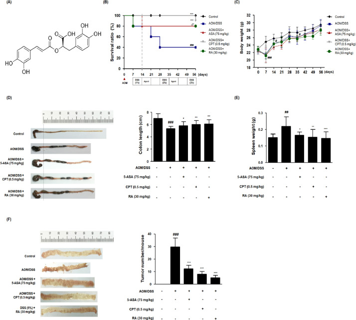
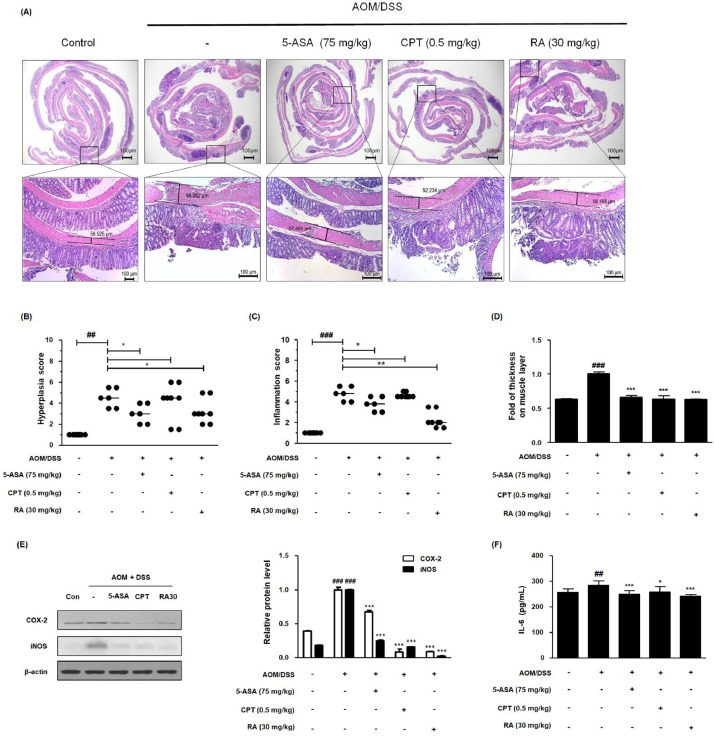
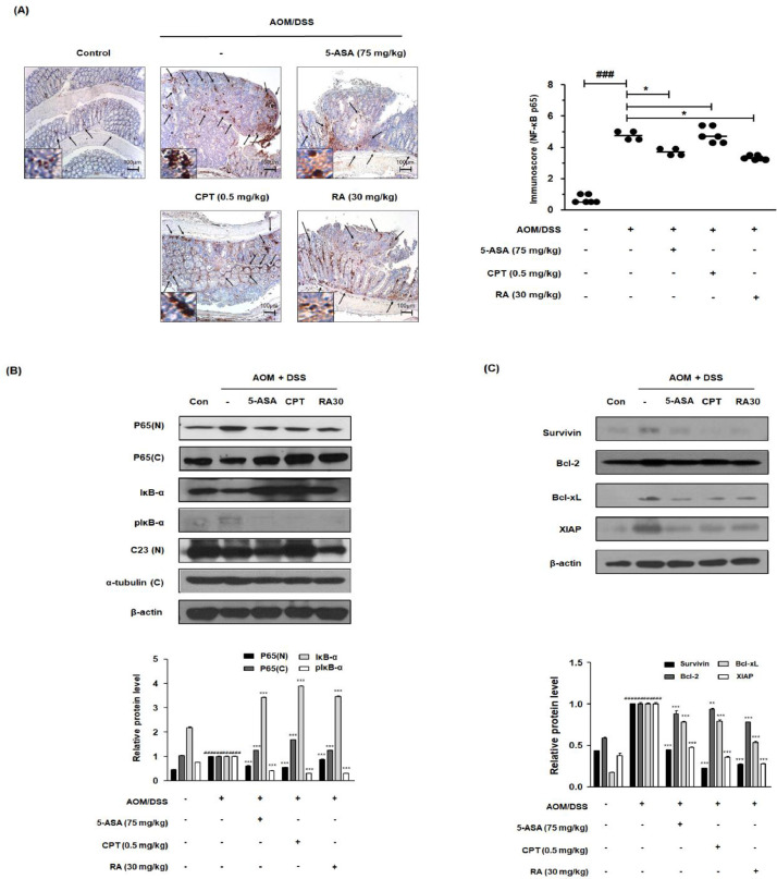
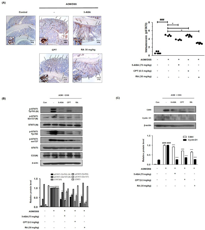
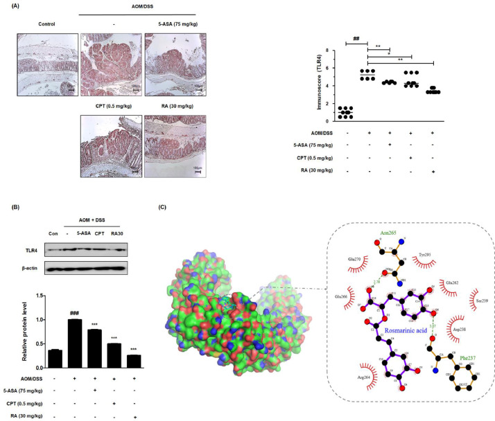
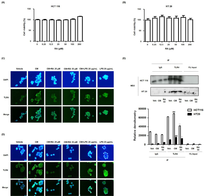
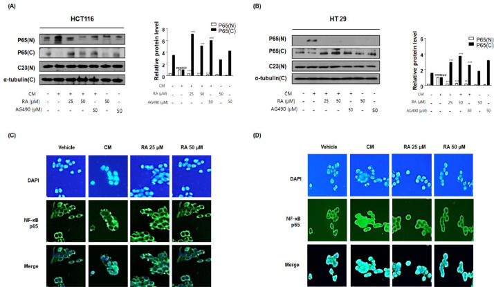
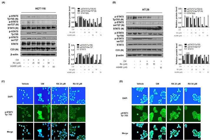
Previously, we found that rosmarinic acid (RA) exerted anti-inflammatory activities in a dextran sulfate sodium (DSS)-induced colitis model. Here, we investigated the anti-tumor effects of RA on colitis-associated colon cancer (CAC) and the underlying molecular mechanisms. We established an azoxymethane (AOM)/DSS-induced CAC murine model for in vivo studies and used a conditioned media (CM) culture system in vitro. H&E staining, immunohistochemistry, western blot assay, enzyme-linked immunosorbent assay, molecular docking, co-immunoprecipitation, and immunofluorescence assay were utilized to investigate how RA prevented colorectal cancer. In the AOM/DSS-induced CAC murine model, RA significantly reduced colitis severity, inflammation-related protein expression, tumor incidence, and colorectal adenoma development. It significantly modulated toll-like receptor-4 (TLR4)-mediated nuclear factor-kappa B (NF-κB) and signal transducer and activator of transcription 3 (STAT3) activation, thus attenuating the expression of anti-apoptotic factors, which mediate transcription factor-dependent tumor growth. In vitro, RA inhibited CM-induced TLR4 overexpression and competitively inhibited TLR4-myeloid differentiation factor 2 complex in an inflammatory microenvironment. Thus, RA suppressed NF-κB and STAT3 activation in colon cancer cells in an inflammatory microenvironment. Therefore, RA suppressed colitis-associated tumorigenesis in the AOM/DSS-induced CAC murine model and abrogated human colon cancer progression in an inflammatory microenvironment by propitiating TLR4-mediated NF-κB and STAT3 activation, pleiotropically.

Keywords: Colitis-associated colon cancer (CAC); Myeloid differentiation factor 2 (MD-2); Nuclear factor-kappa B (NF-κB); Rosmarinic acid (RA); Signal transducer and activator of transcription 3 (STAT3); Toll-like receptor-4 (TLR4).


Conflict of interest statement

Competing interest The authors declare no conflict of interest.

Figures

Fig 1
Fig. 1
Effects of RA on the development of the AOM/DSS-induced CAC in vivo model. (A) Molecular structure of RA. (B) Kaplan–Meier survival analysis shows the effect of RA on the survival ratio of AOM/DSS-induced CAC mice. (C) Body weight was evaluated every week during the experimental period. (D) A representative appearance and data statics of colon length is presented. The length of the colon of each animal was measured between the caecum and proximal rectum. (E) The spleen was weighed in each experimental group. (F) Using a blinded method, all tumor masses in each colonic tissue were numbered and the mean values were calculated for each group. Numerical values are presented as means ± standard deviations (n = 8); #P < 0.05, ##P < 0.01, ###P < 0.001 when compared with the control group; *P < 0.05, **P < 0.01, ***P < 0.001 when compared with the AOM/DSS-induced CAC group; significances between each experimental group were determined by analysis of variance and Dunnett's post hoc test.
Fig 2
Fig. 2
Effect of RA on the inflammatory response in the AOM/DSS-induced CAC in vivo model. (A) Whole colonic tissues were stained using H&E. (B) The hyperplasia score and (C) inflammation score in AOM/DSS-induced CAC mice were estimated. (D) Colonic muscle thickness was determined using the LAS software. Slide sections were scrutinized by microscopy. Magnification × 40 and × 100 inset. (E) The expression of COX-2 and iNOS was estimated by western blotting in triplicate, and relative protein level was measured by densitometric analysis using Image J. (F) Production of IL-6 was determined using an ELISA kit. Values are means ± standard deviations (n = 8); ##P < 0.01, ###P < 0.001 compared with the control group; *P < 0.05, ⁎⁎P < 0.01, ⁎⁎⁎P < 0.001 compared with the AOM/DSS-induced CAC group; significances between treated groups were determined by analysis of variance and Dunnett's post hoc test.
Fig 3
Fig. 3
Effect of RA on NF-κB activation and its relative proteins in the AOM/DSS-induced CAC in vivo model. (A) The nuclear manifestation and translocation of NF-κB p65 in colon tissues were observed using IHC. Immunoscore of NF-κB p65 in the colon of AOM/DSS-induced mice was estimated. (B) Nuclear (N) and cytosolic (C) extracts were prepared from colon tissues. NF-κB p65 translocation to the nucleus and phosphorylation of IκB were estimated by western blotting in triplicate. (C) The expression of NF-κB-related proteins was determined by western blotting in triplicate. C23, α-tubulin, and β-actin were used as internal controls. The relative ratio level was normalized to internal controls and determined by densitometric analysis. Values are presented as means ± standard deviations (n = 8); ###P < 0.001 vs the control group; *P < 0.05, ⁎⁎P < 0.01, ⁎⁎⁎P < 0.001 vs the AOM/DSS-induced CAC group; significances between treated groups were determined using an analysis of variance and Dunnett's post hoc test.
Fig 4
Fig. 4
Effect of RA on constitutive activation of STAT3 and its relative proteins in the AOM/DSS-induced CAC in vivo mouse model. (A) The manifestation and translocation of pSTAT3 (Tyr705) in colon tissues were identified using IHC. Immunoreactivity of pSTAT3 in AOM/DSS-induced mice was assessed. (B) Nuclear (N) protein and total protein were extracted from the colon tissues of AOM/DSS-induced CAC mice. The phosphorylation of STAT3 at Tyr705 and Ser727 and the nuclear translocation of pSTAT3 (Try705) were estimated by western blotting in triplicate. (C) The expression of STAT3-relative proteins was confirmed by western blotting in triplicate. The relative ratio level was determined by densitometric analysis and normalized to internal controls. Numerical values are presented as means ± standard deviations (n = 8); ###P < 0.001 compared with the control group; *P < 0.05, ⁎⁎⁎P < 0.001 compared with the AOM/DSS-induced CAC group; significances between treated groups were determined by analysis of variance and Dunnett's post hoc test.
Fig 5
Fig. 5
Effect of RA on TLR4 in the AOM/DSS-induced CAC in vivo model and molecular docking for the binding of RA to TLR4. (A) The expression of TLR4 in colon tissues of AOM/DSS-induced mice was identified using IHC. Immunoreactivity of TLR4 was assessed based on stained slides. (B) The expression of TLR4 in AOM/DSS-induced CAC mice colon tissue was estimated by western blotting in triplicate. Values are the means ± standard deviations (n = 8); ##P < 0.01, ###P < 0.001 vs the control group; *P < 0.05, ⁎⁎P < 0.01, ⁎⁎⁎P < 0.001 vs the AOM/DSS-induced CAC group; significances between treated groups were determined using an analysis of variance and Dunnett's post hoc test. (C) Molecular docking simulation was conducted to determine whether RA binds to TLR4 using the AutoDock Vina program.
Fig 6
Fig. 6
Effect of RA on binding of TLR4-MD-2 complex in human colon cancer cells exposed to inflammatory microenvironments. (A) MTT assay was used to detect the cytotoxic effect of RA on HCT116 and HT29 cells. Experiments were performed in triplicate in a parallel manner, and the values were represented as means ± standard deviations. (C-E) Every group was treated with CM. In the conditioned culture system, the ratio of the colon cancer cell media to the THP-1 supernatant was 1:5. The vehicle and LPS 20 ug/mL groups were treated with CM from the nonstimulated THP-1 cell, and received no treatment or were treated with LPS 20 ug/mL. The cells exposed to CM from PMA-activated THP-1 cells received no treatment or were treated with 25, 50 μM RA or 20 μg/mL LPS. Then, the cells were further incubated for 5 min. (C-D) Immunofluorescence staining of TLR4 (green) and 4’6-diamidino-2-phenylindole (DAPI; blue) in CM-treated (C) HCT116 cells and (D) HT29 cells. LPS was used as a positive control to evaluate the effects of CM on HT29 and HCT116 cells via TLR-4 mediated NF-κB/STAT3 pathways. (E) Binding activity of TLR4 and MD-2 complexes was analyzed using HT 29 and HCT116 cell lysates using co-immunoprecipitation in triplicate. Values are presented as means ± standard deviations; ###P < 0.001 vs Veh (Vehicle) group where TLR4 was pulled down from cancer cell; ⁎⁎P < 0.01 and ⁎⁎⁎P < 0.001 vs CM group where TLR4 was pulled down from cancer cell; significances between groups were determined by analysis of variance and Dunnett's post hoc test.
Fig 7
Fig. 7
Effect of RA on NF-κB activation in human colon cancer cells exposed to inflammatory microenvironments. Every group was treated with CM. In the conditioned culture system, the ratio of the colon cancer cell media to the THP-1 supernatant was 1:5. The vehicle groups were treated with CM from the non-stimulated THP-1 cell group. The cells exposed to CM from PMA-activated THP-1 cells received no treatment or were treated with 25 and 50 μM RA and 50 μM AG 490, and the cells were incubated for an additional 30 min. (A, B) NF-κB p65 translocation to the nucleus was estimated by western blotting in triplicate. Total protein extracts and nuclear and cytosol extracts were prepared from HCT116 and HT29 cells. C23, α-tubulin, and β-actin were used as internal controls. Values are presented as means ± standard deviations; ###P < 0.001 vs the vehicle group; ⁎⁎⁎P < 0.001 vs CM from PMA-activated THP-1 cell-treated group; significances between groups were determined by analysis of variance and Dunnett's post hoc test. (C-D) Immunofluorescence staining of NF-κB p65 (green) and 4’6-diamidino-2-phenylindole (DAPI; blue) in CM-treated (C) HCT116 and (D) HT29 cells treated with or without RA25 and 50 μM.
Fig 8
Fig. 8
Effect of RA on STAT3 activation in human colon cancer cells exposed to inflammatory microenvironments. HCT116 and HT29 cells exposed to CM received no treatment or were treated with 25, 50 μM RA or 50 μM AG 490, and the cells were further incubated for 30 min. (A, B) The expression of pSTAT3 and translocation to the nucleus were estimated by western blotting in triplicate. C23 and β-actin were used as internal controls. Total protein extracts and nuclear and cytosol extracts were prepared from HCT116 and HT29 cells. Values are presented as means ± standard deviations; ###P < 0.001 vs the vehicle group; ⁎⁎⁎P < 0.001 vs the CM from the PMA-activated THP-1 cells treated group; significances between treated groups were determined using an analysis of variance and Dunnett's post hoc test. (C-D) Immunofluorescence staining of pSTAT3 Tyr 705 (green) and 4’6-diamidino-2-phenylindole (DAPI; blue) in CM-treated (C) HCT116 and (D) HT29 cells treated with or without RA 25 and 50 μM. Note that the merged regions indicate the co-localization of the target molecules in the bottom panels.

Similar articles

Cited by

References

    1. Siegel RL, Miller KD, Jemal A. Cancer statistics, 2016. CA: a cancer j for clin. 2016;66:7–30. - PubMed
    1. Eaden J, Abrams K, McKay H, Denley H, Mayberry J. Inter-observer variation between general and specialist gastrointestinal pathologists when grading dysplasia in ulcerative colitis. The J pathol. 2001;194:152–157. - PubMed
    1. Nadeem MS, Kumar V, Al-Abbasi FA, Kamal MA, Anwar F. Risk of colorectal cancer in inflammatory bowel diseases. Seminars in cancer biololgy 64, 51–60. - PubMed
    1. Sica A, Allavena P, Mantovani A. Cancer related inflammation: the macrophage connection. Cancer Lett. 2008;267:204–215. - PubMed
    1. Keibel A, Singh V, Sharma MC. Inflammation, microenvironment, and the immune system in cancer progression. Curr Pharm Des. 2009;15:1949–1955. - PubMed

Publication types

MeSH terms