Clinical Trial
. 2021 Aug 15;27(16):4491-4499.
doi: 10.1158/1078-0432.CCR-21-0611. Epub 2021 Jun 2.

Vorasidenib, a Dual Inhibitor of Mutant IDH1/2, in Recurrent or Progressive Glioma; Results of a First-in-Human Phase I Trial

Affiliations
Clinical Trial

Vorasidenib, a Dual Inhibitor of Mutant IDH1/2, in Recurrent or Progressive Glioma; Results of a First-in-Human Phase I Trial

Ingo K Mellinghoff et al. Clin Cancer Res. .

Abstract

Purpose: Lower grade gliomas (LGGs) are malignant brain tumors. Current therapy is associated with short- and long-term toxicity. Progression to higher tumor grade is associated with contrast enhancement on MRI. The majority of LGGs harbor mutations in the genes encoding isocitrate dehydrogenase 1 or 2 (IDH1/IDH2). Vorasidenib (AG-881) is a first-in-class, brain-penetrant, dual inhibitor of the mutant IDH1 and mutant IDH2 enzymes.

Patients and methods: We conducted a multicenter, open-label, phase I, dose-escalation study of vorasidenib in 93 patients with mutant IDH1/2 (mIDH1/2) solid tumors, including 52 patients with glioma that had recurred or progressed following standard therapy. Vorasidenib was administered orally, once daily, in 28-day cycles until progression or unacceptable toxicity. Enrollment is complete; this trial is registered with ClinicalTrials.gov, NCT02481154.

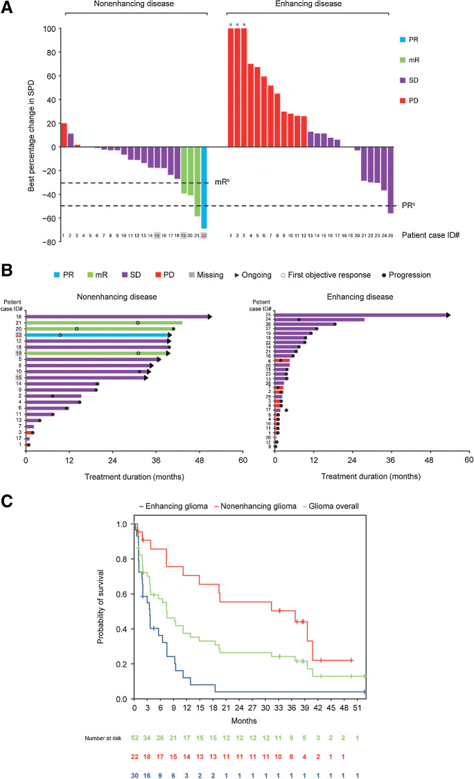
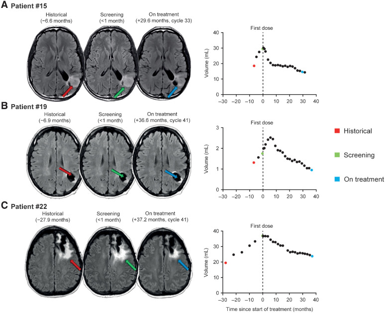
Results: Vorasidenib showed a favorable safety profile in the glioma cohort. Dose-limiting toxicities of elevated transaminases occurred at doses ≥100 mg and were reversible. The protocol-defined objective response rate per Response Assessment in Neuro-Oncology criteria for LGG in patients with nonenhancing glioma was 18% (one partial response, three minor responses). The median progression-free survival was 36.8 months [95% confidence interval (CI), 11.2-40.8] for patients with nonenhancing glioma and 3.6 months (95% CI, 1.8-6.5) for patients with enhancing glioma. Exploratory evaluation of tumor volumes in patients with nonenhancing glioma showed sustained tumor shrinkage in multiple patients.

Conclusions: Vorasidenib was well tolerated and showed preliminary antitumor activity in patients with recurrent or progressive nonenhancing mIDH LGG.


Figures

Figure 1. Clinical activity and efficacy of vorasidenib in patients with glioma. A, Best response in evaluable patients with measurable disease (25 enhancing and 22 nonenhancing) expressed as the percentage change in SPD of target lesions from the start of treatment. Among the 52 patients, 4 patients with enhancing disease had evaluable but nonmeasurable disease, and 1 withdrew from the study before tumor response evaluations. B, left, Treatment duration and best response for patients with nonenhancing glioma; 8 patients remained on treatment. B, right, Treatment duration and best response for patients with enhancing glioma; 1 patient remained on treatment. In A and B, shaded patient case ID numbers (#) written in bold brown font indicate patients with nonenhancing glioma for whom brain MRI images and volumetric growth curves are shown in Fig. 2. C, One patient with nonenhancing disease had a >50% reduction from baseline that was not confirmed with subsequent scan and is therefore categorized as mR. aLesion growth >100%. bAn mR is defined as a ≥25% but ≤50% decrease in tumor measurements relative to baseline. cA >50% decrease in tumor measurements relative to baseline corresponds to a PR. A >50% reduction from baseline was not confirmed with subsequent scan in 1 patient with nonenhancing disease and was therefore categorized as mR. One patient with enhancing disease had a >50% reduction that was not confirmed and was categorized as SD. PD, progressive disease; SD, stable disease; SPD, sum of products of the diameters.
Figure 1.
Clinical activity and efficacy of vorasidenib in patients with glioma. A, Best response in evaluable patients with measurable disease (25 enhancing and 22 nonenhancing) expressed as the percentage change in SPD of target lesions from the start of treatment. Among the 52 patients, 4 patients with enhancing disease had evaluable but nonmeasurable disease, and 1 withdrew from the study before tumor response evaluations. B, left, Treatment duration and best response for patients with nonenhancing glioma; 8 patients remained on treatment. B, right, Treatment duration and best response for patients with enhancing glioma; 1 patient remained on treatment. In A and B, shaded patient case ID numbers (#) written in bold brown font indicate patients with nonenhancing glioma for whom brain MRI images and volumetric growth curves are shown in Fig. 2. C, One patient with nonenhancing disease had a >50% reduction from baseline that was not confirmed with subsequent scan and is therefore categorized as mR. aLesion growth >100%. bAn mR is defined as a ≥25% but ≤50% decrease in tumor measurements relative to baseline. cA >50% decrease in tumor measurements relative to baseline corresponds to a PR. A >50% reduction from baseline was not confirmed with subsequent scan in 1 patient with nonenhancing disease and was therefore categorized as mR. One patient with enhancing disease had a >50% reduction that was not confirmed and was categorized as SD. PD, progressive disease; SD, stable disease; SPD, sum of products of the diameters.
Figure 2. Brain MRI and volume growth curves in 3 patients with nonenhancing glioma treated with vorasidenib. Visual inspection of the images, as well as tumor size and volume measurements, showed tumor shrinkage after vorasidenib treatment. A, Patient #15 is a 47-year-old male with an anaplastic astrocytoma that was initially treated with surgery, radiotherapy, and procarbazine/CCNU/vincristine. Best response as of data cutoff: stable disease. B, Patient #19 is a 40-year-old female with an oligoastrocytoma that was initially treated with surgery and temozolomide. Best response as of data cutoff: mR. C, Patient #22 is a 49-year-old female with an oligodendroglioma that was initially treated with surgery and no other treatment. Best response as of data cutoff: PR. Scan collection time points relative to first dose and corresponding on-treatment cycle numbers are shown.
Figure 2.
Brain MRI and volume growth curves in 3 patients with nonenhancing glioma treated with vorasidenib. Visual inspection of the images, as well as tumor size and volume measurements, showed tumor shrinkage after vorasidenib treatment. A, Patient #15 is a 47-year-old male with an anaplastic astrocytoma that was initially treated with surgery, radiotherapy, and procarbazine/CCNU/vincristine. Best response as of data cutoff: stable disease. B, Patient #19 is a 40-year-old female with an oligoastrocytoma that was initially treated with surgery and temozolomide. Best response as of data cutoff: mR. C, Patient #22 is a 49-year-old female with an oligodendroglioma that was initially treated with surgery and no other treatment. Best response as of data cutoff: PR. Scan collection time points relative to first dose and corresponding on-treatment cycle numbers are shown.

Comment in

  • Clin Cancer Res. 27:4457.

References

    1. GBD 2016 Brain and Other CNS Cancer Collaborators. Global, regional, and national burden of brain and other CNS cancer, 1990–2016: a systematic analysis for the Global Burden of Disease Study 2016. Lancet Neurol 2019;18:376–93. - PMC - PubMed
    1. Louis DN, Perry A, Reifenberger G, von Deimling A, Figarella-Branger D, Cavenee WK, et al. The 2016 World Health Organization Classification of Tumors of the Central Nervous System: a summary. Acta Neuropathol 2016;131:803–20. - PubMed
    1. Cancer Genome Atlas Research Network, Brat DJ, Verhaak RGW, Aldape KD, Yung WKA, Salama SR, Cooper LAD, et al. Comprehensive, integrative genomic analysis of diffuse lower-grade gliomas. N Engl J Med 2015;372:2481–98. - PMC - PubMed
    1. Pallud J, Capelle L, Taillandier L, Fontaine D, Mandonnet E, Guillevin R, et al. Prognostic significance of imaging contrast enhancement for WHO grade II gliomas. Neuro Oncol 2009;11:176–82. - PMC - PubMed
    1. van den Bent MJ, Smits M, Kros JM, Chang SM. Diffuse infiltrating oligodendroglioma and astrocytoma. J Clin Oncol 2017;35:2394–401. - PubMed

Publication types

MeSH terms

Associated data