. 2021 Jun 2;13(596):eabg4535.
doi: 10.1126/scitranslmed.abg4535.

First exposure to the pandemic H1N1 virus induced broadly neutralizing antibodies targeting hemagglutinin head epitopes

Affiliations

First exposure to the pandemic H1N1 virus induced broadly neutralizing antibodies targeting hemagglutinin head epitopes

Jenna J Guthmiller et al. Sci Transl Med. .

Abstract

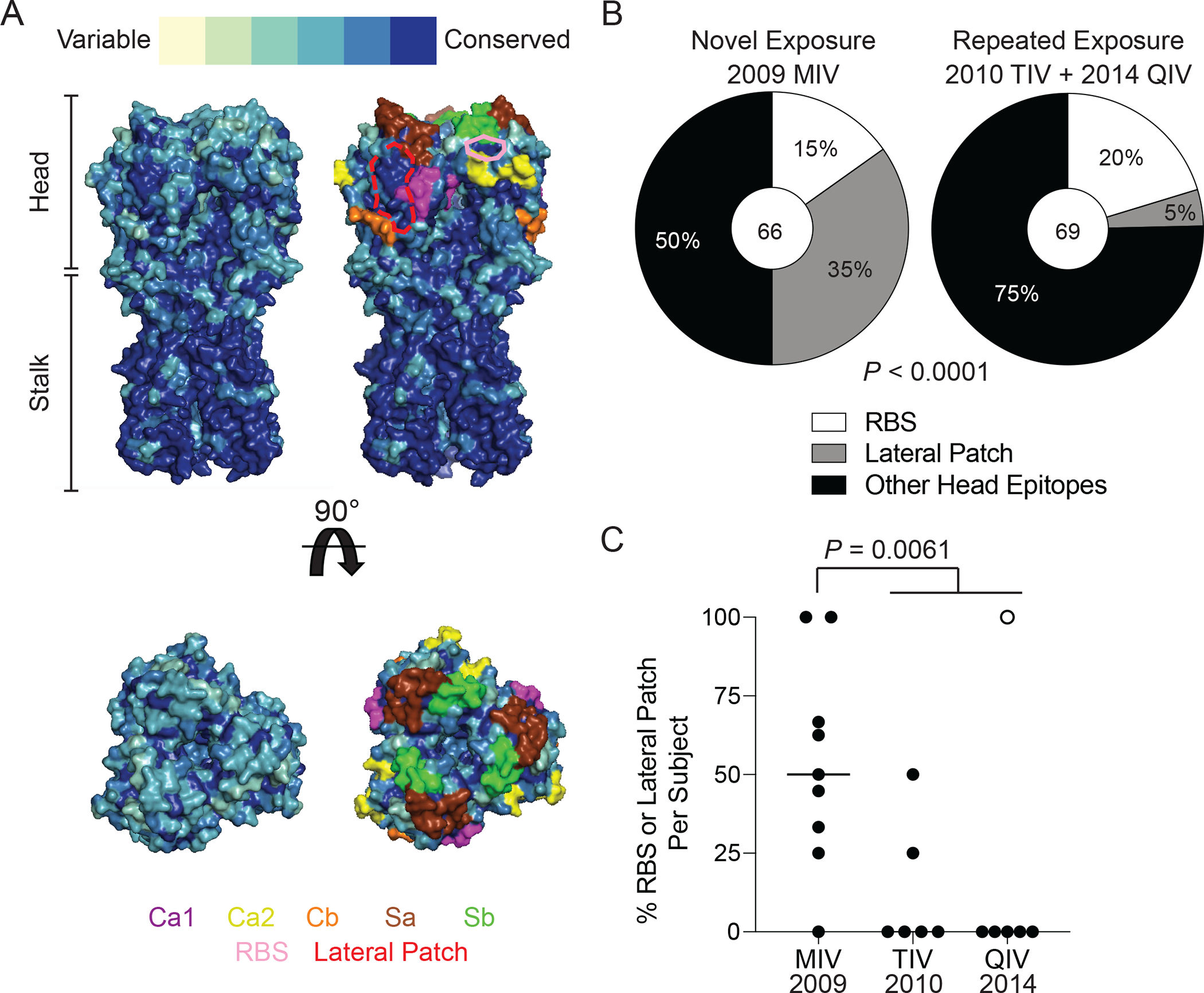
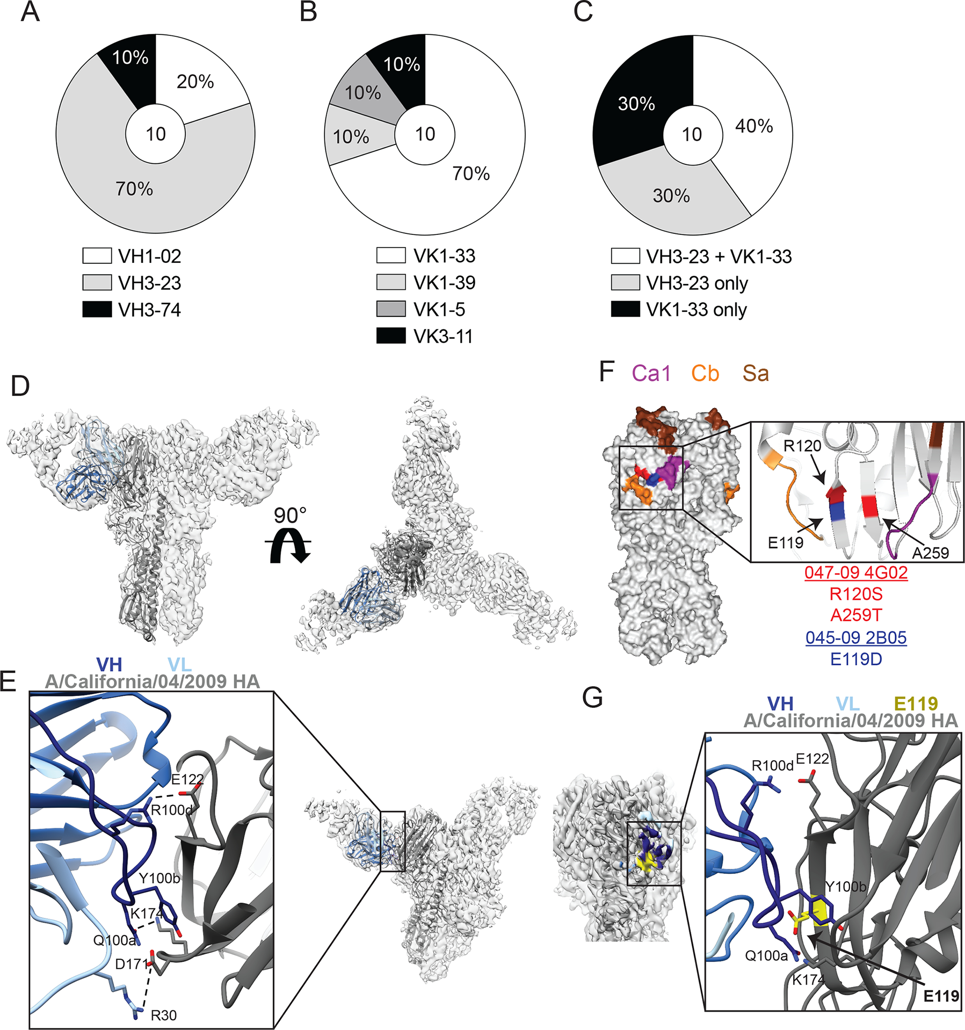
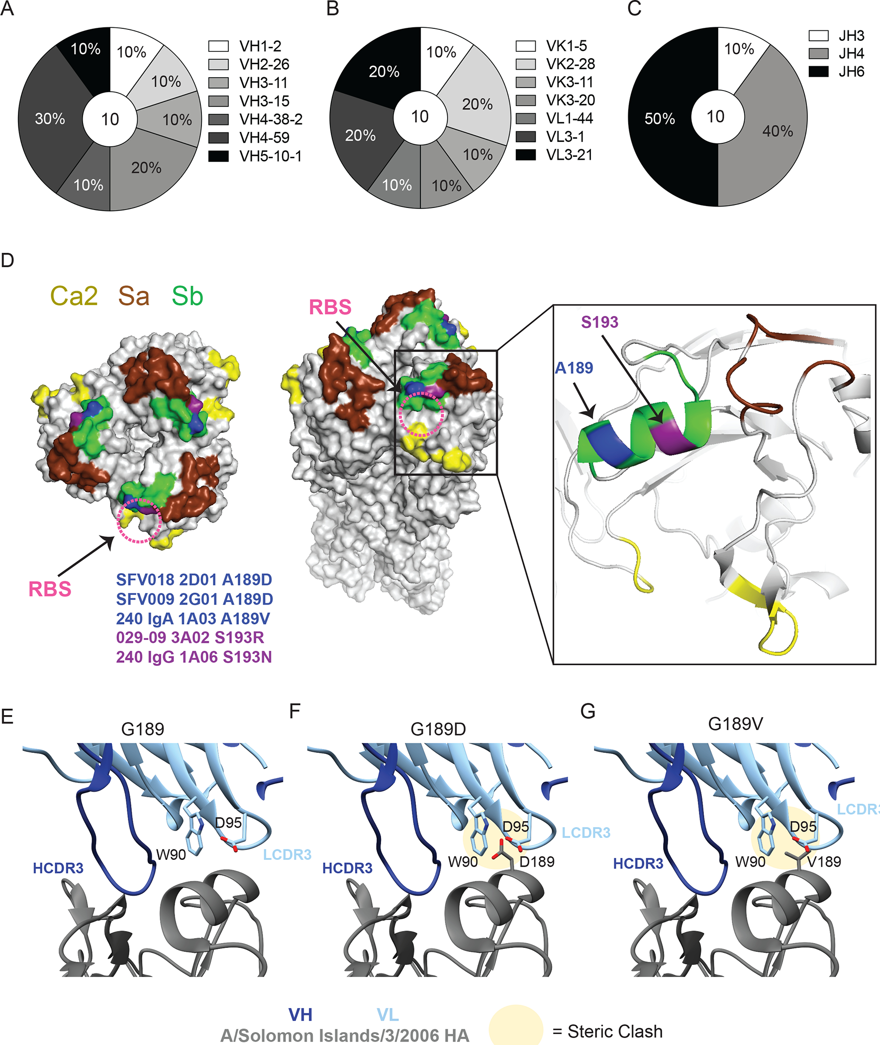
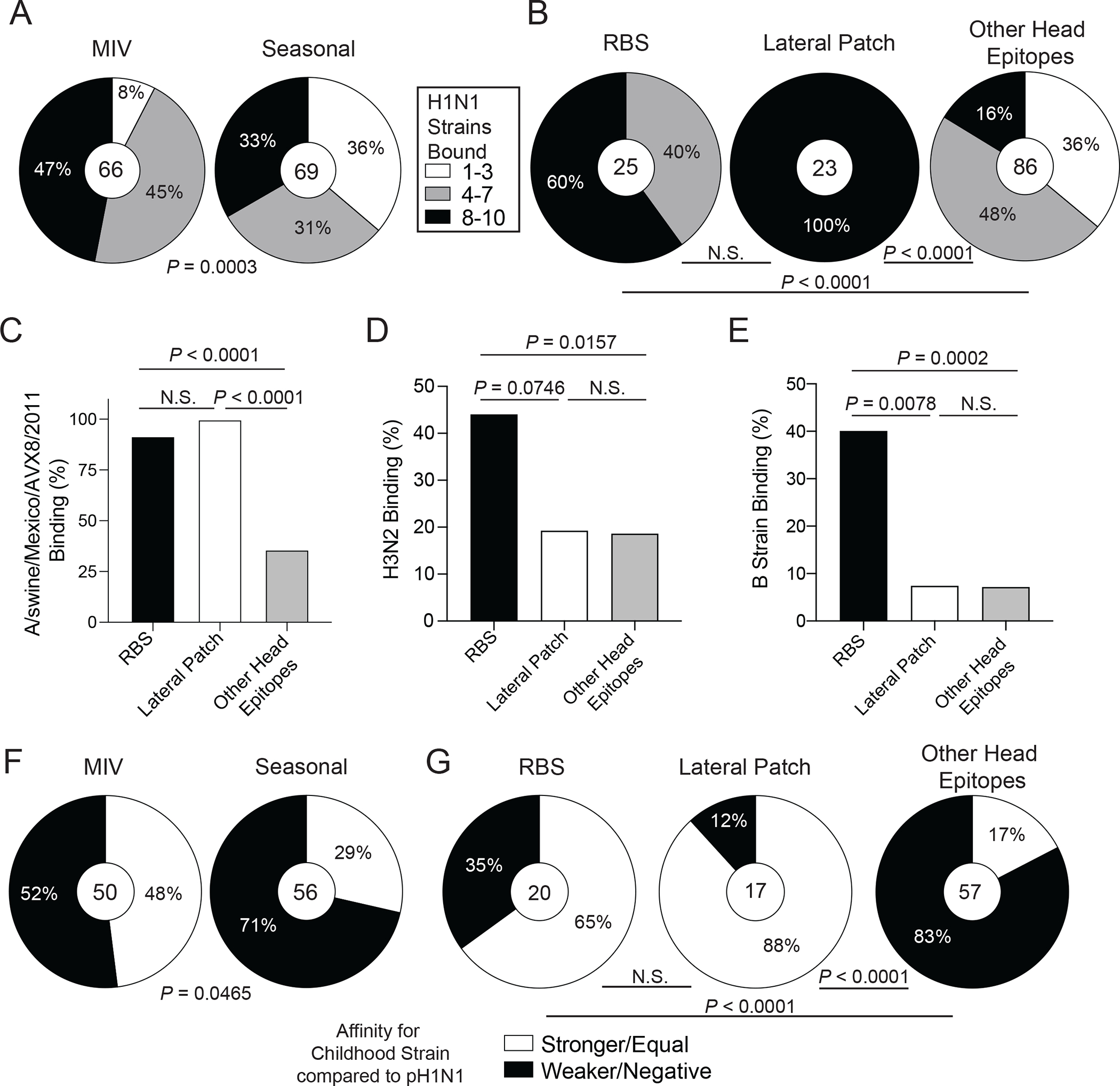
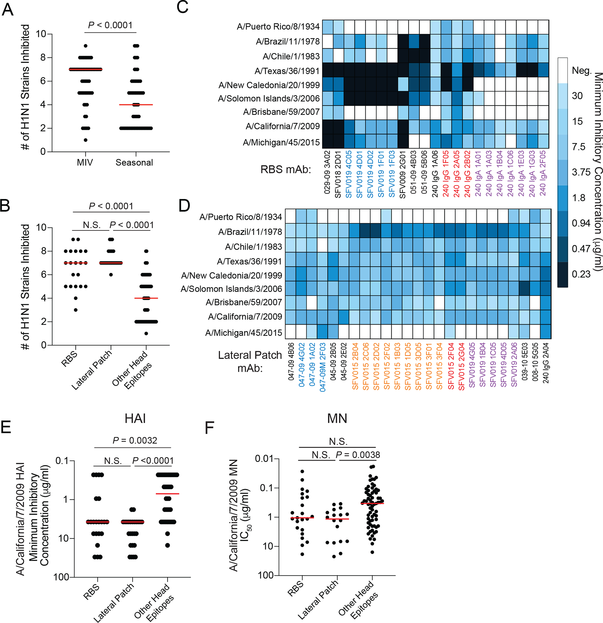
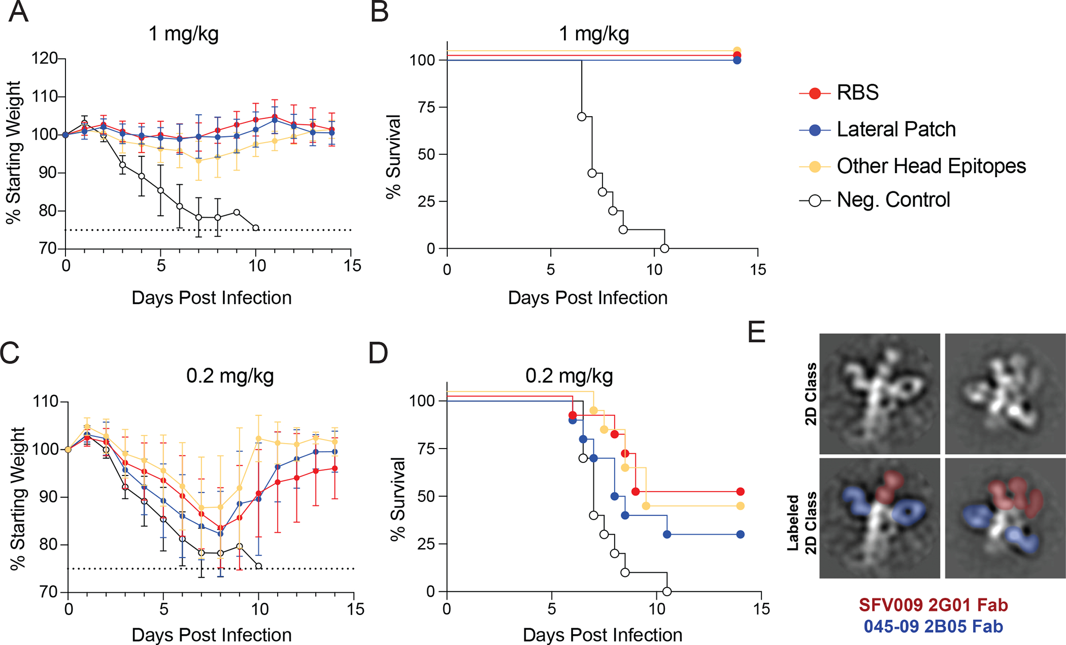
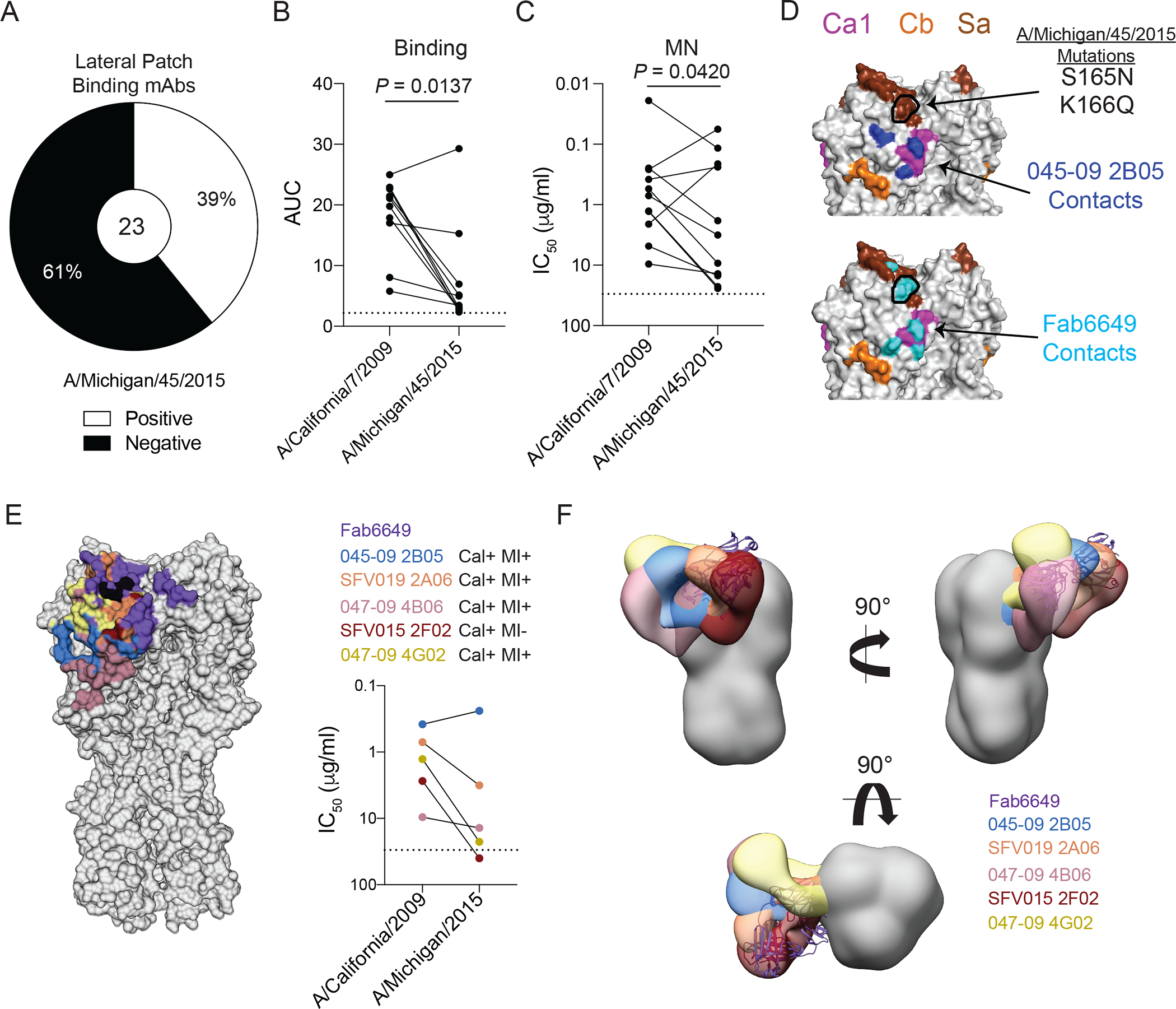
Broadly neutralizing antibodies are critical for protection against both drifted and shifted influenza viruses. Here, we reveal that first exposure to the 2009 pandemic H1N1 influenza virus recalls memory B cells that are specific to the conserved receptor-binding site (RBS) or lateral patch epitopes of the hemagglutinin (HA) head domain. Monoclonal antibodies (mAbs) generated against these epitopes are broadly neutralizing against H1N1 viruses spanning 40 years of viral evolution and provide potent protection in vivo. Lateral patch-targeting antibodies demonstrated near universal binding to H1 viruses, and RBS-binding antibodies commonly cross-reacted with H3N2 viruses and influenza B viruses. Lateral patch-targeting mAbs were restricted to expressing the variable heavy-chain gene VH3-23 with or without the variable kappa-chain gene VK1-33 and often had a Y-x-R motif within the heavy-chain complementarity determining region 3 to make key contacts with HA. Moreover, lateral patch antibodies that used both VH3-23 and VK1-33 maintained neutralizing capability with recent pH1N1 strains that acquired mutations near the lateral patch. RBS-binding mAbs used a diverse repertoire but targeted the RBS epitope similarly and made extensive contacts with the major antigenic site Sb. Together, our data indicate that RBS- and lateral patch-targeting clones are abundant within the human memory B cell pool, and universal vaccine strategies should aim to drive antibodies against both conserved head and stalk epitopes.


Conflict of interest statement

Competing interests: F.K. reports consulting for Curevac, Merck, Pfizer, Seqirus, and Avimex on topics unrelated to this manuscript, and consulting for Dynavax and GlaxoSmithKline on the development of influenza virus vaccines. A.F., F.K., P.P., R.N. are inventors on a patent application (63002682) submitted by the University of Pennsylvania regarding influenza virus vaccines. F.K., P.P., and R.N. are listed as inventors on a patent (20190125859) submitted by the Icahn School of Medicine at Mount Sinai regarding influenza virus vaccines. A.F. and R.N. are currently employed by Moderna, Inc.

Figures

Fig. 1:
Fig. 1:. First exposure to pH1N1 induces antibodies against the RBS and lateral patch epitopes of the HA head.
(A) The conservation of H1 over 102 years of H1N1 evolution with the major antigenic sites highlighted and the RBS epitope outlined in pink and lateral patch epitopes outlined in dashed red line projected onto the structure of A/California/04/2009 (side and top views; PDB:4jtv). (B) The proportion of HAI+ mAbs induced by the 2009 MIV or seasonal influenza virus vaccines (2010 TIV and 2014 QIV) binding the RBS, lateral patch, or other head epitopes. (C) The proportion of RBS- and lateral patch-binding mAbs out of all HAI+ mAbs per individual isolated from the 2009 MIV, 2010 TIV, and 2014 QIV vaccine cohorts. Each symbol represents one participant. The hollow symbol represents participant 240. Statistical significance was determined using Chi-square test (B) and an unpaired two-sided non-parametric Mann-Whitney Test (C). Bar in (C) represents the median. Numbers in center of pie graphs represent number of individual mAbs analyzed.
Fig. 2:
Fig. 2:. Lateral patch-binding antibodies utilize a restricted repertoire.
(A to C) The repertoire of unique B cell clones targeting the lateral patch including VH gene usage (A), VK gene usage (B), and proportion of clones utilizing VH3–23 or VK1–33 (C). Numbers in center of pie graphs represents number of distinct clones analyzed. (D and E) Cryo-EM structure of 045–09 2B05 in complex with A/California/7/2009 HA. Side and top views of 045–09 2B05 binding HA (D) and zoomed in view of paratope:epitope interactions (E). Dashed lines represent contacts between 045–09 2B05 and HA. (F and G) Virus escape mutants for 045–09 2B05 and 047–09 4G02 were generated using A/Netherlands/602/2009 H1N1. (F) Location of mutations (arrows) driven by 045–09 2B05 (blue; E119D) and 047–09 4G02 (red; R120S and A259T) shown on A/California/04/2009 HA (PDB: 4jtv). (G) The location of E119 (arrow) within the 045–09 2B05 and HA paratope:epitope interactions.
Fig. 3:
Fig. 3:. RBS-binding antibodies utilize a diverse repertoire and are susceptible to mutations in Sb.
(A-C) Repertoire of unique B cell clones targeting the RBS including VH gene usage (A), VK or VL gene usage (B), and JH gene usage (C). Numbers in center of pie graphs represents number of distinct clones analyzed. (D) Location of virus escape mutations at A189 (blue; A189D or A189V) and S193 (purple; S193R or S193N) shown on A/California/04/2009 HA (PDB: 4jtv) for SFV018 2D01, SFV009 2G01, 240 IgA 1A03, 029–09 3A02, and 240 IgG 1A06, generated using A/Netherlands/602/2009 H1N1. The RBS is shown with a pink dashed circle. (E to G) CH65 binding to WT G189 (E), and steric clashes between L-CDR3 and HA resulting from G189D (F) and G189V (G) mutations in A/Solomon Islands/3/2006 HA (PDB:5ugy).
Fig. 4:
Fig. 4:. Recalled RBS- and lateral patch-binding B cells are broadly reactive and were likely initially induced during childhood.
(A-B) HAI+ mAbs induced by the 2009 MIV or seasonal vaccines (2010 TIV and 2014 QIV; A) or binding distinct head epitopes (B) were tested for binding to seasonal and pandemic H1N1 strains. Data are shown as percent of mAbs binding to different numbers of strains. (C to E) Proportion of mAbs targeting the RBS, lateral patch, or other head epitopes binding a swine H1N2 virus (C), H3N2 viruses (D), or influenza B viruses (E). (F and G) Percent of MIV- or seasonal vaccine-induced HAI+ mAbs (F) and RBS, lateral patch, and other head epitopes binding mAbs (G) with equal or greater binding affinity to childhood strains relative to A/California/7/2009. Statistical significance was determined using Chi-square tests (A and B) and Fisher’s exact tests (C-G). Numbers in center of pie graphs (A, B, F, and G) represent number of individual mAbs analyzed. All mAbs were tested in duplicate and each ELISA was performed twice.
Fig. 5:
Fig. 5:. Recalled RBS- and lateral patch-binding B cells are broadly neutralizing against H1N1 strains.
(A) Number of H1N1 strains inhibited by HAI+ mAbs induced by the 2009 MIV or seasonal vaccines (2010 TIV and 2014 QIV). (B) Number of H1N1 strains inhibited in an HAI assay by mAbs targeting the RBS, lateral patch, or other head epitopes. (C and D) Heat maps of HAI activity of RBS (C) and lateral patch (D) binding antibodies. MAb names of non-black colors represent mAbs within a clone. (E) HAI potency of mAbs targeting the RBS, lateral patch, or other head epitopes against A/California/7/2009 strain. (F) MN potency of mAbs targeting the RBS, lateral patch, and other head antibodies against A/California/7/2009. Statistical significance was determined by an unpaired two-sided non-parametric Mann-Whitney test (A) and unpaired two-sided non-parametric Kruskal-Wallis test (B, E, and F). Bars (A, B, E, and F) represent the median. Each symbol (A, B, E, and F) represents a distinct mAb. All mAbs were tested in duplicate and each HAI and MN assay was performed twice.
Fig. 6:
Fig. 6:. MAbs targeting the lateral patch and RBS are potently protective in vivo.
(A to D) Epitope mAb cocktails (5 mAbs per cocktail) were administered prophylactically by intraperitoneal injection, and mice were infected 2 hours later with 10 LD50 of A/Netherlands/602/2009 H1N1 virus intranasally. Weight loss (A) and survival (B) were measured for mice treated with 1 mg/kg of mAb cocktails. Weight loss (C) and survival (D) were measured for mice treated with 0.2 mg/kg of mAb cocktails (E) 2D class averages of a RBS clone (SFV009 2G01; red) and lateral patch clone (045–09 2B05; blue) binding the same HA. Data in A and C are the mean ± standard deviation (n=10 mice per group), and data from A-D are pooled from two independent experiments. Dashed lines in A and C represent the humane end point of 25% weight loss.
Fig. 7:
Fig. 7:. VH3–23/VK1–33 lateral patch-binding antibodies can bind and neutralize a natural escape mutant.
(A) Proportion of lateral patch-targeting mAbs binding A/Michigan/45/2015. (B and C) Binding strength as represented as area under the curve (AUC) from virus-specific ELISAs (B) and MN potency (C) of A/Michigan/45/2015 binding lateral patch-targeting mAbs against A/California/7/2009 and A/Michigan/45/2015 viruses. (D) Binding contacts of 045–09 2B05 (blue) and Fab6649 (cyan) relative to the S165N and K166Q mutations (outlined in black) found in A/Michigan/45/2015 projected on A/California/04/2009 (PDB: 4jtv). (E) Footprints of 6 lateral patch-binding antibodies in reference to the S165N and K166Q mutations (black) and their respective MN potencies against A/California/7/2009 and A/Michigan/45/2015 viruses. (F) Side and top view of negative stain EM reconstructions of lateral patch-targeting mAbs binding HA relative to Fab6649 (based on PDB: 5w6g). Statistical significance was determined by a paired two-sided non-parametric Wilcoxon matched-pairs signed rank tests (B-C). Dashed lines in B, C, and E represent the limit of detection (LOD). Each line in B, C, and E connects a single mAb (paired symbols) binding to each virus. All mAbs were tested in duplicate and each ELISA and MN was performed twice.

References

    1. Ekiert DC, Kashyap AK, Steel J, Rubrum A, Bhabha G, Khayat R, Lee JH, Dillon MA, O’Neil RE, Faynboym AM, Horowitz M, Horowitz L, Ward AB, Palese P, Webby R, Lerner RA, Bhatt RR, Wilson IA, Cross-neutralization of influenza A viruses mediated by a single antibody loop. Nature 489, 526–532 (2012); published online EpubSep 27 (10.1038/nature11414). - DOI - PMC - PubMed
    1. Whittle JR, Zhang R, Khurana S, King LR, Manischewitz J, Golding H, Dormitzer PR, Haynes BF, Walter EB, Moody MA, Kepler TB, Liao HX, Harrison SC, Broadly neutralizing human antibody that recognizes the receptor-binding pocket of influenza virus hemagglutinin. Proc Natl Acad Sci U S A 108, 14216–14221 (2011); published online EpubAug 23 (10.1073/pnas.1111497108). - DOI - PMC - PubMed
    1. Raymond DD, Bajic G, Ferdman J, Suphaphiphat P, Settembre EC, Moody MA, Schmidt AG, Harrison SC, Conserved epitope on influenza-virus hemagglutinin head defined by a vaccine-induced antibody. Proc Natl Acad Sci U S A 115, 168–173 (2018); published online EpubJan 2 (10.1073/pnas.1715471115). - DOI - PMC - PubMed
    1. Linderman SL, Chambers BS, Zost SJ, Parkhouse K, Li Y, Herrmann C, Ellebedy AH, Carter DM, Andrews SF, Zheng NY, Huang M, Huang Y, Strauss D, Shaz BH, Hodinka RL, Reyes-Teran G, Ross TM, Wilson PC, Ahmed R, Bloom JD, Hensley SE, Potential antigenic explanation for atypical H1N1 infections among middle-aged adults during the 2013–2014 influenza season. Proc Natl Acad Sci U S A 111, 15798–15803 (2014); published online EpubNov 4 (10.1073/pnas.1409171111). - DOI - PMC - PubMed
    1. Huang KY, Rijal P, Schimanski L, Powell TJ, Lin TY, McCauley JW, Daniels RS, Townsend AR, Focused antibody response to influenza linked to antigenic drift. J Clin Invest 125, 2631–2645 (2015); published online EpubJul 1 (10.1172/JCI81104). - DOI - PMC - PubMed

Publication types