. 2021 Jun 2;12(1):3284.
doi: 10.1038/s41467-021-23591-1.

Through bonds or contacts? Mapping protein vibrational energy transfer using non-canonical amino acids

Affiliations

Through bonds or contacts? Mapping protein vibrational energy transfer using non-canonical amino acids

Erhan Deniz et al. Nat Commun. .

Abstract

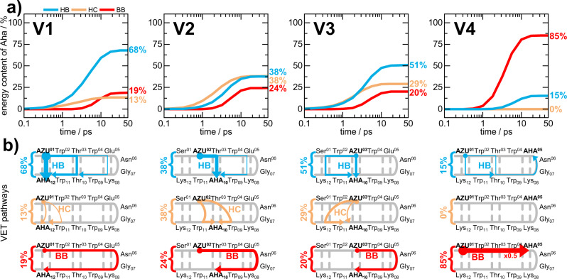
Vibrational energy transfer (VET) is essential for protein function. It is responsible for efficient energy dissipation in reaction sites, and has been linked to pathways of allosteric communication. While it is understood that VET occurs via backbone as well as via non-covalent contacts, little is known about the competition of these two transport channels, which determines the VET pathways. To tackle this problem, we equipped the β-hairpin fold of a tryptophan zipper with pairs of non-canonical amino acids, one serving as a VET injector and one as a VET sensor in a femtosecond pump probe experiment. Accompanying extensive non-equilibrium molecular dynamics simulations combined with a master equation analysis unravel the VET pathways. Our joint experimental/computational endeavor reveals the efficiency of backbone vs. contact transport, showing that even if cutting short backbone stretches of only 3 to 4 amino acids in a protein, hydrogen bonds are the dominant VET pathway.


Conflict of interest statement

The authors declare no competing interests.

Figures

Fig. 1
Fig. 1. Principles of our VET study on TrpZip2.
a Vibrational energy is injected into the system via Azu and propagates along various possible pathways to Aha (gray arrows). b (Left): TRIR spectrum of the TrpZip2 variant above (the spectra of all variants are shown in Supplementary Fig. 1). The spectral VET signature consists of an induced (red) and reduced (blue) absorption, respectively. The shape is due to the shift of the azide band to lower wavenumbers upon arrival of vibrational energy. (Right) transient of the total absorption change of the VET signal. Arrow indicates the peak time. c Time evolution of residue energies of the variant above obtained from non-equilibrium MD simulations (gray), from the master equation model (ME, red) and from the quantum corrected model (QME, blue). Vertical blue lines indicate the peak times according to the QME model. Source data are provided as a Source Data file.
Fig. 2
Fig. 2. Overview of the TrpZip2 variants and their measured and computed VET signals.
a Scheme of our four TrpZip2 variants V1–V4. The systematically varied positions of VET donor-sensor pairs in peptide scaffolds are highlighted with the corresponding color. The shortest pathways along and across strands are marked with colored arrows. b VET transients have the same color coding as the TrpZip2 schemes on the left. Arrows indicate the peak times. The panels on the left display the experimentally measured VET transients, while the right panels show the theoretically determined transients using the rate model. Source data are provided as a Source Data file.
Fig. 3
Fig. 3. Calculated VET pathways from the heater Azu to the sensor Aha in our TrpZip2 variants, obtained from Markov chain Monte Carlo simulations.
a Time-dependent contributions to the energy arriving at Aha on pathways including interstrand H-bonds (HB, blue), heater contacts (HC, orange) and solely backbone (BB, red). Numbers indicate the energy contribution of the corresponding class of pathway in percent at 50 ps. b Scheme of spatially disentangled VET pathways according to Monte Carlo calculations. Color coding as in the top panel. Arrow thickness scales with the contribution of the corresponding pathway. The VET pair is highlighted with bold capitalized letters. For a better comparability the numbers from the top panel indicating the energy contribution of the corresponding class of pathway are shown. Source data underlying Fig. 3a are provided as a Source Data file.

Similar articles

Cited by

References

    1. Rubtsov IV, Burin AL. Ballistic and diffusive vibrational energy transport in molecules. J. Chem. Phys. 2019;150:20901. doi: 10.1063/1.5055670. - DOI - PubMed
    1. Leitner, D. M. & Straub, J. E. eds. Proteins. Energy, Heat and Signal Flow (CRC Press, 2010).
    1. Riedel C, et al. The heat released during catalytic turnover enhances the diffusion of an enzyme. Nature. 2015;517:227–230. doi: 10.1038/nature14043. - DOI - PMC - PubMed
    1. Champion PM. Following the flow of energy in biomolecules. Science. 2005;310:980. doi: 10.1126/science.1120280. - DOI - PMC - PubMed
    1. Davarifar A, Antoniou D, Schwartz SD. The promoting vibration in human heart lactate dehydrogenase is a preferred vibrational channel. J. Phys. Chem. B. 2011;115:15439–15444. doi: 10.1021/jp210347h. - DOI - PMC - PubMed

Publication types