. 2021 Jun 2;12(1):3289.
doi: 10.1038/s41467-021-23492-3.

Outcome after acute ischemic stroke is linked to sex-specific lesion patterns

Anna K Bonkhoff  1 Markus D Schirmer  2   3 Martin Bretzner  2   4 Sungmin Hong  2 Robert W Regenhardt  2 Mikael Brudfors  5 Kathleen L Donahue  2 Marco J Nardin  2 Adrian V Dalca  6   7 Anne-Katrin Giese  8 Mark R Etherton  2 Brandon L Hancock  7 Steven J T Mocking  7 Elissa C McIntosh  7 John Attia  9   10 Oscar R Benavente  11 Stephen Bevan  12 John W Cole  13 Amanda Donatti  14 Christoph J Griessenauer  15   16 Laura Heitsch  17   18 Lukas Holmegaard  19   20 Katarina Jood  19   20 Jordi Jimenez-Conde  21 Steven J Kittner  13 Robin Lemmens  22   23 Christopher R Levi  10   24 Caitrin W McDonough  25 James F Meschia  26 Chia-Ling Phuah  18 Arndt Rolfs  27 Stefan Ropele  28 Jonathan Rosand  2   7   29 Jaume Roquer  21 Tatjana Rundek  30 Ralph L Sacco  30 Reinhold Schmidt  28 Pankaj Sharma  31   32 Agnieszka Slowik  33 Martin Söderholm  34   35 Alessandro Sousa  14 Tara M Stanne  36 Daniel Strbian  37 Turgut Tatlisumak  38   20 Vincent Thijs  39   40 Achala Vagal  41 Johan Wasselius  42   43 Daniel Woo  44 Ramin Zand  45 Patrick F McArdle  46 Bradford B Worrall  47 Christina Jern  36   48 Arne G Lindgren  49   50 Jane Maguire  51 Danilo Bzdok  52   53 Ona Wu  7 MRI-GENIE and GISCOME Investigators and the International Stroke Genetics ConsortiumNatalia S Rost  2
Affiliations

Outcome after acute ischemic stroke is linked to sex-specific lesion patterns

Anna K Bonkhoff et al. Nat Commun. .

Abstract

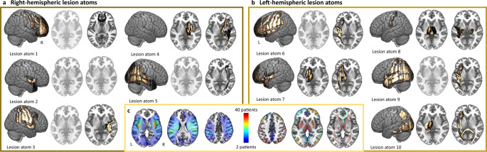
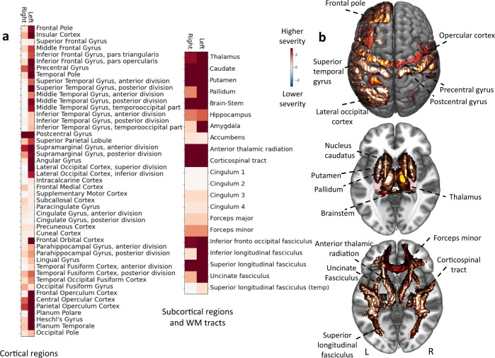
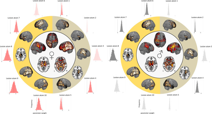
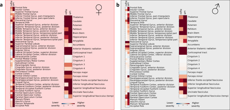
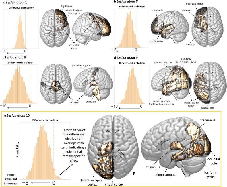
Acute ischemic stroke affects men and women differently. In particular, women are often reported to experience higher acute stroke severity than men. We derived a low-dimensional representation of anatomical stroke lesions and designed a Bayesian hierarchical modeling framework tailored to estimate possible sex differences in lesion patterns linked to acute stroke severity (National Institute of Health Stroke Scale). This framework was developed in 555 patients (38% female). Findings were validated in an independent cohort (n = 503, 41% female). Here, we show brain lesions in regions subserving motor and language functions help explain stroke severity in both men and women, however more widespread lesion patterns are relevant in female patients. Higher stroke severity in women, but not men, is associated with left hemisphere lesions in the vicinity of the posterior circulation. Our results suggest there are sex-specific functional cerebral asymmetries that may be important for future investigations of sex-stratified approaches to management of acute ischemic stroke.


Conflict of interest statement

M.E. has received personal fees for consulting from Astra Zeneca and WorldCare Clinical Group. C.G. has received consulting honoraria from Microvention and Strykere, and research funding from Medtronic and Penumbra. A.V. has received research funding from Cerenovus. A.G.L. has received personal fees from Bayer, Astra Zeneca, BMS Pfizer, and Portola. N.S.R. has received compensation as scientific advisory consultant from Omniox, Sanofi Genzyme, and AbbVie Inc. All other authors declare no competing interests.

Figures

Fig. 1
Fig. 1. Archetypical stroke patterns, lesion atoms, as resulting from non-negative matrix factorization-based dimensionality reduction.
A data-driven pattern discovery framework enabled the derivation of coherent patterns of stroke lesion topographies directly from the segmented high-resolution brain scans from 555 stroke patients. This unsupervised, multi-to-multi mapping approach led to unique, predominantly right-hemispheric (a) or left-hemispheric (b) lesion patterns. In case of either one hemisphere, individual lesion atoms represented anatomically coherent cortical and subcortical brain regions and respective white matter tracts, and had varying emphases on more anterior, medial, and posterior regions. While subcortical basal ganglia lesions and cortical lesions in anterior and insular regions were combined in a single lesion atom on the left side of the brain, these patterns were characterized by two separate lesion atoms on the right side of the brain. Lesion atom 8 comprised several brain regions in the left hemisphere; however, also comprised the brainstem and, to a lesser degree, the right thalamus. This pattern likely arose since we did not exclude patients with bilateral stroke (n = 21, 38% female). Some lesion atoms did not comprise any substantial contributions from subcortical brain regions and are shown in transparent. The anatomical plausibility of our derived lesion pattern may particularly stem from the positivity constraint of the non-negative matrix factorization algorithm, an advantageous quality that motivated our choice of dimensionality reduction technique. Conversely, alternative matrix factorization algorithms, such as principal component analysis, would have hampered a straightforward interpretation of lesion pattern effects by encoding individual lesions through more incomprehensible additions and subtractions of low-dimensional lesion pattern. c Similarity of lesion patterns across patients. A voxel-wise lesion overlap is visualized on the left-hand side, while the right-hand side presents region-wise frequencies, i.e., the number of how often a specific region was affected. The maximum lesion overlap was localized subcortically and in the proximity of insular regions in the left and right vascular supply territory of the middle cerebral artery. Significant region-wise differences in lesion loads and frequencies did neither arise between men and women nor between left and right hemispheres (Supplementary Data 1 and 2). Source data are provided as a Source data file.
Fig. 2
Fig. 2. Local brain regions explaining NIHSS-based stroke severity across 555 patients.
a Relevant cortical and subcortical gray matter regions, as well as relevant white matter (WM) tracts. Shows collection of marginal Bayesian posterior distributions from the hierarchical model to explain high vs. low symptom severity (NIHSS). Lesions affecting pre- and postcentral gyri, as well as opercular regions of both hemispheres explained a higher stroke severity. Further brain-behavior effects were left-lateralized: multiple regions, including left middle and inferior frontal gyrus, as well as superior and middle temporal explained a higher stroke severity only when affecting the left hemisphere. While bilateral subcortical regions, in general, had substantial effects on stroke severity, the highest weights were assigned to the putamen and caudate, as well as anterior thalamic radiation, corticospinal tract, and inferior fronto-occipital fasciculus of the left hemispheres. b Brain renderings of region-wise relevances in the explanation of NIHSS-based stroke severity. Source data are provided as a Source data file.
Fig. 3
Fig. 3. Sex-specific Bayesian posterior distributions of all ten lesion atoms and overall whole-brain region-wise relevance to explain stroke severity in women (left) and men (right).
Our Bayesian framework was purpose-designed to enable fully probabilistic estimations of the parameters that quantify the associations of the ten lesion atoms with stroke severity. These Bayesian posterior parameter distributions are shown in outer circles, corresponding lesion atom renderings are presented in the subjacent circle (right-hemispheric lesion atoms: shaded in yellow-olive, left-hemispheric lesion atoms: orange-yellow; distributions that substantially diverged from zero are nontransparent). Lesion atoms 7 and 8 of the left hemisphere and lesion atom 3 of the right hemisphere had the highest weights, implying a high relevance in explaining stroke severity, in both men and women. In view of seven relevant lesion atoms in women (lesion atoms: 1, 4, 5, 7–10), yet only five of such relevant lesion atoms in men (lesion atoms: 2, 4, 6–8), lesion patterns were more widespread in women compared to men. This female-specific more widespread pattern becomes additionally apparent in whole-brain renderings of region-wise relevances explaining stroke severity, as visualized in circle centers (c.f., Fig. 4 for details). Source data are provided as a Source data file.
Fig. 4
Fig. 4. Local brain regions explaining stroke severity.
a Female-specific results (208 women) and b male-specific results (347 men). In both men and women, subcortical lesions affecting gray matter regions and white matter tracts explained higher stroke severity. Similarly, cortical presumptive bilateral motor and left-lateralized language regions (e.g., especially bilateral precentral and postcentral gyri, left-sided inferior frontal and superior, middle temporal gyri) also explained higher stroke severity. In difference to men, women featured more widespread and also more pronounced lesion pattern, including a greater range of cortical regions contributing to stroke severity, e.g., the left superior, middle and inferior temporal gyrus, left angular gyrus and lateral occipital cortex, lingual gyrus, as well as precuneus and parahippocampal cortex gyrus. These differences in eloquent lesion patterns arose despite comparable total lesion volumes for men and women (two-sided t test: p > 0.05). Source data are provided as a Source data file.
Fig. 5
Fig. 5. Five lesion atoms showed substantially higher relevance for explaining stroke-induced symptom severity in women than men.
a Lesion atom 1. Less than 5% of samples of the difference Bayesian posterior distribution between men and women (i.e., posterior distribution of lesion atom 1 in men—posterior distribution of lesion atom 1 in women) overlapped with zero, suggesting a substantially larger lesion atom 1 effect in women. This specific lesion pattern represented right-hemispheric lesions in frontal, insular, and precentral regions (difference distribution: mean = −1.2, HPDI = −1.96 to −0.31). bd Lesion atoms 7–9. Almost all lesion atoms of the left hemisphere, but the one representing precentral cortex and middle and inferior frontal gyrus regions, indicated more pronounced effects on stroke severity in women. Lesion atom 7 highlighted left-sided insular cortex and subcortical regions (difference distribution: mean = −1.61, HPDI = −3.12 to −0.02). Lesion atom 8 comprised brainstem lesions, as well bilateral, left-hemispherically more distinct thalamus lesions (difference distribution: mean = −4.67, HPDI = −8.55 to −1.08) and lesion atom 9 widespread left-sided cortical lesions relating to the superior, middle and inferior temporal gyri, postcentral cortex, angular and supramarginal gyrus, and latero-occipital cortex (difference distribution: mean = −3.42, HPDI = −6.15 to −0.63). e Lesion atom 10. Lesions in left-sided brain regions of the presumed posterior circulation were associated to more severe strokes specifically in women (difference distribution: mean = −1.99, HPDI = −3.90 to −0.22). The sex difference for lesion atom 10 can be considered the most pronounced and robust one, given it was reliably observable in all ancillary analyses and was replicated in an independent dataset of stroke patients. All analyses were corrected for overall sex effects, i.e., lesion atom-independent effects (e.g., due to potential female-specific delayed hospital arrival). Of note, this correction was independent of the explicit knowledge or measurement of the causes of these global sex differences. Source data are provided as a Source data file.

Similar articles

  • Association of Infarct Topography and Outcome After Endovascular Thrombectomy in Patients With Acute Ischemic Stroke.
    Regenhardt RW, Bonkhoff AK, Bretzner M, Etherton MR, Das AS, Hong S, Alotaibi NM, Vranic JE, Dmytriw AA, Stapleton CJ, Patel AB, Leslie-Mazwi TM, Rost NS. Regenhardt RW, et al. Neurology. 2022 Mar 15;98(11):e1094-e1103. doi: 10.1212/WNL.0000000000200034. Epub 2022 Jan 31. Neurology. 2022. PMID: 35101908 Free PMC article.
  • Association of Stroke Lesion Pattern and White Matter Hyperintensity Burden With Stroke Severity and Outcome.
    Bonkhoff AK, Hong S, Bretzner M, Schirmer MD, Regenhardt RW, Arsava EM, Donahue K, Nardin M, Dalca A, Giese AK, Etherton MR, Hancock BL, Mocking SJT, McIntosh E, Attia J, Benavente O, Cole JW, Donatti A, Griessenauer C, Heitsch L, Holmegaard L, Jood K, Jimenez-Conde J, Kittner S, Lemmens R, Levi C, McDonough CW, Meschia J, Phuah CL, Rolfs A, Ropele S, Rosand J, Roquer J, Rundek T, Sacco RL, Schmidt R, Sharma P, Slowik A, Soederholm M, Sousa A, Stanne TM, Strbian D, Tatlisumak T, Thijs V, Vagal A, Wasselius J, Woo D, Zand R, McArdle P, Worrall BB, Jern C, Lindgren AG, Maguire J, Golland P, Bzdok D, Wu O, Rost NS. Bonkhoff AK, et al. Neurology. 2022 Sep 27;99(13):e1364-e1379. doi: 10.1212/WNL.0000000000200926. Epub 2022 Jul 8. Neurology. 2022. PMID: 35803717 Free PMC article.
  • The relevance of rich club regions for functional outcome post-stroke is enhanced in women.
    Bonkhoff AK, Schirmer MD, Bretzner M, Hong S, Regenhardt RW, Donahue KL, Nardin MJ, Dalca AV, Giese AK, Etherton MR, Hancock BL, Mocking SJT, McIntosh EC, Attia J, Cole JW, Donatti A, Griessenauer CJ, Heitsch L, Holmegaard L, Jood K, Jimenez-Conde J, Kittner SJ, Lemmens R, Levi CR, McDonough CW, Meschia JF, Phuah CL, Ropele S, Rosand J, Roquer J, Rundek T, Sacco RL, Schmidt R, Sharma P, Slowik A, Sousa A, Stanne TM, Strbian D, Tatlisumak T, Thijs V, Vagal A, Wasselius J, Woo D, Zand R, McArdle PF, Worrall BB, Jern C, Lindgren AG, Maguire J, Wu O, Rost NS; MRI-GENIE and GISCOME Investigators and the International Stroke Genetics Consortium. Bonkhoff AK, et al. Hum Brain Mapp. 2023 Mar;44(4):1579-1592. doi: 10.1002/hbm.26159. Epub 2022 Nov 28. Hum Brain Mapp. 2023. PMID: 36440953 Free PMC article. Clinical Trial.
  • Effect of Sex on Clinical Outcome and Imaging after Endovascular Treatment of Large-Vessel Ischemic Stroke.
    Demeestere J, Christensen S, Mlynash M, Federau C, Albers GW, Lemmens R, Lansberg MG. Demeestere J, et al. J Stroke Cerebrovasc Dis. 2021 Feb;30(2):105468. doi: 10.1016/j.jstrokecerebrovasdis.2020.105468. Epub 2020 Nov 21. J Stroke Cerebrovasc Dis. 2021. PMID: 33227604 Free PMC article.
  • Abnormal dynamic functional connectivity is linked to recovery after acute ischemic stroke.
    Bonkhoff AK, Schirmer MD, Bretzner M, Etherton M, Donahue K, Tuozzo C, Nardin M, Giese AK, Wu O, D Calhoun V, Grefkes C, Rost NS. Bonkhoff AK, et al. Hum Brain Mapp. 2021 May;42(7):2278-2291. doi: 10.1002/hbm.25366. Epub 2021 Mar 2. Hum Brain Mapp. 2021. PMID: 33650754 Free PMC article.

Cited by

References

    1. Feigin VL, et al. Global and regional burden of stroke during 1990–2010: findings from the Global Burden of Disease Study 2010. Lancet. 2014;383:245–255. doi: 10.1016/S0140-6736(13)61953-4. - DOI - PMC - PubMed
    1. Kelly-Hayes M, et al. The influence of gender and age on disability following ischemic stroke: the Framingham study. J. Stroke Cerebrovasc. Dis. 2003;12:119–126. doi: 10.1016/S1052-3057(03)00042-9. - DOI - PubMed
    1. Benjamin EJ, et al. Heart disease and stroke statistics-2017 update: a report from the American Heart Association. Circulation. 2017;135:e146–e603. doi: 10.1161/CIR.0000000000000485. - DOI - PMC - PubMed
    1. Bushnell C, et al. Sex differences in the evaluation and treatment of acute ischaemic stroke. Lancet Neurol. 2018;17:641–650. doi: 10.1016/S1474-4422(18)30201-1. - DOI - PubMed
    1. Carcel C, Woodward M, Wang X, Bushnell C, Sandset EC. Sex matters in stroke: a review of recent evidence on the differences between women and men. Front. Neuroendocrinol. 2020;59:100870. doi: 10.1016/j.yfrne.2020.100870. - DOI - PubMed

Publication types

MeSH terms