. 2021 Jun 2;12(1):3292.
doi: 10.1038/s41467-021-23599-7.

NEK9 regulates primary cilia formation by acting as a selective autophagy adaptor for MYH9/myosin IIA

Affiliations

NEK9 regulates primary cilia formation by acting as a selective autophagy adaptor for MYH9/myosin IIA

Yasuhiro Yamamoto et al. Nat Commun. .

Abstract

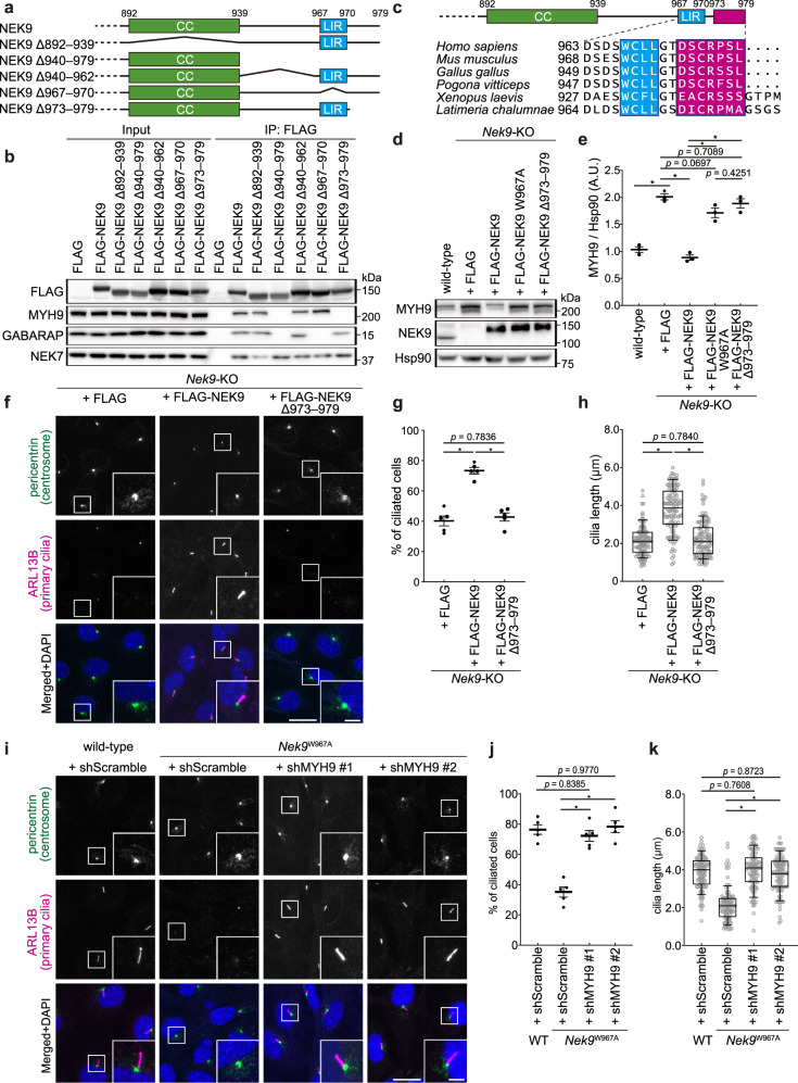
Autophagy regulates primary cilia formation, but the underlying mechanism is not fully understood. In this study, we identify NIMA-related kinase 9 (NEK9) as a GABARAPs-interacting protein and find that NEK9 and its LC3-interacting region (LIR) are required for primary cilia formation. Mutation in the LIR of NEK9 in mice also impairs in vivo cilia formation in the kidneys. Mechanistically, NEK9 interacts with MYH9 (also known as myosin IIA), which has been implicated in inhibiting ciliogenesis through stabilization of the actin network. MYH9 accumulates in NEK9 LIR mutant cells and mice, and depletion of MYH9 restores ciliogenesis in NEK9 LIR mutant cells. These results suggest that NEK9 regulates ciliogenesis by acting as an autophagy adaptor for MYH9. Given that the LIR in NEK9 is conserved only in land vertebrates, the acquisition of the autophagic regulation of the NEK9-MYH9 axis in ciliogenesis may have possible adaptive implications for terrestrial life.


Conflict of interest statement

The authors declare no competing interests.

Figures

Fig. 1
Fig. 1. Differential interactome screen identified NEK9 as a GABARAP-interacting protein.
a Scheme of the differential interactome screen to identify substrates or adaptors of selective autophagy using GABARAPL1 and its LIR docking site mutant GABARAPL1Y49A/L50A. b Results of the differential interactome screen. Four independent immunoprecipitation and mass spectrometry (MS) analyses were conducted. The number of times each protein was detected is shown as #GABARAPL1-IP or #GABARAPL1Y49A/L50A-IP. The x- and y-axes represent GABARAPL1 binding intensity and the #GABARAPL1-IP / (#GABARAPL1-IP + #GABARAPL1 Y49A/L50A -IP) ratio, respectively. The area defined by x < 10 or y < 0.5 is colored gray. See also Supplementary Data 1. c Structures of Homo sapiens and Danio rerio NEK9 and a multiple sequence alignment of NEK9 proteins in vertebrates. Identical and similar residues are colored in red and yellow, respectively. LIR was predicted by iLIR search. KD, kinase-domain; RCC1, RCC1-repeats; CC, coiled-coil. d Immunoprecipitation of FLAG-ATG8s in HEK293T cells. e Co-immunoprecipitation of FLAG-GABARAP and wild-type or mutant GFP-NEK9 in HEK293T cells. In GFP-NEK9 LIR4A, the LIR residues (WCLL) were substituted by four alanines. Data are representative of three independent experiments in (d) and (e).
Fig. 2
Fig. 2. NEK9 is degraded by selective autophagy.
a Immunofluorescence microscopy of MEFs expressing GFP-NEK9 and mRuby3-GABARAP under nutrient-rich conditions and amino acid and serum starvation (2 h) conditions with or without 100 nM bafilomycin A1 (baf A1). b Quantification of the number of NEK9 puncta in (a); p values correspond to a Tukey’s multiple comparisons test. c Immunofluorescence microscopy of wild-type and Fip200-KO MEFs expressing GFP-NEK9 (top), and wild-type MEFs expressing GFP-NEK9 W967A (LIR-mutant) (bottom) after starvation (2 h). d, e Quantification of the number of NEK9 puncta in (c); p values correspond to two-tailed Mann–Whitney tests. f Wild-type and Fip200-KO MEFs were incubated under starvation conditions with or without 100 nM bafilomycin A1 for the indicated time. Whole-cell lysates were subjected to immunoblotting. g Quantification of the intensity of the NEK9 bands in (f). Data represent the mean ± SEM values of three independent experiments. h Immunoblotting of indicated organs of three-month-old Atg5+/+ (WT) and Atg5−/−;NSE-Atg5 (KO) mice. Data are representative of three biologically independent replicates. For (b, d, and e), data were collected from 100 cells for each condition. Solid bars indicate the medians, boxes the interquartile range (25th to 75th percentile), and whiskers the 10th to 90th percentile. Scale bars, 10 µm and 3 µm (insets).
Fig. 3
Fig. 3. Selective autophagy of NEK9 is required for primary cilia formation.
a Generation of homozygous Nek9W967A (LIR-mutant) cell lines by CRISPR-mediated recombination using a donor plasmid harboring short homology arms. b Immunoblotting of wild-type or Nek9W967A MEFs (two independent clones, #7 and #13) cultured in nutrient-rich medium. c, Quantification of the intensity of the NEK9 bands in (b). Data represent the mean ± SEM of three independent experiments. d Immunofluorescence microscopy of wild-type or Nek9W967A MEFs after serum starvation (24 h). Centrosomes and primary cilia were stained with anti-pericentrin and anti-ARL13B antibodies, respectively. e The frequency of ciliated cells in (d). f Quantification of cilia length in (d). Data were collected from 100 ciliated cells for each cell-type. g Immunofluorescence microscopy of wild-type or Fip200-KO MEFs after serum starvation (24 h). h The frequency of ciliated cells in (g), as in (e). i Quantification of cilia length in (g), as in (f). Data were collected from 100 ciliated cells for each cell-type. j Immunofluorescence microscopy of wild-type or Nek9-KO MEFs stably expressing the indicated constructs. k The frequency of ciliated cells in (j), as in (e). l Quantification of cilia length in (j), as in (f). Data were collected from 100 ciliated cells for each cell-type. p values correspond to Tukey’s multiple comparisons tests in (c, e, f, k, and l) and two-tailed Mann–Whitney tests in (h) and (i); *p < 0.0001. Scale bars, 10 µm and 3 µm (insets). Data represent the mean ± SEM of five independent experiments (300 cells were counted in each experiment) in (e, h, k). Solid bars indicate the medians, boxes the interquartile range (25th–75th percentile), and whiskers the 10th–90th percentile in (f, i, and l).
Fig. 4
Fig. 4. Selective autophagy of NEK9 is required for primary cilia formation in mouse kidneys.
a Immunoblotting of the indicated organs of five-month-old wild-type (WT) and Nek9W967A/W967A mice (KI). Data are representative of three biologically independent replicates. b Immunohistochemistry of the cortical region of kidneys from five-month-old wild-type and Nek9W967A/W967A mice using LTL-FITC (the lumen of proximal-tubular cells) and anti-ARL13B antibody (primary cilia). Scale bars, 40 µm and 5 µm (insets). c Frequency of ciliated cells in LTL-FITC positive cells in (b). Data represent the mean ± SEM of three mice (300 cells were counted in each experiment). d Quantification of cilia length in LTL-FITC positive cells in (b). Data were collected from 100 ciliated cells for each genotype. Solid bars indicate the medians, boxes the interquartile range (25th–75th percentile), and whiskers the 10th–90th percentile. e Hematoxylin and eosin staining of the cortical region of kidneys from five-month-old wild-type and Nek9W967A/W967A mice. Scale bars, 100 µm and 10 µm (insets). f Measurement of the surface area of tubular cells in (e). Examples of measured areas are shown with broken lines in (e). Data represent the mean ± SEM of five mice (300 cells were counted in each experiment); p values correspond to two-tailed Mann–Whitney tests in (c, d, and f).
Fig. 5
Fig. 5. NEK9 is a selective autophagy adaptor for MYH9.
a Results of mass spectrometry analysis of FLAG-NEK9 or FLAG immunoprecipitates. The x- and y-axes represent Peptide Spectrum Match (PSM) and abundance ratio (FLAG-NEK9 / FLAG), respectively. Proteins with an abundance ratio above five were tested for actual interaction with NEK9. Proteins above the dotted line were detected only in FLAG-NEK9 immunoprecipitates. See also Supplementary Data 2. b Immunoprecipitation using MEFs stably expressing FLAG or FLAG-NEK9 after serum starvation (4 h). Data are representative of three independent experiments. c Immunofluorescence microscopy of wild-type and Nek9W967A MEFs stably expressing GFP-MYH9 after serum-starvation (4 h). Cells were stained with anti-NEK9 and anti-LC3 antibodies. Scale bars, 10 µm and 3 µm (insets). d Quantification of the number of GFP-MYH9 puncta in (c). e Colocalization between GFP-MYH9 and endogenous LC3 in (c) was determined by calculating Pearson’s correlation coefficient between intensities within each cell. Data were collected from 100 cells for each cell-type in (d, e). Solid bars indicate the medians, boxes the interquartile range (25th–75th percentile), and whiskers the 10th to 90th percentile. f Immunoblotting of wild-type and Nek9W967A MEFs. g Quantification of the intensity of the MYH9 bands in (f). Data represent the mean ± SEM of three independent experiments. h Immunoblotting of the indicated organs of five-month-old wild-type (WT) and Nek9W967A/W967A mice (KI). Asterisks (*) indicate non-specific bands in skeletal muscles. Data are representative of three biologically independent replicates; p values correspond to Tukey’s multiple comparisons tests.
Fig. 6
Fig. 6. NEK9-mediated selective autophagy of MYH9 is required for primary cilia formation.
a Schematic representation of the C-terminal regions of Homo sapiens NEK9 and deletion mutants. CC, coiled-coil. b Immunoprecipitation using MEFs stably expressing wild-type or deletion mutation NEK9 constructs after serum starvation (4 h). Data are representative of three independent experiments. c The C-terminal region of Homo sapiens NEK9. The putative MYH9-binding region is colored in magenta (top). Multiple sequence alignment of NEK9 from terrestrial vertebrates (bottom). See also Fig. 1c. d Immunoblotting of wild-type or Nek9-KO MEFs stably expressing indicated constructs. e, Quantification of the intensity of the MYH9 bands in (d). Data represent the mean ± SEM of three independent experiments. f Immunofluorescence microscopy of Nek9-KO MEFs stably expressing indicated constructs after serum starvation (24 h). g Frequency of ciliated cells in f. Data represent the mean ± SEM of five independent experiments (300 cells were counted in each experiment). h Quantification of cilia length in (f). Data were collected from 100 ciliated cells for each cell-type. i Immunofluorescence microscopy of Nek9W967A MEFs in which MYH9 was depleted by two independent shRNAs (#1 and #2). See Supplementary Fig. 6e, f for the knockdown efficiency of MYH9 in these cells. j Frequency of ciliated cells in (i), as in (g). Data represent the mean ± SEM of five independent experiments (300 cells were counted in each experiment). k Quantification of cilia length in (i), as in (h). Data were collected from 100 ciliated cells for each cell-type; p values correspond to a Tukey’s multiple comparisons test; *p < 0.0001. Scale bars, 10 µm and 3 µm (insets). Solid bars indicate the medians, boxes the interquartile range (25th–75th percentile), and whiskers the 10th–90th percentile in (h, k).
Fig. 7
Fig. 7. Selective autophagy of MYH9 promotes ciliogenesis by increasing actin dynamics.
FRAP analysis of GFP-actin in wild-type or Nek9W967A MEFs after serum starvation (24 h). MYH9 was depleted by shRNA-mediated knockdown (#1). Images were recorded at 5-s intervals following photobleaching of the indicated area, and fluorescence recovery at different time points was quantified. Data represent the mean ± SEM of 10 cells. Similar results were obtained using shMYH9 #2 (not shown). See Supplementary Fig. 6e, f for the knockdown efficiency. Scale bars, 10 µm and 3 µm (insets).
Fig. 8
Fig. 8. Autophagic degradation of NEK9–MYH9 and OFD1 is required for primary cilia formation.
a FRAP analysis of GFP-actin in wild-type MEFs stably expressing FLAG or FLAG-OFD1 after serum starvation (24 h). Images were recorded at 5-s intervals following photobleaching of the indicated area, and fluorescence recovery at different time points was quantified. Data represent the mean ± SEM of 10 cells. b Immunofluorescence microscopy of wild-type or Nek9W967A MEFs after serum starvation (24 h). OFD1 or MYH9 was depleted by shRNA-mediated knockdown (shMYH9 #1 was used). Similar results were obtained using shMYH9 #2 and two independent shOFD1 (not shown). See Supplementary Fig. 8b–d for the knockdown efficiency. c Percentage of cells with centriolar satellites OFD1 in (b). Data represent the mean ± SEM of three independent experiments (100 cells were counted in each experiment). d Frequency of ciliated cells in (b). Data represent the mean ± SEM of five independent experiments (300 cells were counted in each experiment). e Quantification of cilia length in (b). Data were collected from 100 ciliated cells for each cell-type. Solid bars indicate the medians, boxes the interquartile range (25th–75th percentile), and whiskers the 10th–90th percentile. f Immunofluorescence microscopy of wild-type or Fip200-KO MEFs after serum starvation (24 h). OFD1 and/or MYH9 were depleted by shRNA-mediated knockdown. See Supplementary Fig. 8e–g for the knockdown efficiency. g Frequency of ciliated cells in (f), as in (d). h Quantification of cilia length in (f), as in (e); p values correspond to a Tukey’s multiple comparisons test; *p < 0.0001. Scale bars, 10 µm and 3 µm (insets). i Model of how autophagy drives primary cilia formation.

Similar articles

Cited by

References

    1. Malicki JJ, Johnson CA. The cilium: cellular antenna and central processing unit. Trends Cell Biol. 2017;27:126–140. doi: 10.1016/j.tcb.2016.08.002. - DOI - PMC - PubMed
    1. Anvarian Z, Mykytyn K, Mukhopadhyay S, Pedersen LB, Christensen ST. Cellular signalling by primary cilia in development, organ function and disease. Nat. Rev. Nephrol. 2019;15:199–219. doi: 10.1038/s41581-019-0116-9. - DOI - PMC - PubMed
    1. Spasic M, Jacobs CR. Primary cilia: Cell and molecular mechanosensors directing whole tissue function. Semin Cell Dev. Biol. 2017;71:42–52. doi: 10.1016/j.semcdb.2017.08.036. - DOI - PMC - PubMed
    1. Berbari NF, O’Connor AK, Haycraft CJ, Yoder BK. The primary cilium as a complex signaling center. Curr. Biol. 2009;19:R526–R535. doi: 10.1016/j.cub.2009.05.025. - DOI - PMC - PubMed
    1. Tummala P, Arnsdorf EJ, Jacobs CR. The role of primary cilia in mesenchymal stem cell differentiation: a pivotal switch in guiding lineage commitment. Cell Mol. Bioeng. 2010;3:207–212. doi: 10.1007/s12195-010-0127-x. - DOI - PMC - PubMed

Publication types

MeSH terms