Review
. 2021 Sep;34(9):1651-1657.
doi: 10.1038/s41379-021-00825-7. Epub 2021 Jun 2.

Counting mitoses: SI(ze) matters!

Affiliations
Review

Counting mitoses: SI(ze) matters!

Ian A Cree et al. Mod Pathol. 2021 Sep.

Abstract

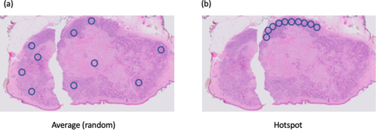
Mitoses are often assessed by pathologists to assist the diagnosis of cancer, and to grade malignancy, informing prognosis. Historically, this has been done by expressing the number of mitoses per n high power fields (HPFs), ignoring the fact that microscope fields may differ substantially, even at the same high power (×400) magnification. Despite a requirement to define HPF size in scientific papers, many authors fail to address this issue adequately. The problem is compounded by the switch to digital pathology systems, where ×400 equivalent fields are rectangular and also vary in the area displayed. The potential for error is considerable, and at times this may affect patient care. This is easily solved by the use of standardized international (SI) units. We, therefore, recommend that features such as mitoses are always counted per mm2, with an indication of the area to be counted and the method used (usually "hotspot" or "average") to obtain the results.


Conflict of interest statement

RAS is supported by a Commonwealth Government of Australia National Health and Medical Research Council of Australia (NHMRC) Practitioner Fellowship.

Figures

Fig. 1
Fig. 1. Diagram of a typical 400× microscope field  (small circle, 0.24 mm2) superimposed on a simple slide micrometer scale, permitting measurement of the diameter of the field of view of a microscope with an objective and eyepiece combination.
High power fields (HPF) conventionally use the ×40 objective, giving an overall magnification of ×400 with a 10× eyepiece, or ×500 with a 12.5× eyepiece. The area is calculated by πr2, where “r” is the radius (half the diameter) of the field of view.
Fig. 2
Fig. 2. Hotspot counting based on square tiles or round microscope fields.
Random counting (a) should use a randomization method to avoid bias, while hotspot counts (b) are usually linear or serpentine. It should be noted that contiguous round fields miss some areas of tumor which may contain mitoses. The method used should be clearly specified.

Similar articles

Cited by

References

    1. Cree IA, Foss AJ, Luthert PJ. Undefined high-power fields. Lancet. 1996;347:273–4. doi: 10.1016/S0140-6736(96)90455-9. - DOI - PubMed
    1. Ellis PS, Whitehead R. Mitosis counting—a need for reappraisal. Hum Pathol. 1981;12:3–4. doi: 10.1016/S0046-8177(81)80235-3. - DOI - PubMed
    1. Sadler DW, Coghill SB. Histopathologists, malignancies, and undefined high-power fields. Lancet. 1989;8641:785–6. doi: 10.1016/S0140-6736(89)92605-6. - DOI - PubMed
    1. Scolyer RA, Shaw HM, Thompson JF, Li LX, Colman MH, Lo SK, et al. Interobserver reproducibility of histopathologic prognostic variables in primary cutaneous melanomas. Am J Surg Pathol. 2003;27:1571–6. doi: 10.1097/00000478-200312000-00011. - DOI - PubMed
    1. McGovern VJ, Cochran AJ, Van der Esch EP, Little JH, MacLennan R. The classification of malignant melanoma, its histological reporting and registration: a revision of the 1972 Sydney classification. Pathology. 1986;18:12–21. doi: 10.3109/00313028609090822. - DOI - PubMed