. 2021 Jul;595(7866):295-302.
doi: 10.1038/s41586-021-03609-w. Epub 2021 Jun 2.

Base editing of haematopoietic stem cells rescues sickle cell disease in mice

Affiliations

Base editing of haematopoietic stem cells rescues sickle cell disease in mice

Gregory A Newby et al. Nature. 2021 Jul.

Abstract

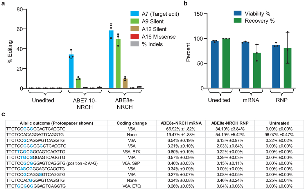
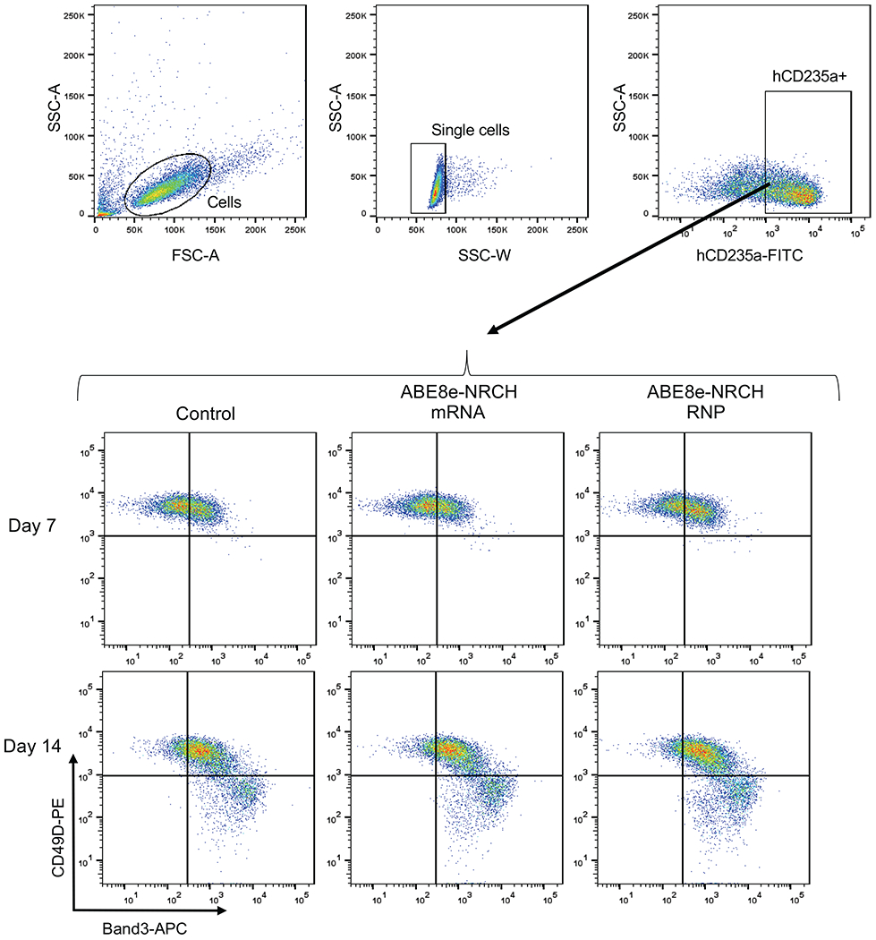
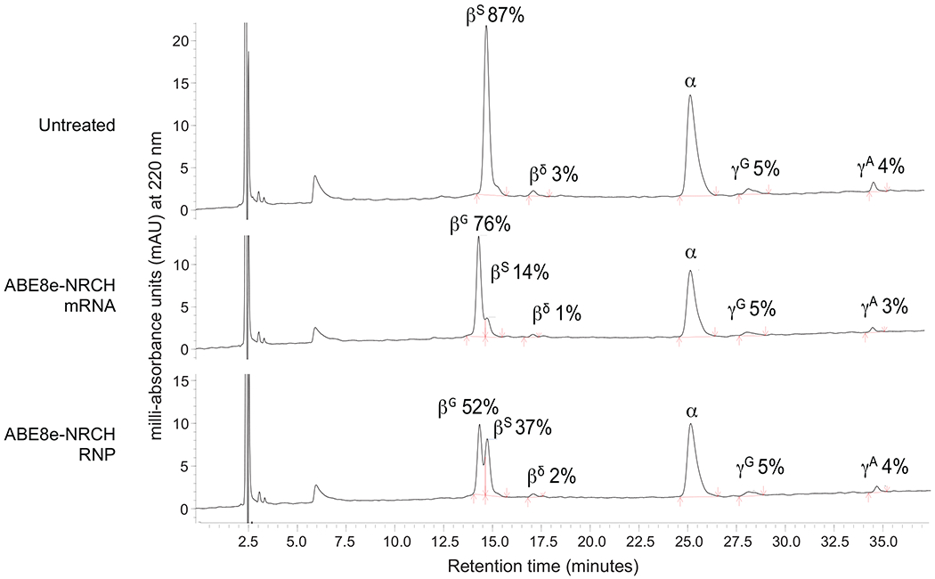
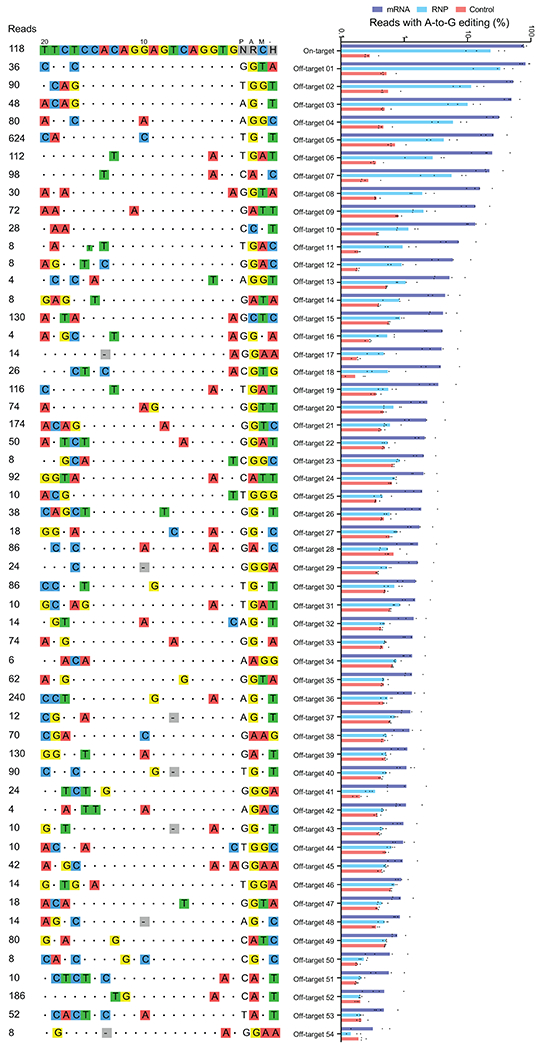
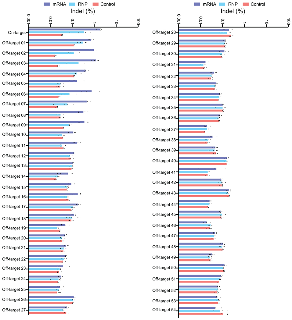
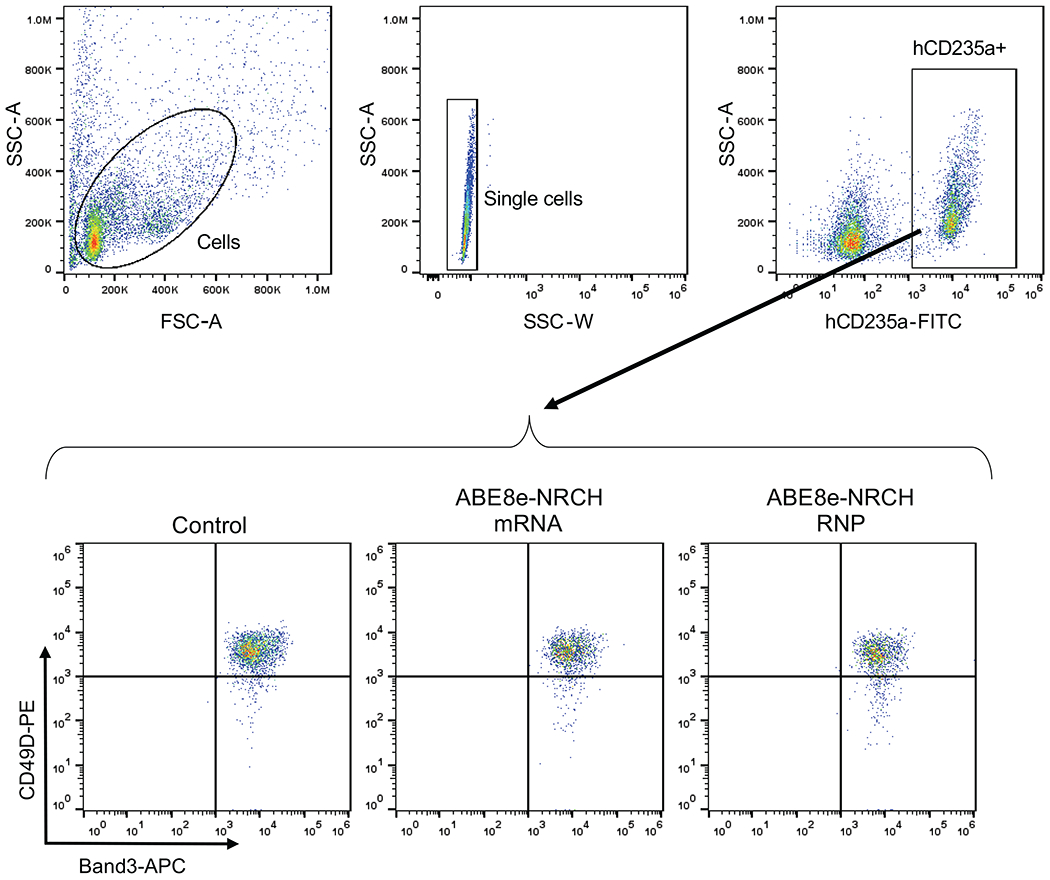
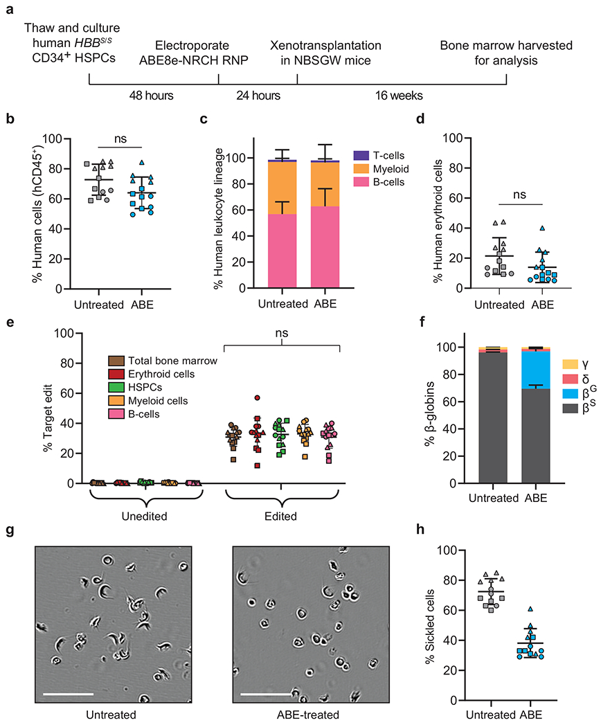
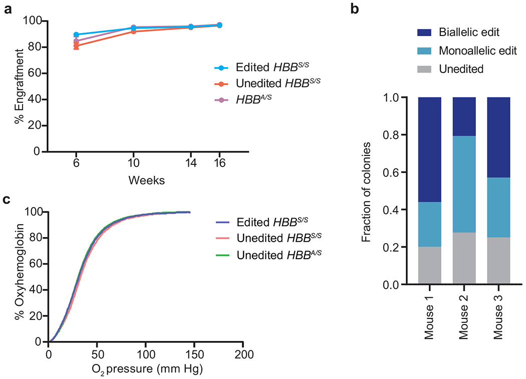
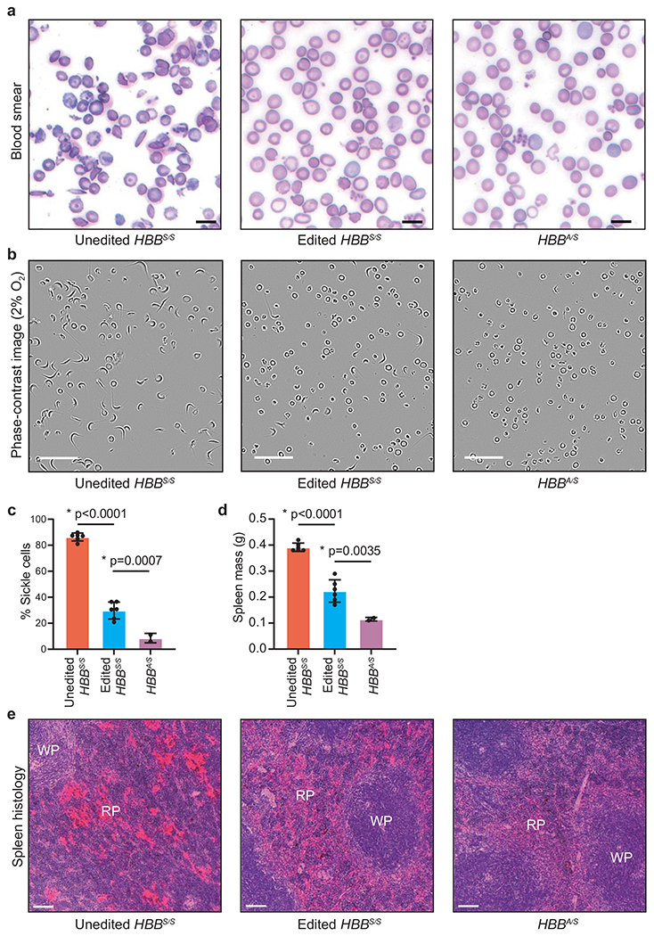
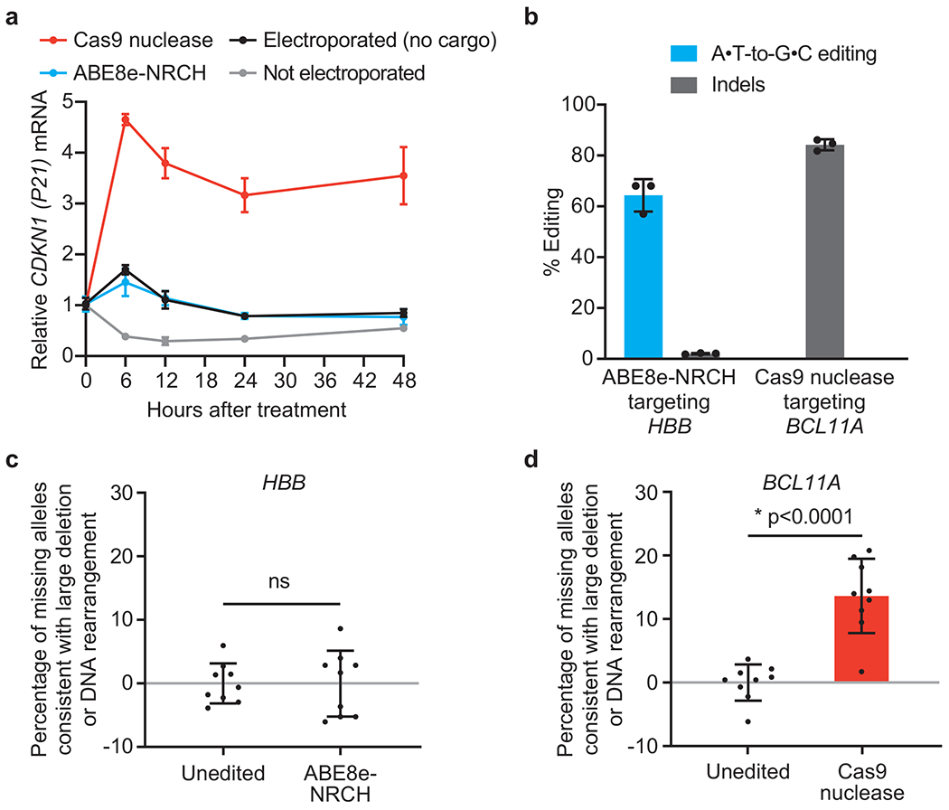
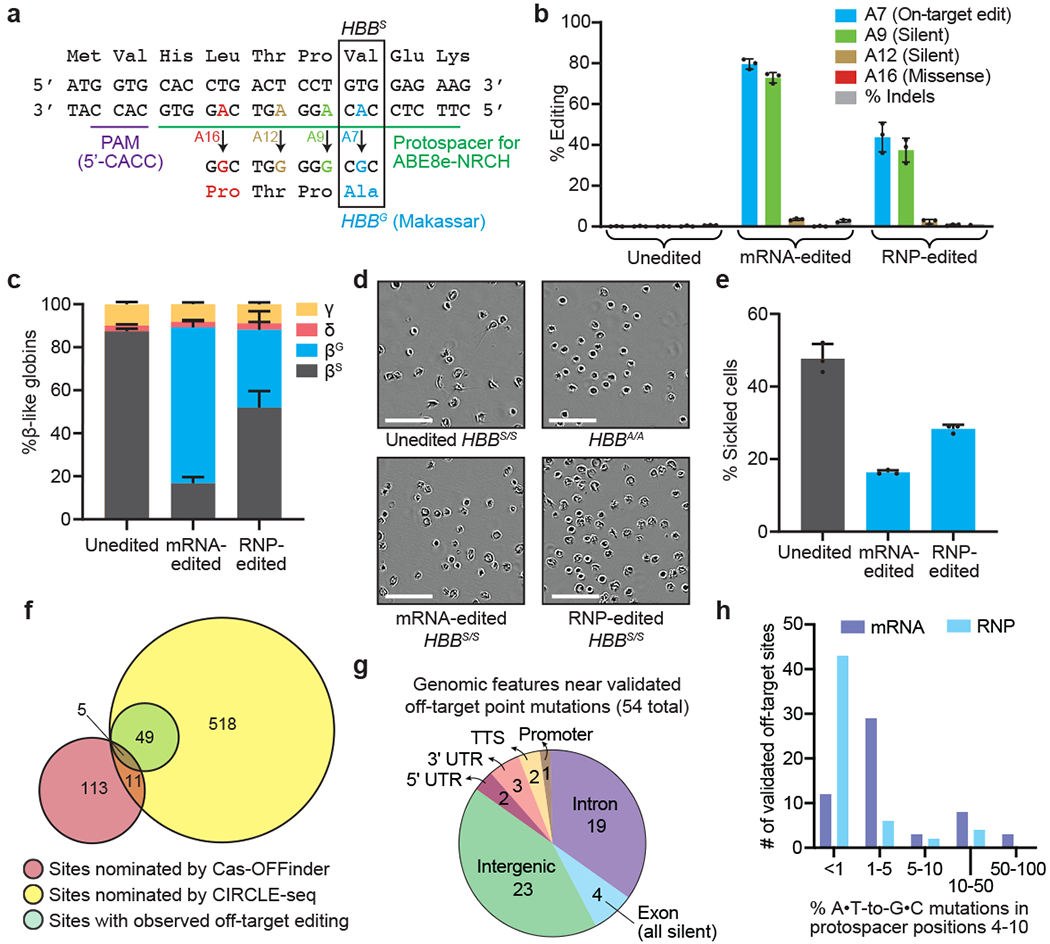
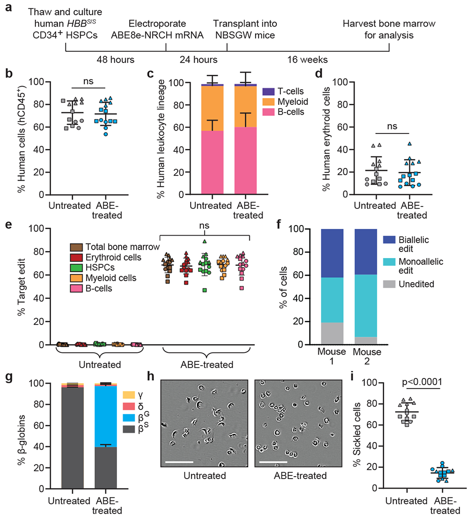
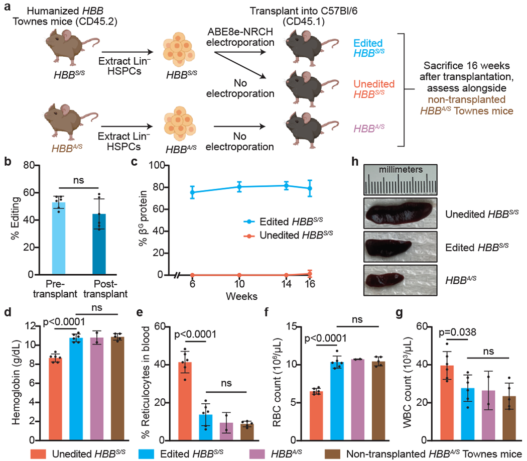
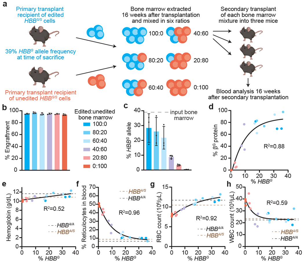
Sickle cell disease (SCD) is caused by a mutation in the β-globin gene HBB1. We used a custom adenine base editor (ABE8e-NRCH)2,3 to convert the SCD allele (HBBS) into Makassar β-globin (HBBG), a non-pathogenic variant4,5. Ex vivo delivery of mRNA encoding the base editor with a targeting guide RNA into haematopoietic stem and progenitor cells (HSPCs) from patients with SCD resulted in 80% conversion of HBBS to HBBG. Sixteen weeks after transplantation of edited human HSPCs into immunodeficient mice, the frequency of HBBG was 68% and hypoxia-induced sickling of bone marrow reticulocytes had decreased fivefold, indicating durable gene editing. To assess the physiological effects of HBBS base editing, we delivered ABE8e-NRCH and guide RNA into HSPCs from a humanized SCD mouse6 and then transplanted these cells into irradiated mice. After sixteen weeks, Makassar β-globin represented 79% of β-globin protein in blood, and hypoxia-induced sickling was reduced threefold. Mice that received base-edited HSPCs showed near-normal haematological parameters and reduced splenic pathology compared to mice that received unedited cells. Secondary transplantation of edited bone marrow confirmed that the gene editing was durable in long-term haematopoietic stem cells and showed that HBBS-to-HBBG editing of 20% or more is sufficient for phenotypic rescue. Base editing of human HSPCs avoided the p53 activation and larger deletions that have been observed following Cas9 nuclease treatment. These findings point towards a one-time autologous treatment for SCD that eliminates pathogenic HBBS, generates benign HBBG, and minimizes the undesired consequences of double-strand DNA breaks.


Conflict of interest statement

Competing interests

Authors have filed patent applications on base editing through the Broad Institute. D.R.L. is a consultant and equity owner of Beam Therapeutics, Prime Medicine, and Pairwise Plants, companies that use genome editing. M.J.W. is on advisory boards for Cellarity Inc., Novartis, and Forma Therapeutics, and is an equity owner of Beam Therapeutics. A.S. is a consultant for Spotlight Therapeutics and his institution receives clinical trial support for the conduct of sickle cell disease gene editing trials from Vertex Pharmaceuticals, CRISPR Therapeutics, and Novartis. J.S.Y is an equity owner of Beam Therapeutics. The authors declare no competing non-financial interests.

Figures

Extended Data Figure 1.
Extended Data Figure 1.. Optimization in HEK293T cells, viability and recovery following human SCD patient HSPC editing, and allelic editing outcomes.
(a) Plasmids encoding the HBBS-targeting sgRNA and either ABE7.10-NRCH or ABE8e-NRCH were transfected by lipofection into HEK293T cells. Editing efficiency was measured after 3 days by high-throughput DNA sequencing (HTS). Unedited cells were not lipofected. (b) Two days after electroporation into human patient HSPCs of base editor mRNA and sgRNA, or electroporation of ribonucleoprotein (RNP), cell number and viability were measured using a Chemometec Nucleocounter-3000. Acridine orange was used to stain the total cell number and DAPI was used to stain dead, permeabilized cells. The percent viability was calculated as the DAPI stained cells divided by the acridine orange cells within each sample. The percent recovery was normalized to the cell count of the unedited sample. Unedited cells were not electroporated, (c) Six days after electroporation of SCD patient HSPCs, genomic DNA was extracted and the target HBB locus was PCR amplified and sequenced using an Illumina instrument. The sequencing analysis program CRIS.py was used to identify and quantify the resulting alleles. All alleles above a threshold of 0.2% frequency are shown. Below this threshold, variant alleles appear with greatest frequency in the untreated control sample, suggesting they do not arise from base editor treatment. Nucleotides altered from the endogenous sequence are shown in blue. Rare cytosine base editing was observed at <1% frequency, as has been previously described as a possible outcome from adenine base editing. Bar values and error bars reflect mean±SD, n=3.
Extended Data Figure 2.
Extended Data Figure 2.. Erythroid differentiation of edited SCD patient CD34+ HSPCs.
Representative, immuno-flow cytometry for erythroid maturation stage markers, at culture days 7 and 14. Top: gating strategy to identify single cells expressing the erythroid marker hCD235a. Bottom: gating strategy to track the progress of erythroid maturation based on expression of CD49D and Band3 in hCD235a+ cells. SSC-A: Side scatter area. SSC-W: Side scatter width. FSC-A: Forward scatter area.
Extended Data Figure 3.
Extended Data Figure 3.. Reverse-phase high performance liquid chromatography (HPLC) analysis of erythroid cells derived from in vitro differentiation of edited SCD patient CD34+ HSPCs.
Reverse-phase HPLC chromatograms of erythroid cell lysates at culture day 18, with β-like globins and their associated fractions marked near the associated peak. Data from the most efficiently edited donor is shown. Red arrows indicate the start and end of globin chain peaks.
Extended Data Figure 4.
Extended Data Figure 4.. Off-target base editing associated with ABE8e-NRCH conversion of HBBS to HBBG Makassar in sickle cell disease patient CD34+ hematopoietic stem and progenitor cells.
CIRCLE-seq read counts obtained for each verified off-target site and the alignment of each site to the guide sequence are shown. Bar graphs show the percentage of sequencing reads containing A•T-to-G•C mutations within protospacer positions 4-10 at on-and off-target sites in genomic DNA samples from patient CD34+ HSPCs treated with ABE8e-NRCH mRNA, protein, or untreated controls (n=4). Note that the mutation frequency shown is summed across all reads with one or more A•T-to-G•C mutations in this window. Sequencing errors therefore accumulate in control samples compared to standard sequencing error frequencies for a single nucleotide. Bar values and error bars reflect mean±SD.
Extended Data Figure 5.
Extended Data Figure 5.. Off-target indel formation associated with ABE8e-NRCH conversion of HBBS to HBBG Makassar in sickle cell disease patient CD34+ hematopoietic stem and progenitor cells.
Bar graph showing the percentage of sequencing reads containing alleles harboring indels at on-and off-target sites in genomic DNA samples from patient CD34+ HSPCs treated with ABE8e-NRCH mRNA, protein, or untreated controls (n=4). Bar values and error bars reflect mean±SD.
Extended Data Figure 6.
Extended Data Figure 6.. Flow cytometry analysis of human donor-derived erythroid CD235a+ cells after transplantation.
(a) Human CD235a+ erythroid cells were purified by immuno-magnetic bead selection and analyzed by flow cytometry for the indicated erythroid maturation markers,, (b) Enucleated reticulocytes were assessed by the cell-permeable DNA stain Hoechst 33342 and the erythroid marker CD235a.
Extended Data Figure 7.
Extended Data Figure 7.. Engraftment of ABE8e-NRCH RNP-treated SCD patient CD34+ HSPCs after transplantation into immunodeficient mice.
CD34+ HSPCs from three HBBS/S SCD patient donors were electroporated with ABE8e-NRCH RNP using a single guide RNA (sgRNA) targeting the SCD mutant codon, followed by transplantation of 2-5x10⁵ treated cells into NBSGW mice via tail-vein injection. Mice were sacrificed and analyzed 16 weeks after transplantation, (a) Experimental workflow, (b) Engraftment measured by the percentage of human donor CD45+ cells (hCD45+ cells) in recipient mouse bone marrow, (c) Human B-cells (hCD19+), myeloid cells (hCD33+), and T-cells (hCD3+) cells in recipient mouse bone marrow, shown as percentages of the total hCD45+ population. (d) Human erythroid precursors (hCD235a+) in recipient mouse bone marrow shown as percentage of total human and mouse CD45cells. (e) On-target (A7, Fig. 1a) editing efficiencies in human donor CD34+ cell-derived lineages purified from recipient bone marrow by fluorescence-activated cell sorting. Erythroid, myeloid, B-cell, and HSPC human lineages were collected using antibodies that recognize hCD235a, hCD33, hCD19, and hCD34+, respectively. Statistical significance was assessed by one-way ANOVA to compare groups; “ns”, not significant. (f) Percentages of β-like globin proteins determined by reverse-phase HPLC analysis of human donor-derived reticulocytes isolated from recipient mouse bone marrow. (g) Representative phase contrast images of human reticulocytes purified from bone marrow and incubated for 8 hours in 2% O2. Nine images of >50 cells per image were collected per sample. Scale bar=50 μm. (h) Quantification of sickled cells calculated by counting images after incubation for 8 hours in 2% O2 such as in (g). More than 300 randomly selected cells per sample were counted by a blinded observer. n=14 total mice analyzed in panels b-f; triangle, square, and circle symbols represent samples from three different SCD CD34+ HSPC donors. Negative control data is shared with Figure 2. Bar values and error bars reflect mean±SD. Statistical significance between treated and untreated samples was assessed by a two-tailed Student’s t-test; “ns”, not significant.
Extended Data Figure 8.
Extended Data Figure 8.. Engraftment of transplanted Townes mouse HSPCs, clonality of editing outcomes, and oxygen binding affinity of blood.
(a) Donor cell engraftment measured by flow cytometry assessing the percentage of CD45.1+ cells among peripheral blood mononuclear cells (PBMCs). (b) Bone marrow from three mice transplanted with edited mouse HSPCs was plated at low density in methylcellulose. After 14 days of culture, 30 to 35 individual colonies per mouse were picked into cell lysis buffer and the edited locus was amplified by PCR and sequenced by HTS. Colonies were categorized by whether they contained no editing, a monoallelic edit, or a biallelic edit. (c) Blood was drawn from mice at week 14 after transplantation. Hemoglobin oxygenation was measured using a Hemox Analyzer (TCS Scientific) across a continuous declining gradient of oxygen pressure to assess whether HBBS-to-HBBG editing led to altered hemoglobin-oxygen binding. Bar values and error bars reflect mean±SD.
Extended Data Figure 9.
Extended Data Figure 9.. Adenine base editing of the sickle cell disease β-globin allele (HBBS) to the Makassar variant (HBBG) reduces erythrocyte sickling and splenic pathologies in mice.
Mice were treated as described in Fig. 3a. Blood and spleen were analyzed sixteen weeks after transplantation of Lin mouse HSPCs containing human HBB alleles, (a) Representative images of blood smears. One blood smear image was collected per mouse. Scale bar=25 μm. (b) Representative phase contrast images of peripheral blood incubated for 8 hours in 2% O2. Nine images of >50 cells per image were collected per sample. Scale bar=50 μm. (c) Quantification of sickled cells. More than 300 randomly selected cells were counted by a blinded observer. (d) Mass of dissected spleens. (e) Histological sections of spleens of recipient mice 16 weeks after transplantation. Splenic pathologies in mice that received unedited donor HBBS/S HSCs include excessive extramedullary erythropoiesis and vascular congestion indicated by RBC pooling (bright red color) resulting in expansion of red pulp (RP), reduction in white pulp sizes (WP), and splenomegaly. Images were taken at 10x magnification and were processed, stained and photographed at the same time under identical conditions. Three images of each spleen were collected from different parts of the organ for each mouse. Scale bar=100 μm. Unedited HBBS/S: n=6 mice; edited HBBS/S: n=6 mice; HBBA/S: n=2 mice. Plotted values and error bars reflect mean±SD, with individual values shown as dots. Statistical significance was assessed using one-way ANOVA, with Šidák’s multiple comparisons test of the edited HBBS/S values compared to each other group to calculate p-values.
Extended Data Figure 10.
Extended Data Figure 10.. Comparison of DNA damage response and loss of target allele amplification consistent with large deletion or DNA rearrangement in HSPCs following treatment with Cas9 nuclease or with adenine base editor.
HSPCs from a healthy human donor were electroporated in triplicate with Cas9 nuclease RNP targeting the BCL11A erythroid-specific enhancer, ABE8e-NRCH mRNA and an sgRNA targeting the wild-type HBB locus, or no cargo as a control. An additional set of control cells was not electroporated, (a) CDKN1 transcription levels, a measure of the p53-mediated DNA damage response, were quantified by droplet digital PCR (ddPCR) after reverse transcription, and were normalized to CDKN1 levels before electroporation (n=3). (b) Editing efficiencies at the targeted genomic loci in HSPCs were measured by HTS 6 days after electroporation. Adenine base editing at the synonymous bystander position 9 of the HBB protospacer is shown for ABE8e-NRCH. (c, d) The indicated target sites were amplified and quantified by ddPCR to measure the fraction of missing alleles consistent with larger deletions, translocations, or other chromosomal rearrangements that result in loss of the ability to be amplified by PCR. PCR amplification of a non-targeted ACTB site was used to normalize each sample. Each DNA sample was assessed in triplicate (n=9). Plotted values and error bars reflect mean±SD, with individual values in bar graphs shown as dots. Statistical significance between edited and unedited samples was assessed by a two-tailed Student’s t-test; “ns”, not significant.
Figure 1.
Figure 1.. Adenine base editing converts sickle cell disease β-globin (HBBS) to benign Makassar β-globin (HBBG) in patient CD34+ HSPCs.
CD34+ cells from three SCD patient donors were electroporated with ABE8e-NRCH mRNA or RNP using an sgRNA targeting the SCD mutant HBB codon. (a) The edited region of HBB with the target A at protospacer position 7 shown in blue along with potential bystander edits in green (silent), brown (silent), and red (non-silent). (b) Editing efficiencies by HTS at target and bystander adenines, and indels after 6 days in stem-cell culture media following electroporation. (c) Proportion of β-like globin proteins by HPLC of reticulocyte lysates after 18 days in differentiation media following electroporation. (d) Representative phase-contrast images of reticulocytes derived from unedited or edited donor HSPCs incubated 8 hours in 2% O2. Nine images of >50 cells each were collected per sample. Scale bar=50 μm. (e) Quantification of sickled reticulocytes from counting >300 randomly selected cells by a blinded observer from images as in (d). (f) Venn diagram showing candidate off-target sites nominated by Cas-OFFinder and CIRCLE-seq, and nominated sites for which off-target editing was observed by targeted DNA sequencing in SCD patient CD34+ cells electroporated with ABE8e-NRCH mRNA. (g) Predicted genomic features of validated off-target sites. TTS, ≤1 kb from the transcription termination site; UTR, untranslated region. (h) ABE8e-NRCH-treated HSPCs from two different SCD patient donors were sequenced at 697 potential off-target sites. The histogram shows the number of validated off-target base editing sites binned by average percentage of sequencing reads for each site with any A•T-to-G•C mutations in protospacer nucleotides 4-10. Bar values in (b), (c), and (e) and error bars reflect mean±SD of three independent biological replicates, with individual values shown as dots.
Figure 2.
Figure 2.. Engraftment of ABE8e-NRCH mRNA-treated SCD patient CD34+ HSPCs after transplantation into immunodeficient mice.
CD34+ HSPCs from three HBBS/S SCD patient donors were electroporated with ABE8e-NRCH mRNA and sgRNA targeting the SCD mutant HBB codon. 2-5x10⁵ treated cells were transplanted into NBSGW mice via tail-vein injection. Mice were analyzed 16 weeks after transplantation. (a) Experimental workflow. (b) Engraftment measured by percentage of human CD45+ (hCD45+) cells in recipient mouse bone marrow. (c) Human B-cells (hCD19+), myeloid cells (hCD33+), and T-cells (hCD3+) cells in recipient mouse bone marrow shown as percentages of the hCD45+ population. (d) Human erythroid precursors (hCD235a+) in recipient mouse bone marrow shown as percentage of human and mouse CD45 cells, (e) HBBS-to-HBBG editing efficiencies in human donor CD34+ cell-derived lineages from recipient bone marrow. Erythroid, myeloid, B-cell, and HSPC human lineages were collected using antibodies that recognize hCD235a, hCD33, hCD19, and hCD34, respectively, (f) Clonal editing outcomes determined by single-cell 5’ RNA-seq in CD235a+ cells from the bone marrow of two edited mice. (g) Proportions of β-like globin proteins by HPLC of human donor-derived reticulocytes isolated from recipient mouse bone marrow. (h) Representative phase-contrast images of human reticulocytes from bone marrow incubated 8 hours in 2% O2. Nine images of >50 cells each were collected per sample. Scale bar=50 μm. (i) Quantification of sickled cells as in Fig. 1e. n=14 mice receiving edited cells and n=13 mice receiving unedited cells in b-e, g, and i. Triangle, square, and circle symbols represent HSPCs from three different SCD donors. Plotted values and error bars reflect mean±SD. Statistical significance was assessed by one-way ANOVA in i and by two-tailed Student’s t-test elsewhere; “ns”, not significant.
Figure 3.
Figure 3.. HBBS-to-HBBG base editing alleviates pathology in a mouse model of SCD.
(a) Lineage negative (Lin) HSPCs from the bone marrow of Townes SCD mice (CD45.2, human HBBS/S) were electroporated with ABE8e-NRCH and sgRNA RNP or not electroporated, then transplanted into irradiated CD45.1 C57BI/6 recipient mice. Unedited HBBA/S HSPCs from Townes sickle-cell trait mice transplanted into irradiated CD45.1 C57BI/6 mice, and non-transplanted HBBA/S Townes mice were used as healthy controls. (b) HBBS-to-HBBG editing efficiency in cells cultured 3 days after electroporation (pre-transplant) or in PBMCs collected 16 weeks post-transplant. ns, not significant by two-tailed Student’s t-test. (c) Percentage of βG among β-like globin proteins by RP-HPLC analysis of blood. (d-g) Hematologic indices 16 weeks post-transplant. Statistical significance was assessed using one-way ANOVA, with Šidák’s multiple comparisons test to calculate p-values. Differences among edited HBBS/S, transplanted HBBA/S, and non-transplanted HBBA/S mice were not significant. Bar values and error bars reflect mean±SD of n=6 mice (unedited HBBS/S, edited HBBS/S), n=2 mice (HBBA/S), or n=5 mice (non-transplanted HBBA/S). (h) Spleens were imaged from each mouse 16 weeks after transplantation with Townes mouse HSPCs. Representative images are shown.
Figure 4.
Figure 4.. Secondary transplantation reveals HBBS-to-HBBG base editing requirements for hematological correction.
(a) Bone marrow from a CD45.1 C57/BI6 mouse 16 weeks after primary transplantation with ABE8e-NRCH RNP-edited Lin HSPCs from Townes SCD mice (CD45.2, HBBS/S) was mixed in varying proportions with bone marrow from a C57/BI6 mouse 16 weeks after transplantation with unedited HBBS/S HSPCs from a Townes SCD mouse. For each of six bone marrow mixtures, secondary transplantations of 2x10⁶ cells were performed into three irradiated CD45.1 C57BI/6 recipient mice. Peripheral blood was analyzed after 16 weeks. (b) Engraftment measured by percentage of PBMCs with CD45.1. (c) HBBS-to-HBBG editing efficiency in PBMCs. (d) Percentage of βG among β-like globin proteins by HPLC and (e-h) hematologic indices plotted against HBBG allele frequency measured for each mouse. Parameters from non-transplanted HBBA/S (brown line) and HBBA/A (black line) Townes mice were assessed as healthy controls. One-phase decay fits are shown in (d), (f), (g), and (h), and a linear fit in (e). Bars values and error bars reflect mean±SD of n=3 mice. Dots represent different mice. Colors in (c-h) match the key in (b).

References

    1. Piel FB, Steinberg MH & Rees DC Sickle Cell Disease. N Engl J Med 377, 305, doi:10.1056/NEJMc1706325 (2017). - DOI - PubMed
    1. Richter MF et al. Phage-assisted evolution of an adenine base editor with improved Cas domain compatibility and activity. Nat Biotechnol 38, 883–891, doi:10.1038/s41587-020-0453-z (2020). - DOI - PMC - PubMed
    1. Miller SM et al. Continuous evolution of SpCas9 variants compatible with non-G PAMs. Nat Biotechnol 38, 471–481, doi:10.1038/s41587-020-0412-8 (2020). - DOI - PMC - PubMed
    1. Sangkitporn S, Rerkamnuaychoke B, Sangkitporn S, Mitrakul C & Sutivigit Y Hb G Makassar (beta 6:Glu-Ala) in a Thai family. J Med Assoc Thai 85, 577–582 (2002). - PubMed
    1. Blackwell RQ, Oemijati S, Pribadi W, Weng MI & Liu CS Hemoglobin G Makassar: beta-6 Glu leads to Ala. Biochim Biophys Acta 214, 396–401 (1970). - PubMed

References cited in Methods and Extended Data figure legends

    1. Komor AC, Kim YB, Packer MS, Zuris JA & Liu DR Programmable editing of a target base in genomic DNA without double-stranded DNA cleavage. Nature 533, 420–424, doi:10.1038/nature17946 (2016). - DOI - PMC - PubMed
    1. Gaudelli NM et al. Programmable base editing of A*T to G*C in genomic DNA without DNA cleavage. Nature 551, 464–471, doi:10.1038/nature24644 (2017). - DOI - PMC - PubMed
    1. Huang TP, Newby GA & Liu DR Precision genome editing using cytosine and adenine base editors in mammalian cells. Nat Protoc 16, 1089–1128, doi:10.1038/s41596-020-00450-9 (2021). - DOI - PubMed
    1. Clement K et al. CRISPResso2 provides accurate and rapid genome editing sequence analysis. Nat Biotechnol 37, 224–226, doi:10.1038/s41587-019-0032-3 (2019). - DOI - PMC - PubMed
    1. Vaidyanathan S et al. Uridine Depletion and Chemical Modification Increase Cas9 mRNA Activity and Reduce Immunogenicity without HPLC Purification. Mol Ther Nucleic Acids 12, 530–542, doi:10.1016/j.omtn.2018.06.010 (2018). - DOI - PMC - PubMed

Publication types