

Dermoscopic Findings and the Clinicopathologic Correlation of Pigmented Purpuric Dermatosis: A Retrospective Review of 60 Cases
- PMID: 34079180
- PMCID: PMC8137322
- DOI: 10.5021/ad.2021.33.3.214
Dermoscopic Findings and the Clinicopathologic Correlation of Pigmented Purpuric Dermatosis: A Retrospective Review of 60 Cases
Abstract
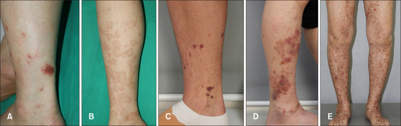
Background: Pigmented purpuric dermatosis (PPD) is known as a chronic recurrent eruption which usually presents with petechiae and pigmented macules on the lower extremities. Dermoscopy is a noninvasive diagnostic tool in identifying pigmented and vascular lesions, which can also be beneficial in the evaluation of PPD.
Objective: We aimed to analyze the common dermoscopic characteristics of PPD, and correlate those findings with the histopathologic features. Additionally, dermoscopic and pathological findings in this study population were compared with other similar studies from the literature review.
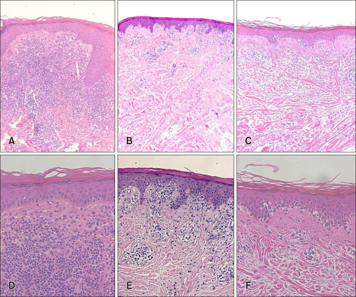
Methods: A retrospective analysis was performed using data of 60 patients who were diagnosed as PPD by skin biopsy and had dermoscopic examination. The pathologic analysis was performed by categorizing the pattern into lichenoid, perivascular, interface, and spongiotic subtype, and the dermoscopic assessment was performed by the three authors independently.
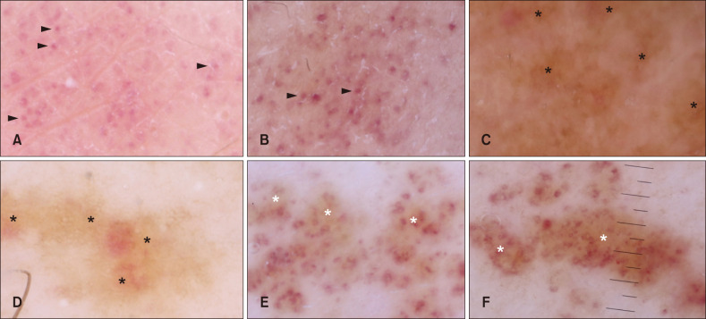
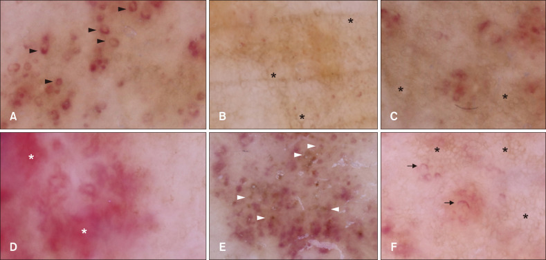
Results: In dermoscopy, 96.7% of the patients showed red globules and dots, followed by brownish patch, coppery-red pigmentation, and annular comma-like vessels. The pathologic pattern analysis revealed statistically significant association of lichenoid pattern with coppery red pigmentation, perivascular pattern with annular/comma-like vessels, and spongiosis pattern with reticular pigmented network and linear vessels. The interrater similarity test showed total kappa value of 0.811 which referred to "very good".
Conclusion: In this study, the prevalence of dermoscopic features in Asian PPD patients was identified, which was similar with previous studies. The dermoscopic-pathologic correlation was found in four dermoscopic features. We suggest that dermoscopic examination is helpful in clinical diagnosis and pathological prediction of PPD.
Keywords: Dermoscopy; Pathology, clinical; Pigmentation disorders; Pigmented purpuric dermatosis; Skin diseases, vascular.
Copyright © 2021 The Korean Dermatological Association and The Korean Society for Investigative Dermatology.
Conflict of interest statement
CONFLICTS OF INTEREST: The authors have nothing to disclose.
Figures
Similar articles
-
Dermoscopic profile of pigmented purpuric dermatosis: new observations.Postepy Dermatol Alergol. 2019 Dec;36(6):687-691. doi: 10.5114/ada.2019.91419. Epub 2019 Dec 30. Postepy Dermatol Alergol. 2019. PMID: 31997996 Free PMC article.
-
Pigmented purpuric dermatoses versus purpuric mycosis fungoides: Clinicopathologic similarities and new insights into dermoscopic features.Australas J Dermatol. 2022 Feb;63(1):81-85. doi: 10.1111/ajd.13759. Epub 2021 Dec 14. Australas J Dermatol. 2022. PMID: 34905635
-
Dermoscopic Findings in Patients with Pigmented Purpuric Dermatoses.Acta Dermatovenerol Croat. 2016 Dec;24(4):291-295. Acta Dermatovenerol Croat. 2016. PMID: 28128081
-
Pigmented Purpuric Dermatosis of the Hand: Clinicopathologic Analysis of Six Cases With Review of the Literature.Am J Dermatopathol. 2022 Aug 1;44(8):553-558. doi: 10.1097/DAD.0000000000002204. Epub 2022 Apr 27. Am J Dermatopathol. 2022. PMID: 35503879 Review.
-
Dermoscopic features of actinic keratosis.J Dtsch Dermatol Ges. 2007 Nov;5(11):970-6. doi: 10.1111/j.1610-0387.2007.06318.x. Epub 2007 Oct 1. J Dtsch Dermatol Ges. 2007. PMID: 17908179 Review. English, German.
Cited by
-
Utility of Dermoscopy in Cutaneous Small Vessel Vasculitis: Preliminary Observations from a Study of 30 Cases.Indian Dermatol Online J. 2023 May 25;14(4):506-509. doi: 10.4103/idoj.idoj_648_22. eCollection 2023 Jul-Aug. Indian Dermatol Online J. 2023. PMID: 37521212 Free PMC article.
-
Clinico Epidemiological Study and Dermoscopic Findings of Pigmented Purpuric Dermatosis.Indian Dermatol Online J. 2022 Sep 21;14(1):107-109. doi: 10.4103/idoj.idoj_150_22. eCollection 2023 Jan-Feb. Indian Dermatol Online J. 2022. PMID: 36776197 Free PMC article. No abstract available.
References
-
- Sardana K, Sarkar R, Sehgal VN. Pigmented purpuric dermatoses: an overview. Int J Dermatol. 2004;43:482–488. - PubMed
-
- Haden A, Peng DH. Pigmented purpuric dermatoses. In: Kang S, Amagai M, Bruckner AL, Enk AH, Margolis DJ, McMichael AJ, et al., editors. Fitzpatrick's dermatology. 9th ed. New York: McGraw-Hill Education; 2019.
LinkOut - more resources
Full Text Sources
