. 2021 May 17:14:665931.
doi: 10.3389/fnmol.2021.665931. eCollection 2021.

Transcriptome Analysis Reveals the Role of Cellular Calcium Disorder in Varicella Zoster Virus-Induced Post-Herpetic Neuralgia

Affiliations

Transcriptome Analysis Reveals the Role of Cellular Calcium Disorder in Varicella Zoster Virus-Induced Post-Herpetic Neuralgia

Songbin Wu et al. Front Mol Neurosci. .

Abstract

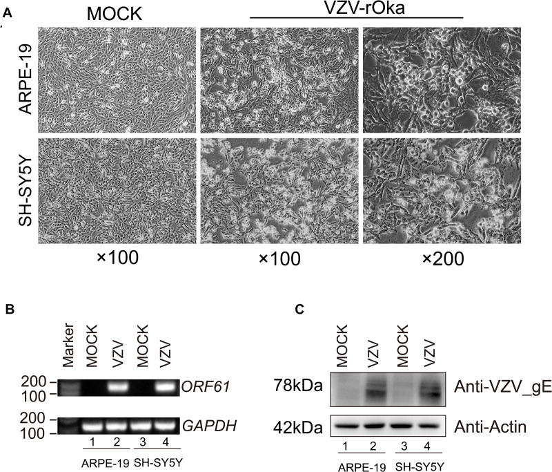
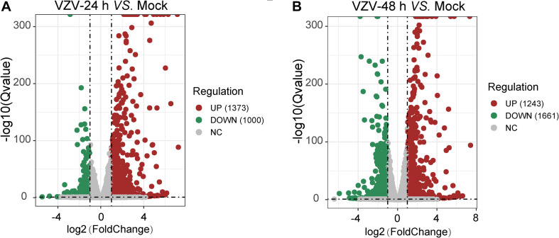
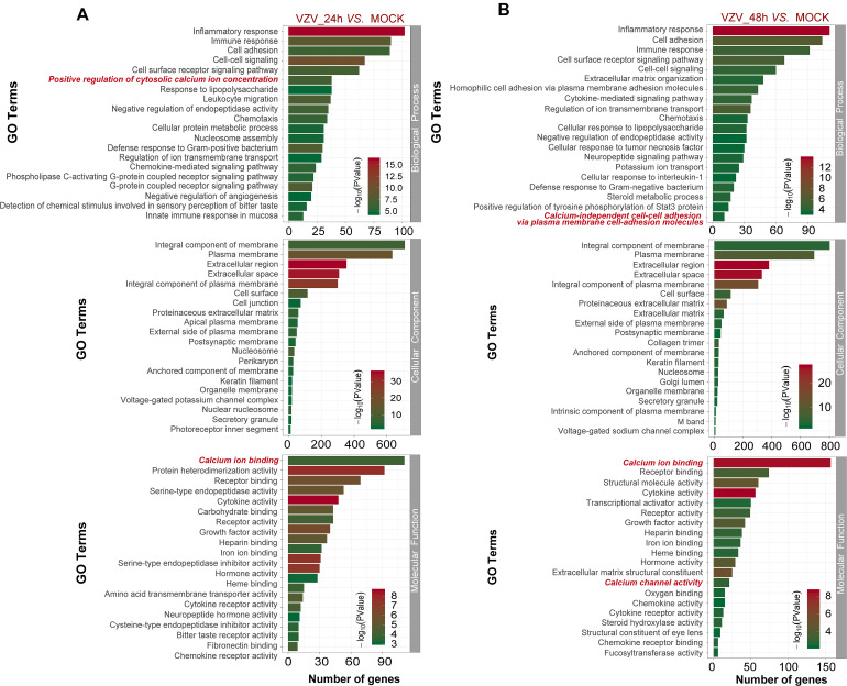
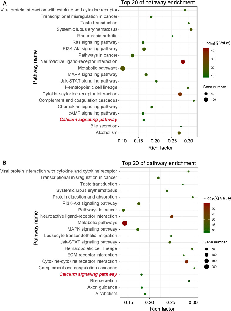
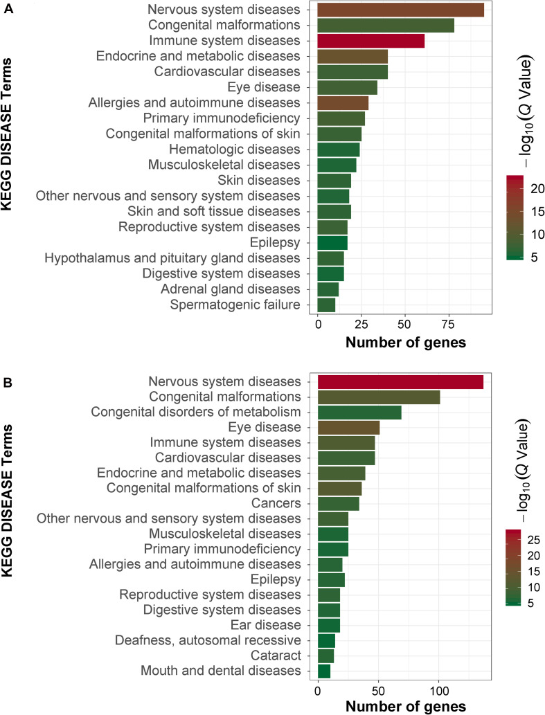
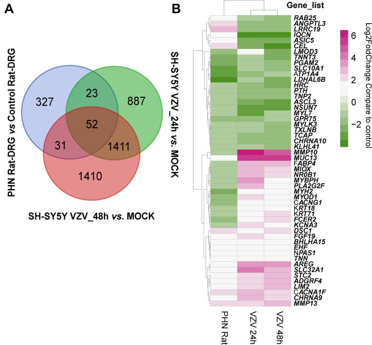
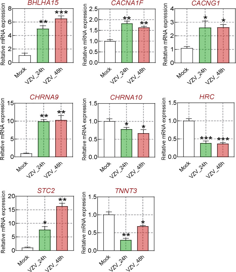
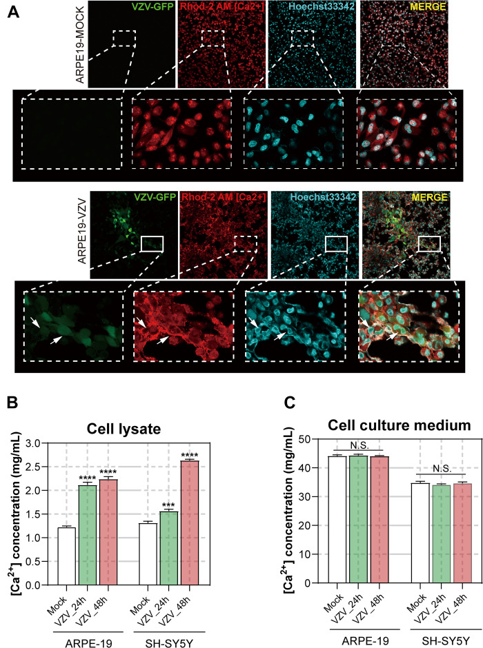
As a typical neuropathic pain, post-herpetic neuralgia (PHN) is a common complication of herpes zoster (HZ), which seriously affects the normal life and work of patients. The unclear pathogenesis and lack of effective drugs make the clinical efficacy of PHN unsatisfactory. Here, we obtained the transcriptome profile of neuroblastoma cells (SH-SY5Y) and DRG in rats infected with varicella zoster virus (VZV) by transcriptome sequencing (RNA-Seq) combined with publicly available gene array data sets. Next, the data processing of the transcriptome map was analyzed using bioinformatics methods, including the screening of differentially expressed genes (DEGs), Gene Ontology (GO), and the Kyoto Encyclopedia of Genes and Genomes (KEGG) analysis. Finally, real-time fluorescent quantitative PCR (qRT-PCR) was used to detect the expression of calcium-related genes, and calcium fluorescent probes and calcium colorimetry were used to evaluate the distribution and content of calcium ions in cells after VZV infection. Transcriptome data analysis (GO and KEGG enrichment analysis) showed that calcium disorder played an important role in SH-SY5Y cells infected by VZV and dorsal root ganglion (DRG) of the PHN rat model. The results of qRT-PCR showed that the expression levels of calcium-related genes BHLHA15, CACNA1F, CACNG1, CHRNA9, and STC2 were significantly upregulated, while the expression levels of CHRNA10, HRC, and TNNT3 were significantly downregulated in SH-SY5Y cells infected with VZV. Our calcium fluorescent probe and calcium colorimetric test results showed that VZV could change the distribution of calcium ions in infected cells and significantly increase the intracellular calcium content. In conclusion, our results revealed that the persistence of calcium disorder caused by VZV in nerve cells might be a crucial cause of herpetic neuralgia, and a potential target for clinical diagnosis and treatment of PHN.

Keywords: Ca2+; RNA-seq; VZV; calcium channel; calcium-related genes; post-herpetic neuralgia.


Conflict of interest statement

The authors declare that the research was conducted in the absence of any commercial or financial relationships that could be construed as a potential conflict of interest.

Figures

FIGURE 1
FIGURE 1
ARPE-19 and SH-SY5Y cells infected with VZV induced obvious cytopathic changes. (A) ARPE-19 and SH-SY5Y cells infected with VZV for 48 h induce obvious cytopathic changes, and cells not infected with (MOCK) virus maintain normal morphology and growth. (B) Real-time quantitative PCR was used to measure the expression of ORF61 encoding the early phosphorylation protein of virus in ARPE-19 cells and SH-SY5Y cells after 48 h of VZV infection. (C) Western blot was used to evaluate the expression of VZV glycoprotein gE in ARPE-19 cells and SH-SY5Y cells.
FIGURE 2
FIGURE 2
A mass number of DEGs were induced by VZV in SH-SY5Y cells. (A) Volcanic map showed the number and distribution of DEGs in SH-SY5Y cells after 24 h of VZV infection. (B) Volcanic map showed the number and distribution of DEGs in SH-SY5Y cells after 48 h of VZV infection. Volcano map showed that VZV group had significant mRNA expression compared with mock without VZV infection. Log2 (Fold change) and log10 (Q-value) of DEGs are expressed as abscissa and ordinate, respectively. The significantly upregulated genes are shown in red and the significantly downregulated genes are shown in green.
FIGURE 3
FIGURE 3
The functional enrichment of DEGs was analyzed by gene ontology classification. (A) GO analysis of DEGs in SH-SY5Y cells 24 h after VZV infection. (B) GO analysis of DEGs in SH-SY5Y cells 48 h after VZV infection. The number of DEGs was plotted as abscissa and GO terms were plotted as ordinate. It shows the top 20 highly representative GO terms enriched in DEGs, including biological processes, cellular components, and molecular functions.
FIGURE 4
FIGURE 4
KEGG enrichment of DEGs induced by VZV in SH-SY5Y cells. (A) The DEGs enriched KEGG pathway was induced by VZV infection in SH-SY5Y cells for 24 h. (B) The DEGs enriched KEGG pathway was induced by VZV infection in SH-SY5Y cells for 48 h. The graph shows the top 20 significantly enriched KEGG pathways by plotting rich factors as abscissa and KEGG terms as ordinates.
FIGURE 5
FIGURE 5
KEGG disease analysis of DEGs in SH-SY5Y cells induced by VZV. (A) KEGG disease analysis of DEGs induced by VZV infection of SH-SY5Y cells for 24 h. (B) KEGG disease analysis of DEGs induced by VZV infection of SH-SY5Y cells for 48 h. The graph shows the first 20 significantly abundant KEGG diseases by using the number of differentially expressed genes as the abscissa and the KEGG term as the ordinate.
FIGURE 6
FIGURE 6
Analysis of the identical DEGs in VZV-infected SH-SY5Y cells and VZV-induced PHN rat DRG. (A) The Draw Venn Diagram online tool was used to obtain the identical DEGs in VZV-infected SH-SY5Y cells and VZV-induced PHN rat DRG. (B) The Pheatmap package was used to map the gene expression heatmap of the identical DEGs in the DRG of VZV-induced PHN rats and SH-SY5Y cells infected by VZV.
FIGURE 7
FIGURE 7
The calcium signal DEGs were verified by qRT-PCR. After VZV-infected SH-SY5Y cells for 24 and 48 h, respectively, qRT-PCR was used to quantify the expression levels of BHLHA15, CACNA1F, CACNG1, CHRNA9, CHRNA10, HRC, STC2, and TNNT3 genes. Data are expressed as the mean ± SD from at least three independent experiments (compared to the Mock group, *P < 0.05, **P < 0.01, ***P < 0.001).
FIGURE 8
FIGURE 8
Effect of VZV on Ca2+ in ARPE-19 and SH-SY5Y cells. (A) The Rhod-2 AM calcium ion probe was used to assess the intracellular calcium ion distribution of ARPE-19 cells 72 h after VZV infection. The calcium colorimetric assay kit was used to evaluate the calcium ion content in the cell lysate (B) and cell culture supernatant (C) after VZV infection of ARPE-19 and SH-SY5Y cells at 24 h and 48 h, respectively. Data are expressed as the mean ± SD from at least three independent experiments (compared to the Mock group, ***P < 0.001, ****P < 0.0001, N.S.; not significant). The white arrows indicate the distribution of calcium ions on the cell membrane surface.

Similar articles

Cited by

References

    1. Almanzar G., Kienle F., Schmalzing M., Maas A., Tony H. P., Prelog M. (2019). Tofacitinib modulates the VZV-specific CD4+ T cell immune response in vitro in lymphocytes of patients with rheumatoid arthritis. Rheumatology (Oxford) 58 2051–2060. 10.1093/rheumatology/kez175 - DOI - PubMed
    1. An J., Wang L., Guo Q., Li L., Xia F., Zhang Z. (2012). Behavioral phenotypic properties of a natural occurring rat model of congenital stationary night blindness with Cacna1f mutation. J. Neurogenet. 26 363–373. 10.3109/01677063.2012.684416 - DOI - PubMed
    1. Bading H. (2013). Nuclear calcium signalling in the regulation of brain function. Nat. Rev. Neurosci. 14 593–608. 10.1038/nrn3531 - DOI - PubMed
    1. Bourinet E., Altier C., Hildebrand M. E., Trang T., Salter M. W., Zamponi G. W. (2014). Calcium-permeable ion channels in pain signaling. Physiol. Rev. 94 81–140. 10.1152/physrev.00023.2013 - DOI - PubMed
    1. Burgess D. L., Davis C. F., Gefrides L. A., Noebels J. L. (1999). Identification of three novel Ca(2+) channel gamma subunit genes reveals molecular diversification by tandem and chromosome duplication. Genome Res. 9 1204–1213. 10.1101/gr.9.12.1204 - DOI - PMC - PubMed