

Galectins in the Pathogenesis of Common Retinal Disease
- PMID: 34079467
- PMCID: PMC8165321
- DOI: 10.3389/fphar.2021.687495
Galectins in the Pathogenesis of Common Retinal Disease
Abstract
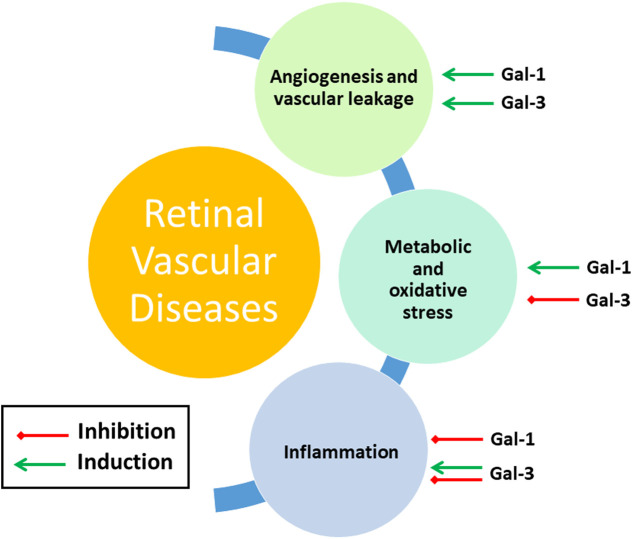
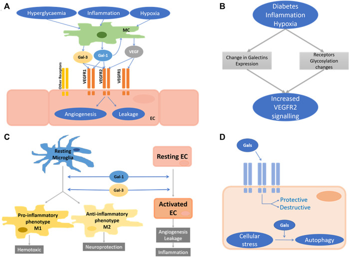
Diseases of the retina are major causes of visual impairment and blindness in developed countries and, due to an ageing population, their prevalence is continually rising. The lack of effective therapies and the limitations of those currently in use highlight the importance of continued research into the pathogenesis of these diseases. Vascular endothelial growth factor (VEGF) plays a major role in driving vascular dysfunction in retinal disease and has therefore become a key therapeutic target. Recent evidence also points to a potentially similarly important role of galectins, a family of β-galactoside-binding proteins. Indeed, they have been implicated in regulating fundamental processes, including vascular hyperpermeability, angiogenesis, neuroinflammation, and oxidative stress, all of which also play a prominent role in retinopathies. Here, we review direct evidence for pathological roles of galectins in retinal disease. In addition, we extrapolate potential roles of galectins in the retina from evidence in cancer, immune and neuro-biology. We conclude that there is value in increasing understanding of galectin function in retinal biology, in particular in the context of the retinal vasculature and microglia. With greater insight, recent clinical developments of galectin-targeting drugs could potentially also be of benefit to the clinical management of many blinding diseases.
Keywords: VEGF; age-related macula degeneration; angiogenesis; diabetic retinopathy; leakage; retina.
Copyright © 2021 Caridi, Doncheva, Sivaprasad and Turowski.
Conflict of interest statement
The authors declare that the research was conducted in the absence of any commercial or financial relationships that could be construed as a potential conflict of interest.
Figures
Similar articles
-
[Aging and retinal vascular diseases].Nippon Ganka Gakkai Zasshi. 2007 Mar;111(3):207-30; discussion 231. Nippon Ganka Gakkai Zasshi. 2007. PMID: 17402563 Review. Japanese.
-
Role of the vascular endothelial growth factor isoforms in retinal angiogenesis and DiGeorge syndrome.Verh K Acad Geneeskd Belg. 2005;67(4):229-76. Verh K Acad Geneeskd Belg. 2005. PMID: 16334858 Review.
-
Vascular permeability in retinopathy is regulated by VEGFR2 Y949 signaling to VE-cadherin.Elife. 2020 Apr 21;9:e54056. doi: 10.7554/eLife.54056. Elife. 2020. PMID: 32312382 Free PMC article.
-
[Cell biology of intraocular vascular diseases].Nippon Ganka Gakkai Zasshi. 1999 Dec;103(12):923-47. Nippon Ganka Gakkai Zasshi. 1999. PMID: 10643294 Review. Japanese.
-
The role of placental growth factor (PlGF) and its receptor system in retinal vascular diseases.Prog Retin Eye Res. 2019 Mar;69:116-136. doi: 10.1016/j.preteyeres.2018.10.006. Epub 2018 Oct 30. Prog Retin Eye Res. 2019. PMID: 30385175 Review.
Cited by
-
Resolution of Inflammation in Retinal Disorders: Briefly the State.Int J Mol Sci. 2022 Apr 19;23(9):4501. doi: 10.3390/ijms23094501. Int J Mol Sci. 2022. PMID: 35562891 Free PMC article. Review.
-
Loss of Pigment Epithelium Derived Factor Sensitizes C57BL/6J Mice to Light-Induced Retinal Damage.bioRxiv [Preprint]. 2024 Dec 4:2024.12.04.626802. doi: 10.1101/2024.12.04.626802. bioRxiv. 2024. PMID: 39679905 Free PMC article. Preprint.
-
Ablation of Htra1 leads to sub-RPE deposits and photoreceptor abnormalities.JCI Insight. 2025 Feb 10;10(3):e178827. doi: 10.1172/jci.insight.178827. JCI Insight. 2025. PMID: 39927462 Free PMC article.
-
The Role of Galectin-3 in Retinal Degeneration and Other Ocular Diseases: A Potential Novel Biomarker and Therapeutic Target.Int J Mol Sci. 2023 Oct 24;24(21):15516. doi: 10.3390/ijms242115516. Int J Mol Sci. 2023. PMID: 37958500 Free PMC article. Review.
-
Inhibition of Galectins and the P2X7 Purinergic Receptor as a Therapeutic Approach in the Neurovascular Inflammation of Diabetic Retinopathy.Int J Mol Sci. 2023 Jun 3;24(11):9721. doi: 10.3390/ijms24119721. Int J Mol Sci. 2023. PMID: 37298672 Free PMC article. Review.
References
Publication types
LinkOut - more resources
Full Text Sources