. 2020 Mar;13(3):736-744.
doi: 10.1007/s12274-020-2684-1. Epub 2020 Feb 21.

PLGA nanodepots co-encapsulating prostratin and anti-CD25 enhance primary natural killer cell antiviral and antitumor function

Affiliations

PLGA nanodepots co-encapsulating prostratin and anti-CD25 enhance primary natural killer cell antiviral and antitumor function

Elizabeth E Sweeney et al. Nano Res. 2020 Mar.

Abstract

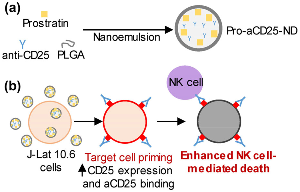
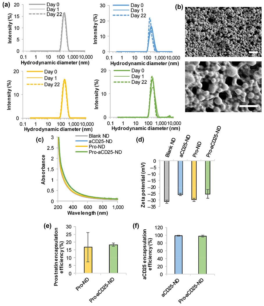
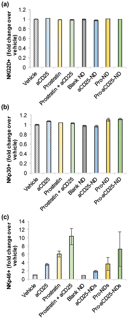
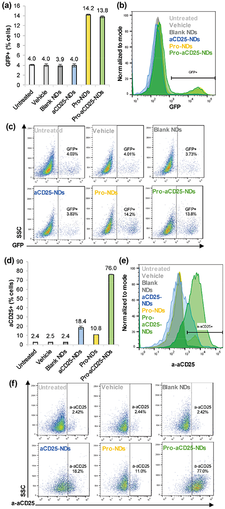
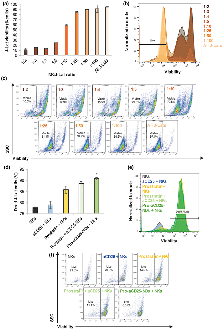
Natural killer (NK) cells are attractive effector cells of the innate immune system against human immunodeficiency virus (HIV) and cancer. However, NK cell therapies are limited by the fact that target cells evade NK cells, for example, in latent reservoirs (in HIV) or through upregulation of inhibitory signals (in cancer). To address this limitation, we describe a biodegradable nanoparticle-based "priming" approach to enhance the cytotoxic efficacy of peripheral blood mononuclear cell-derived NK cells. We present poly(lactic-co-glycolic acid) (PLGA) nanodepots (NDs) that co-encapsulate prostratin, a latency-reversing agent, and anti-CD25 (aCD25), a cell surface binding antibody, to enhance primary NK cell function against HIV and cancer. We utilize a nanoemulsion synthesis scheme to encapsulate both prostratin and aCD25 within the PLGA NDs (termed Pro-aCD25-NDs). Physicochemical characterization studies of the NDs demonstrated that our synthesis scheme resulted in stable and monodisperse Pro-aCD25-NDs. The NDs successfully released both active prostratin and anti-CD25, and with controllable release kinetics. When Pro-aCD25-NDs were administered in an in vitro model of latent HIV and acute T cell leukemia using J-Lat 10.6 cells, the NDs were observed to prime J-Lat cells resulting in significantly increased NK cell-mediated cytotoxicity compared to free prostratin plus anti-CD25, and other controls. These findings demonstrate the feasibility of using our Pro-aCD25-NDs to prime target cells for enhancing the cytotoxicity of NK cells as antiviral or antitumor agents.

Keywords: antibody; cancer; human immunodeficiency virus (HIV); latency-reversing agent; natural killer (NK) cells; poly(lactic-co-glycolic acid) (PLGA) nanodepots.


Figures

Figure 1
Figure 1
Schematic overview of our PLGA ND-based approach for enhancing NK cell cytotoxic function, (a) PLGA NDs encapsulating prostratin, a latency-reversing agent, and aCD25 were synthesized using a nanoemulsion scheme, (b) J-Lat 10.6 cells, a model of latent HIV and leukemia, treated with Pro-aCD25-ND increased CD25 expression in response to released prostratin, which enables increased aCD25 binding (also released from the NDs). These effects function in concern to prime the targeted cells for enhanced NK cell-mediated killing.
Figure 2
Figure 2
Stable and monodisperse PLGA NDs co-encapsulate prostratin and aCD25. (a) ND hydrodynamic diameters were measured by DLS; gray: Blank NDs, blue: aCD25-NDs, yellow: Pro-NDs, green: Pro-aCD25-NDs. (b) SEM images of Pro-aCD25-NDs at increasing magnifications (top to bottom). Scale bar = 2 μm (top) and 1 μm (bottom), (c) Absorbance spectra were analyzed using UV–Vis–NIR spectroscopy, (d) Zeta potential was measured by electrophoretic light scattering. The encapsulation efficiency of (e) prostratin was determined by absorbance spectroscopy and that of (f) aCD25 was determined by BCA protein assay.
Figure 3
Figure 3
Pro-aCD25-NDs exhibit tunable release kinetics of both prostratin and aCD25. (a) and (b) Pro-aCD25-NDs were incubated in PBS at 37 °C. Cumulative release of (a) prostratin and (b) aCD25 into the solution was analyzed at the listed time periods by UV–Vis spectroscopy and BCA protein assay, respectively, (c) Pro-aCD25-NDs were incubated in PBS at 37 °C for 24 h. At indicated timepoints, release of aCD25 from NDs into the solution was measured by BCA assay. Fresh PBS was used for replacement at each timepoint in the replacement group (shaded circles). No fresh PBS was added to the no replacement group (empty circles), (d) and (e) After synthesis, Pro-aCD25-NDs were incubated at 4 °C. After 24 h, Pro-aCD25-NDs were incubated in PBS at 37 °C for six days. At each listed timepoint, an aliquot was centrifuged, and released (d) prostratin and (e) aCD25 was measured by UV-Vis spectroscopy and BCA assay, respectively. Fresh PBS was replaced at every timepoint measured (i.e. 24, 48, and 72 h). Values represent means ± standard deviation (n = 3/group).
Figure 4
Figure 4
PLGA NDs and their encapsulated contents do not significantly alter NK cell phenotype. Primary NK cells were cultured with NDs, their constituent components, or controls. After 24 h, (a) NKG2D, (b) NKp30, and (c) NKp46 expressions were measured by flow cytometry. Values represent means ± standard deviation (n = 2/group).
Figure 5
Figure 5
NDs function to generate activation of antibody binding to J-Lat cells, (a) J-Lat cells were cultured with NDs and controls. After 24 h> GFP expression was analyzed as a measure of J-Lat cell activation by flow cytometry, (b) Representative histograms of J-Lat cell GFP expression after 24 h culture with NDs and controls, (c) Representative scatter plots of J-Lat cell GFP expression after 24 h culture with NDs and controls, (d) J-Lat cells were cultured with NDs and controls. After 24 h, aCD25 binding to J-Lat cells was analyzed by detecting secondary antibody binding via flow cytometry, (e) Representative histograms of aCD25 binding to J-Lat cells after 24 h culture with NDs and controls, (f) Representative scatter plots of aCD25 binding to J-Lat cells after 24 h culture with NDs and controls. Values represent means ± standard deviation (n = 3/group).
Figure 6
Figure 6
NDs enhance NK cell cytotoxic function on J-Lat cells, (a) NK cells were co-cultured with J-Lat cells at listed E:T ratios for 24 h. J-Lat cell viability was measured by flow cytometry, (b) Representative histograms of J-Lat cell viability after 24 h co-culture with NK cells at varied E:T ratios, (c) Representative scatter plots of J-Lat cell viability after 24 h co-culture with NK cells at varied E:T ratios, (d) NK cells, in combination with the listed additional agents, were co-cultured with J-Lat cells for 24 h at a 1:1 E:T ratio; J-Lat cell death was measured by flow cytometry, (e) Representative histograms of J-Lat cell death after 24 h co-culture with NK cells + additional agents, (f) Representative scatter plots of J-Lat cell death after 24 h co-culture with NK cells + additional agents. Values represent means ± standard deviation (n = 3/group), *p < 0.05 (versus all other groups).

References

    1. Mikulak J; Oriolo F; Zaghi E; Di Vito C; Mavilio D Natural killer cells in HIV-1 infection and therapy. AIDS 2017, 31, 2317–2330. - PMC - PubMed
    1. Guillerey C; Huntington ND; Smyth MJ Targeting natural killer cells in cancer immunotherapy. Nat. Immunol 2016, 17, 1025–1036. - PubMed
    1. Breunig M; Lungwitz U; Liebl R; Goepferich A Breaking up the correlation between efficacy and toxicity for nonviral gene delivery. Proc. Natl. Acad. Sci. USA 2007, 104, 14454–14459. - PMC - PubMed
    1. Daher M; Rezvani K Next generation natural killer cells for cancer immunotherapy: The promise of genetic engineering. Curr. Opin. Immunol 2018, 51, 146–153. - PMC - PubMed
    1. Florea BI; Meaney C; Junginger HE; Borchard G Transfection efficiency and toxicity of polyethylenimine in differentiated Calu-3 and nondifferentiated COS-1 cell cultures. AAPS PharmSci 2002, 4, 1–11. - PMC - PubMed