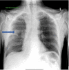
Ectopic Cushing Syndrome in Adenocarcinoma of the Lung: Case Report and Literature Review
- PMID: 34079679
- PMCID: PMC8162053
- DOI: 10.7759/cureus.14733
Ectopic Cushing Syndrome in Adenocarcinoma of the Lung: Case Report and Literature Review
Abstract
Paraneoplastic syndromes are rare disorders that occur with many types of tumors. Ectopic cushing syndrome (ECS) is the second most common paraneoplastic syndrome that is only seen in 1-5% of all small cell lung cancers (SCLC), with limited papers reporting this syndrome since it was first described by Brown in 1928 or in carcinoid tumors. It is also found to be associated to a lesser extent with pheochromocytoma, thymic tumors, pancreatic carcinoma, and anaplastic thyroid carcinoma. While lung adenocarcinoma is the most common histological type of lung neoplasms, it is seldom associated with Cushing syndrome. In this article, we describe a patient who initially presented with Cushing syndrome and found to have adenocarcinoma of the lung.
Keywords: adenocarcinoma; ectopic cushing’s syndrome; paraneoplastic syndromes.
Copyright © 2021, Al-Zakhari et al.
Conflict of interest statement
The authors have declared that no competing interests exist.
Figures


References
-
- Myers DJ, Wallen JM. StatPearls [Internet] Treasure Island: StatPearls Publishing; 2021. Lung Adenocarcinoma. [Updated 2020 Jun 26] - PubMed
-
- Adenocarcinoma of the lung causing ectopic adrenocorticotropic hormone syndrome. Myers EA, Hardman JM, Worsham GF, Eil C. Arch Intern Med. 1982;142:1387–1389. - PubMed
-
- Paraneoplastic syndromes: the way to an early diagnosis of lung cancer. Paraschiv B, Diaconu CC, Toma CL, Bogdan MA. https://www.researchgate.net/profile/Bianca-Paraschiv/publication/280086... Pneumologia. 2015;64:14–19. - PubMed
Publication types
LinkOut - more resources
Full Text Sources
