

Divergent effects of irradiation on brain cortical morphology in patients with nasopharyngeal carcinoma: one-year follow-up study using structural magnetic resonance imaging
- PMID: 34079703
- PMCID: PMC8107334
- DOI: 10.21037/qims-20-662
Divergent effects of irradiation on brain cortical morphology in patients with nasopharyngeal carcinoma: one-year follow-up study using structural magnetic resonance imaging
Abstract
Background: Increasing evidence indicates that radiotherapy (RT)-induced brain cortical deficits may play a critical role in developing radiation encephalopathy in patients with nasopharyngeal carcinoma (NPC). However, the evolutional processes of RT-induced cortical injury have not been sufficiently investigated. This study investigates RT-induced effects on cortical morphology using longitudinal structural magnetic resonance imaging (MRI) in NPC patients.
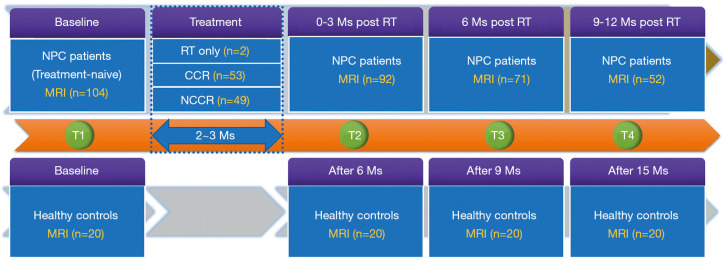
Methods: Using MRI-based morphometry with surface-based measures, we evaluated the longitudinal alterations of cortical volume (CV), cortical thickness (CT), and cortical surface area (CSA) in 104 NPC patients at pre-RT (n=104), within 3 months post-RT (n=92), 6 months post-RT (n=71), and 9-12 months post-RT (n=52). Twenty healthy controls were also evaluated in parallel. Linear mixed models were used to investigate the trajectories of RT-related changes in cortical brain morphology and its association with irradiation dose, with healthy controls data being used to construct a normal age-related benchmark. The level of statistical significance was set at P<0.05, corrected for multiple comparisons.
Results: The results showed that RT-related longitudinal alterations in cortical morphology underwent two diverse patterns during the first year of follow up in NPC patients. The temporal cortices (including the bilateral superior temporal gyrus, middle temporal gyrus, temporal pole, parahippocampal and fusiform gyrus, and the right inferior temporal and right transverse temporal gyrus), the basal occipital cortices (the right lingual gyrus and lateral occipital gyrus), and the basal frontal cortices (the right lateral orbitofrontal gyrus) showed time-dependent attenuation in cortical morphology indices. Furthermore, these effects on multiple cortices were dose-dependent, suggesting they were RT-associated. In contrast, in the left rostral middle frontal gyrus, there was a time-dependent increase in CT.
Conclusions: Our preliminary findings revealed divergent effects of irradiation on cortical brain morphology. These results contribute to a more comprehensive understanding of the underlying neural mechanisms of irradiation-related neurotoxic effects on cortical brain morphology and will help guide the investigation of critically neuroprotective strategies.
Keywords: Cortical thickness (CT); cortical surface area (CSA); cortical volume (CV); nasopharyngeal carcinoma (NPC); radiotherapy (RT).
2021 Quantitative Imaging in Medicine and Surgery. All rights reserved.
Conflict of interest statement
Conflicts of Interest: All authors have completed the ICMJE uniform disclosure form (available at http://dx.doi.org/10.21037/qims-20-662). The authors have no conflicts of interest to declare.
Figures


Similar articles
-
Radiation-induced abnormal cortical thickness in patients with nasopharyngeal carcinoma after radiotherapy.Neuroimage Clin. 2017 Mar 2;14:610-621. doi: 10.1016/j.nicl.2017.02.025. eCollection 2017. Neuroimage Clin. 2017. PMID: 28348952 Free PMC article.
-
Irradiation-related longitudinal white matter atrophy underlies cognitive impairment in patients with nasopharyngeal carcinoma.Brain Imaging Behav. 2021 Oct;15(5):2426-2435. doi: 10.1007/s11682-020-00441-0. Epub 2021 Jan 21. Brain Imaging Behav. 2021. PMID: 33474681
-
Radiation-induced changes in normal-appearing gray matter in patients with nasopharyngeal carcinoma: a magnetic resonance imaging voxel-based morphometry study.Neuroradiology. 2014 May;56(5):423-30. doi: 10.1007/s00234-014-1338-y. Epub 2014 Mar 8. Neuroradiology. 2014. PMID: 24609527
-
Radiation-induced hippocampal atrophy in patients with nasopharyngeal carcinoma early after radiotherapy: a longitudinal MR-based hippocampal subfield analysis.Brain Imaging Behav. 2019 Aug;13(4):1160-1171. doi: 10.1007/s11682-018-9931-z. Brain Imaging Behav. 2019. PMID: 30054872
-
Impairment mechanism of nasal mucosa after radiotherapy for nasopharyngeal carcinoma.Front Oncol. 2022 Dec 15;12:1010131. doi: 10.3389/fonc.2022.1010131. eCollection 2022. Front Oncol. 2022. PMID: 36591522 Free PMC article. Review.
Cited by
-
Case report: intracranial lesions in a patient with anxiety and depression: tumor recurrence or radiation encephalopathy?Front Oncol. 2024 Aug 15;14:1422765. doi: 10.3389/fonc.2024.1422765. eCollection 2024. Front Oncol. 2024. PMID: 39211558 Free PMC article.
-
Radiation-induced aberrant structural covariance networks in patients with nasopharyngeal carcinoma: a source-based morphometry study.Quant Imaging Med Surg. 2024 Dec 5;14(12):9126-9136. doi: 10.21037/qims-24-270. Epub 2024 Nov 29. Quant Imaging Med Surg. 2024. PMID: 39698681 Free PMC article.
References
LinkOut - more resources
Full Text Sources