. 2021 May 17:9:658101.
doi: 10.3389/fcell.2021.658101. eCollection 2021.

hTERT Promotes CRC Proliferation and Migration by Recruiting YBX1 to Increase NRF2 Expression

Affiliations

hTERT Promotes CRC Proliferation and Migration by Recruiting YBX1 to Increase NRF2 Expression

Chunli Gong et al. Front Cell Dev Biol. .

Abstract

High human telomerase reverse transcriptase (hTERT) expression is related to severe Colorectal Cancer (CRC) progression and negatively related to CRC patient survival. Previous studies have revealed that hTERT can reduce cancer cellular reactive oxygen species (ROS) levels and accelerate cancer progression; however, the mechanism remains poorly understood. NFE2-related factor 2 (NRF2) is a molecule that plays a significant role in regulating cellular ROS homeostasis, but whether there is a correlation between hTERT and NRF2 remains unclear. Here, we showed that hTERT increases CRC proliferation and migration by inducing NRF2 upregulation. We further found that hTERT increases NRF2 expression at both the mRNA and protein levels. Our data also revealed that hTERT primarily upregulates NRF2 by increasing NRF2 promoter activity rather than by regulating NRF2 mRNA or protein stability. Using DNA pull-down/MS analysis, we found that hTERT can recruit YBX1 to upregulate NRF2 promoter activity. We also found that hTERT/YBX1 may localize to the P2 region of the NRF2 promoter. Taken together, our results demonstrate that hTERT facilitates CRC proliferation and migration by upregulating NRF2 expression through the recruitment of the transcription factor YBX1 to activate the NRF2 promoter. These results provide a new theoretical basis for CRC treatment.

Keywords: NRF2; YBX1; colorectal cancer; hTERT; progression.


Conflict of interest statement

The authors declare that the research was conducted in the absence of any commercial or financial relationships that could be construed as a potential conflict of interest.

Figures

FIGURE 1
FIGURE 1
Human telomerase reverse transcriptase (hTERT) and NRF2 are highly expressed in CRC tissues and associated with poor diagnosis. (A) hTERT expression in CRC tissues and adjacent normal tissues was investigated in Oncomine database (Cohort1). (B) NRF2 expression in CRC tissues and adjacent normal tissues was investigated in Oncomine database (Cohort2). (C) hTERT mRNA expression in CRC tissues and paired adjacent normal tissues was identified by qRT-PCR (Cohort3). (D) NRF2 mRNA expression in CRC tissues and paired adjacent normal tissues was identified by qRT-PCR (Cohort3). (E) Regulation analysis of the correlation between hTERT and NRF2 (Cohort3). Each point represents one cancer sample. (F) Representative immunohistochemical staining and expression level statistics of hTERT and NRF2 in CRC tissues and paired adjacent normal tissues (Cohort4). (G) Regulation analysis of the correlation between hTERT and NRF2 (Cohort4). Each point represents one cancer sample. (H) Kaplan–Meier analysis of the overall survival of CRC patients with different hTERT expression levels (p < 0.05, log-rank test). (I) Kaplan–Meier analysis of the overall survival of CRC patients with different NRF2 expression levels (p < 0.05, log-rank test). (J) Kaplan–Meier analysis of the overall survival of CRC patients with different hTERT and NRF2 expression levels (p < 0.05, log-rank test). *p < 0.05; **p < 0.01; ***p < 0.001; ****p < 0.0001; ns, no significance.
FIGURE 2
FIGURE 2
Human telomerase reverse transcriptase (hTERT) upregulates NRF2 expression by promoting NRF2 transcriptional activity. (A) hTERT-knockdown cells were constructed by transfecting sh1-hTERT and sh2-hTERT lenviral respectively into HCT 116 cells. The hTERT and NRF2 mRNA expression level after downregulation of hTERT were identified by qRT-PCR. (B) hTERT and NRF2 protein expression levels after downregulation of hTERT were detected by western blotting (Left). Statistical analysis of western blotting (Right). (C) hTERT-overexpressed cell was constructed by transfecting hTERT lenviral into SW620 cells. The hTERT and NRF2 mRNA expression level after upregulation of hTERT was identified by qRT-PCR. (D) hTERT and NRF2 protein expression levels after overexpression of hTERT were detected by western blotting (Left). Statistical analysis of western blotting (Right). (E) Luciferase activity of the NRF2 promoter was detected after downregulation of hTERT. (F) Luciferase activity of NRF2 was detected after overexpression of hTERT. *p < 0.05; **p < 0.01; ***p < 0.001; ns, no significance.
FIGURE 3
FIGURE 3
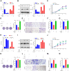
Human telomerase reverse transcriptase (hTERT) promotes colorectal cancer cell proliferation and metastasis by upregulating NRF2. (A) CCK8 assays were performed to detect cell proliferation after downregulation of hTERT but an increase in NRF2 expression. (B) Colony formation assays were performed after downregulation of hTERT but an increase in NRF2 expression. (C) Statistical analysis of the colony numbers. (D) Migration and invasion assays were performed after downregulation of hTERT but an increase in NRF2 expression. (E) Statistical analysis of the migration cell numbers. (F) Statistical analysis of the invasion cell numbers. *p < 0.05; **p < 0.01; ***p < 0.001; ns, no significance.
FIGURE 4
FIGURE 4
Human telomerase reverse transcriptase (hTERT) increases NRF2 expression by recruiting YBX1 to bind to the NRF2 promoter. (A) Flow chart for screening of potential hTERT-recruited NRF2 transcription factors. (B) The YBX1 and NRF2 mRNA expression level was identified by qRT-PCR after downregulation of YBX1. (C) Luciferase activity of the NRF2 promoter was detected after downregulation of YBX1. (D) NRF2 promoter with a 5′ biotin label was used to pull-down YBX1. Mock beads were used as a negative control. (E) Diagrammatic drawing of NRF2 P2 fragment. The P2 fragment was divided into 5 fragments. (F) YBX1 antibody was used to immunoprecipitate binding fragments of the NRF2 promoter under the condition of hTERT overexpression and fragments were identified by ChIP-qPCR with six primers (Left). Statistical analysis of ChIP-qPCR. (G) Luciferase activity of P2 fragment containing different mutant sites was detected after YBX1 overexpression. (H) HCT116 cell lysates were prepared for separate IP with hTERT and YBX1 antibody and then evaluated via western blotting. (I) The subcellular localization and the colocalization of hTERT and YBX1 were examined in SW620 cells via dual immunofluorescence using confocal microscopy. (J) YBX1 in cell nuclei and cytoplasm was identified by western blotting after overexpression of Flag-hTERT. (K) The NRF2 mRNA expression level was identified by qRT-PCR after overexpression of hTERT and simultaneous knockdown of YBX1. (L) Luciferase activity of the NRF2 promoter was detected after overexpression of hTERT and simultaneous knockdown of YBX1. (M) The NRF2 protein level was identified by western blotting after overexpression of hTERT and simultaneous knockdown of YBX1 (Left). Statistical analysis of western blotting (Right). *p < 0.05; **p < 0.01; ***p < 0.001; ns, no significance.
FIGURE 5
FIGURE 5
YBX1 is responsible for upregulation of NRF2 expression and CRC proliferation and migration. (A) The YBX1 and NRF2 mRNA expression level were identified by qRT-PCR after downregulation of YBX1 and simultaneous overexpression of NRF2. (B) The YBX1 and NRF2 protein expression level were identified via western blotting after downregulation of YBX1 and simultaneous overexpression of NRF2 (Left). Statistical analysis of western blotting (Right). (C) CCK8 assays were performed to detect cell proliferation after downregulation of YBX1 but an increase in NRF2 expression. (D) Colony formation assays were performed after downregulation of YBX1 but an increase in NRF2 expression (Left). Statistical analysis of the colony numbers (Right). (E) Migration and invasion assays were performed after downregulation of YBX1 but an increase in NRF2 expression. (F) Statistical analysis of the migration cell numbers. (G) Statistical analysis of the invasion cell numbers. (H) The YBX1 and NRF2 mRNA expression level were identified by qRT-PCR after overexpression of YBX1 and simultaneous downregulation of NRF2. (I) The YBX1 and NRF2 protein expression level were identified via western blotting after overexpression of YBX1 and simultaneous downregulation of NRF2 (Left). Statistical analysis of western blotting (Right). (J) CCK8 assays were performed to detect cell proliferation after overexpression of YBX1 and downregulation of NRF2. (K) Colony formation assays were performed after overexpression of YBX1 and downregulation of NRF2 (Left). Statistical analysis of the colony numbers (Right). (L) Migration and invasion assays were performed after overexpression of YBX1 and downregulation of NRF2. (M) Statistical analysis of the migration cell numbers. (N) Statistical analysis of the invasion cell numbers. *p < 0.05; **p < 0.01; ***p < 0.001; ns, no significance.
FIGURE 6
FIGURE 6
YBX1 is highly expressed in CRC and associated with poor prognosis. (A) YBX1 expression in CRC tissues and adjacent normal tissues was investigated in Oncomine database. (B) YBX1 mRNA expression in CRC tissues and paired adjacent normal tissues was identified by qRT-PCR. (C) Regulation analysis of the correlation between YBX1 and NRF2. Each point represents one cancer sample. (D) Representative immunohistochemical staining and expression level statistics of YBX1 in CRC tissues and paired adjacent normal tissues. (E) Regulation analysis of the correlation between YBX1 and NRF2. Each point represents one cancer sample. (F) Kaplan–Meier analysis of the overall survival of CRC patients with different YBX1 expression levels (p < 0.05, log-rank test). (G) Kaplan–Meier analysis of the overall survival of CRC patients with different YBX1 and NRF2 expression levels (p < 0.05, log-rank test). *p < 0.05;**p < 0.01; ***p < 0.001; ****p < 0.0001; ns, no significance.
FIGURE 7
FIGURE 7
Model for transcriptional regulation of NRF2 by hTERT via recruitment of YBX1 in CRC proliferation and migration. In this model, hTERT recruits YBX1 to form a transcriptional complex, which binds to the NRF2 promoter to promote NRF2 expression, thus promoting CRC proliferation and migration.

Similar articles

Cited by

References

    1. Chen K. G., Sale S., Tan T., Ermoian R. P., Sikic B. I. (2004). CCAAT/enhancer-binding protein beta (nuclear factor for interleukin 6) transactivates the human MDR1 gene by interaction with an inverted CCAAT box in human cancer cells. Mole. Pharm. 65 906–916. 10.1124/mol.65.4.906 - DOI - PubMed
    1. Cui G., Zhao H., Li L. (2020). Long noncoding RNA PRKCQ-AS1 promotes CRC cell proliferation and migration via modulating miR-1287-5p/YBX1 axis. J. Cell. Biochem. 121 4166–4175. 10.1002/jcb.29712 - DOI - PubMed
    1. Dilshara M. G., Jayasooriya R., Choi Y. H., Kim G. Y. (2019). Camptothecin induces c-Myc- and Sp1-mediated hTERT expression in LNCaP cells: Involvement of reactive oxygen species and PI3K/Akt. Food Chem. Toxicol. 127 53–60. 10.1016/j.fct.2019.03.001 - DOI - PubMed
    1. Fan W., Tang Z., Chen D., Moughon D., Ding X., Chen S., et al. (2010). Keap1 facilitates p62-mediated ubiquitin aggregate clearance via autophagy. Autophagy 6 614–621. 10.4161/auto.6.5.12189 - DOI - PMC - PubMed
    1. Ge W., Zhao K., Wang X., Li H., Yu M., He M., et al. (2017). iASPP Is an Antioxidative Factor and Drives Cancer Growth and Drug Resistance by Competing with Nrf2 for Keap1 Binding. Cancer Cell 32 56:e566. - PubMed

LinkOut - more resources