

Do endosonographers agree on the presence of bile duct sludge and the subsequent need for intervention?
- PMID: 34079877
- PMCID: PMC8159618
- DOI: 10.1055/a-1452-8919
Do endosonographers agree on the presence of bile duct sludge and the subsequent need for intervention?
Abstract
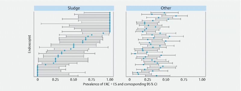
Background and study aims Endoscopic ultrasonography (EUS) is a tool widely used to diagnose bile duct lithiasis. In approximately one out of five patients with positive findings at EUS, sludge is detected in the bile duct instead of stones. The objective of this study was to establish the agreement among endosonographers regarding: 1. presence of common bile duct (CBD) stones, microlithiasis and sludge; and 2. the need for subsequent treatment. Patients and methods 30 EUS videos of patients with an intermediate probability of CBD stones were evaluated by 41 endosonographers. Experience in EUS and endoscopic retrograde cholangiopancreatography, and the endosonographers' type of practices were recorded. Fleiss' kappa statistics were used to quantify the agreement. Associations between levels of experience and both EUS ratings and treatment decisions were investigated using mixed effects models. Results A total of 1230 ratings and treatment decisions were evaluated. The overall agreement on EUS findings was fair (Fleiss' κ 0.32). The agreement on presence of stones was moderate (κ 0.46). For microlithiasis it was fair (κ 0.25) and for sludge it was slight (κ 0.16). In cases with CBD stones there was an almost perfect agreement for the decision to subsequently perform an ERC + ES. In case of presumed microlithiasis or sludge an ERC was opted for in 78 % and 51 % of cases, respectively. Differences in experience and types of practice appear unrelated to the agreement on both EUS findings and the decision for subsequent treatment. Conclusions There is only slight agreement among endosonographers regarding the presence of bile duct sludge. Regarding the need for subsequent treatment of bile duct sludge there is no consensus.
The Author(s). This is an open access article published by Thieme under the terms of the Creative Commons Attribution-NonDerivative-NonCommercial License, permitting copying and reproduction so long as the original work is given appropriate credit. Contents may not be used for commercial purposes, or adapted, remixed, transformed or built upon. (https://creativecommons.org/licenses/by-nc-nd/4.0/).
Conflict of interest statement
Competing interests The authors declare that they have no conflict of interest.
Figures


References
-
- Petrov M S, Savides T J. Systematic review of endoscopic ultrasonography versus endoscopic retrograde cholangiopancreatography for suspected choledocholithiasis. Br J Surg. 2009;96:967–974. - PubMed
-
- Tse F, Liu L, Barkun A N et al.EUS: a meta-analysis of test performance in suspected choledocholithiasis. Gastrointest Endosc. 2008;67:235–244. - PubMed
-
- Manes G, Paspatis G, Aabakken L et al.Endoscopic management of common bile duct stones: European Society of Gastrointestinal Endoscopy (ESGE) guideline. Endoscopy. 2019;51:472–491. - PubMed
-
- Fusaroli P, Lisotti A, Syguda A et al.Reliability of endoscopic ultrasound in predicting the number and size of common bile duct stones before endoscopic retrograde cholangiopancreatography. Dig Liver Dis. 2016;48:277–282. - PubMed
-
- Quispel R, van Driel L M, Veldt B J et al.The utility and yield of endoscopic ultrasonography for suspected choledocholithiasis in common gastroenterology practice. Eur J Gastroenterol Hepatol. 2016;28:1473–1476. - PubMed
LinkOut - more resources
Full Text Sources