Review
. 2021 Dec;206(3):251-265.
doi: 10.1111/cei.13630. Epub 2021 Jul 6.

Magnetic resonance imaging in neuromyelitis optica spectrum disorder

Affiliations
Review

Magnetic resonance imaging in neuromyelitis optica spectrum disorder

Laura Clarke et al. Clin Exp Immunol. 2021 Dec.

Abstract

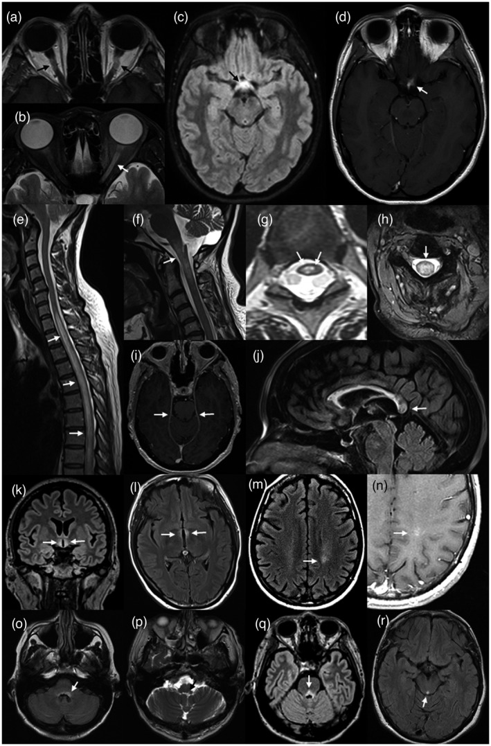
Neuromyelitis optica spectrum disorder (NMOSD) is an inflammatory disease of the central nervous system (CNS) associated with antibodies to aquaporin-4 (AQP4), which has distinct clinical, radiological and pathological features, but also has some overlap with multiple sclerosis and myelin oligodendrocyte glycoprotein (MOG) antibody associated disease. Early recognition of NMOSD is important because of differing responses to both acute and preventive therapy. Magnetic resonance (MR) imaging has proved essential in this process. Key MR imaging clues to the diagnosis of NMOSD are longitudinally extensive lesions of the optic nerve (more than half the length) and spinal cord (three or more vertebral segments), bilateral optic nerve lesions and lesions of the optic chiasm, area postrema, floor of the IV ventricle, periaqueductal grey matter, hypothalamus and walls of the III ventricle. Other NMOSD-specific lesions are denoted by their unique morphology: heterogeneous lesions of the corpus callosum, 'cloud-like' gadolinium (Gd)-enhancing white matter lesions and 'bright spotty' lesions of the spinal cord. Other lesions described in NMOSD, including linear periventricular peri-ependymal lesions and patch subcortical white matter lesions, may be less specific. The use of advanced MR imaging techniques is yielding further useful information regarding focal degeneration of the thalamus and optic radiation in NMOSD and suggests that paramagnetic rim patterns and changes in normal appearing white matter are specific to MS. MR imaging is crucial in the early recognition of NMOSD and in directing testing for AQP4 antibodies and guiding immediate acute treatment decisions. Increasingly, MR imaging is playing a role in diagnosing seronegative cases of NMOSD.

Keywords: diagnosis; magnetic resonance imaging; multiple sclerosis; neuromyelitis optica.


Conflict of interest statement

Simon A. Broadley has received honoraria for attendance at advisory boards and travel sponsorship from Bayer‐Schering, Biogen‐Idec, Merck‐Serono, Novartis and Sanofi‐Genzyme, has received speaker’s honoraria from Biogen‐Idec and Genzyme, is an investigator in clinical trials sponsored by Biogen Idec, Novartis and Genzyme and was the recipient of an unencumbered research grant from Biogen‐Idec. Laura Clarke, Simon Arnett, Kate Lilley, Jacky Liao and Sandeep Bhuta report no disclosures.

Figures

FIGURE 1
FIGURE 1
Perivascular distribution of aquaporin‐4 (AQP4) antibodies and complement in neuromyelitis optica spectrum disorder (NMOSD). AQP4 antibody positive immunofluorescence (green) in mouse cerebellum [serum dilution 1:40, goat anti‐human immunoglobulin (Ig)G F(ab)2 fluorescein isothiocyanate, ×200 magnification], showing microvessel staining of the granular layer, molecular layer and white matter (a). Section of early white matter lesion in NMOSD immunostained (brown) for Cd3 showing typical perivascular complement deposition around small blood vessels (b)
FIGURE 2
FIGURE 2
Schematic representation of the pathogenesis of neuromyelitis optica spectrum disorder (NMOSD). (1) Presentation of aquaporin‐4 (AQP4) antigen via an antigen‐presenting cell (APC) in a peripheral lymph node with promotion from T helper cell leads to activation of autoreactive B cells. (2) Maturation of antibody producing plasma cells results in free circulating autoreactive antibodies to AQP4. (3) Break‐down of the blood–brain barrier (BBB) leads to escape of autoreactive antibodies and extravasation of immune cells in response to chemokines. (4) Opsonization of autoreactive antibodies on AQP4 rafts situated on the astrocyte foot process leads to activation of complement and cell‐mediated lysis of astrocytes. (5) Indiscriminatory inflammation mediated via chemokines, activated complement and degranulation products, as well as loss of trophic support from astrocytes, leads to lysis of oligodendrocytes and neurons. (6) Macrophages attracted by chemokines released by leukocytes and astrocytes produce further proinflammatory products and phagocytose cellular debris and myelin
FIGURE 3
FIGURE 3
Magnetic resonance (MR) imaging of lesions (arrows) associated with aquaporin‐4 (AQP4)‐positive neuromyelitis optica spectrum disorder (NMOSD). Bilateral Gd‐enhancing retro‐orbital lesions of the optic nerves on axial T1 weighted image of the orbits (a). Longitudinally extensive (more than half the length of the optic nerve) high signal lesion of the left optic nerve on axial T2 image of the orbits (b). High signal lesion of the optic chiasm on volumetric, axial FLAIR image of the brain (c). Asymmetric gadolinium (Gd)‐enhancement of the same lesion (d). Longitudinally extensive high signal lesion of the thoracic spinal cord, associated with cord swelling, on sagittal T2 imaging of the spine (e). High cervical cord high signal lesion extending into the medulla on sagittal T2 image of the cervical cord (f). ‘Bright spotty lesions’ of the thoracic spinal cord on axial T2 imaging of the spine (g). Central, whole cord high signal lesion on axial T2 image of the cervical cord (h). Leptomeningeal Gd‐enhancement on axial T1 weighted imaging of the brain at the level of the upper pons (i). Heterogeneous rounded lesion of the corpus callosum on sagittal fluid‐attenuated inversion recovery (FLAIR) imaging of the brain (j). Hypothalamic high signal lesion on volumetric coronal FLAIR imaging of the brain (k). High signal change in the wall and adjacent parenchyma of the III ventricle on axial FLAIR imaging of the brain (l). High signal lesion of the left posterior frontal white matter on axial FLAIR imaging (m) showing ‘cloud‐like’ enhancement on axial T1 imaging of the same region (n). Bilateral area postrema lesions (more prominent on the left) on axial FLAIR image at the level of the medulla (o). Left nucleus solitarius lesion on axial T2 image at the level of the medulla (p). High signal FLAIR lesion of the pons, involving the floor of the IV ventricle (q). High signal lesion of the peri‐aqueductal grey matter on axial FLAIR image through the mid‐brain (r)

References

    1. Devic E. Myélite subaiguë compliquée de névrite optique. Bull Med. 1894;8:1033–4.
    1. Devic E. Acute lumbar spinal myelitis with optic neuritis, autopsy. French Cong Med. 1895;1:434–9.
    1. Albutt FC. On the ophthalmoscopic signs of spinal cord disease. Lancet 1870;i:76–8.
    1. Lennon VA, Wingerchuk DM, Kryzer TJ, Pittock SJ, Lucchinetti CF, Fujihara K, et al. A serum autoantibody marker of neuromyelitis optica: distinction from multiple sclerosis. Lancet 2004;364:2106–12. - PubMed
    1. Paul F, Jarius S, Aktas O, Bluthner M, Bauer O, Appelhans H, et al. Antibody to aquaporin 4 in the diagnosis of neuromyelitis optica. PLOS Med. 2007;4:e133. - PMC - PubMed

Publication types

MeSH terms