Phytochemical library screening reveals betulinic acid as a novel Skp2-SCF E3 ligase inhibitor in non-small cell lung cancer
- PMID: 34080260
- PMCID: PMC8353894
- DOI: 10.1111/cas.15005
Phytochemical library screening reveals betulinic acid as a novel Skp2-SCF E3 ligase inhibitor in non-small cell lung cancer
Erratum in
-
Correction to: Phytochemical library screening reveals betulinic acid as a novel Skp2-SCF E3 ligase inhibitor in non-small cell lung cancer.Cancer Sci. 2024 Jun;115(6):2083-2085. doi: 10.1111/cas.16163. Epub 2024 Mar 20. Cancer Sci. 2024. PMID: 38507574 Free PMC article. No abstract available.
Abstract
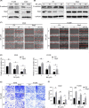
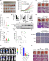
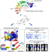
Skp2 is overexpressed in multiple cancers and plays a critical role in tumor development through ubiquitin/proteasome-dependent degradation of its substrate proteins. Drugs targeting Skp2 have exhibited promising anticancer activity. Here, we identified a plant-derived Skp2 inhibitor, betulinic acid (BA), via high-throughput structure-based virtual screening of a phytochemical library. BA significantly inhibited the proliferation and migration of non-small cell lung cancer (NSCLC) through targeting Skp2-SCF E3 ligase both in vitro and in vivo. Mechanistically, BA binding to Skp2, especially forming H-bonds with residue Lys145, decreases its stability by disrupting Skp1-Skp2 interactions, thereby inhibiting the Skp2-SCF E3 ligase and promoting the accumulation of its substrates; that is, E-cadherin and p27. In both subcutaneous and orthotopic xenografts, BA significantly inhibited the proliferation and metastasis of NSCLC through targeting Skp2-SCF E3 ligase and upregulating p27 and E-cadherin protein levels. Taken together, BA can be considered a valuable therapeutic candidate to inhibit metastasis of NSCLC.
Keywords: E-cadherin; NSCLC; Skp2; betulinic acid; metastasis.
© 2021 The Authors. Cancer Science published by John Wiley & Sons Australia, Ltd on behalf of Japanese Cancer Association.
Conflict of interest statement
The authors declare that they have no competing interests.
Figures


Similar articles
-
Brusatol has therapeutic efficacy in non-small cell lung cancer by targeting Skp1 to inhibit cancer growth and metastasis.Pharmacol Res. 2022 Feb;176:106059. doi: 10.1016/j.phrs.2022.106059. Epub 2022 Jan 5. Pharmacol Res. 2022. PMID: 34998973
-
Centipeda minima extracts and the active sesquiterpene lactones have therapeutic efficacy in non-small cell lung cancer by suppressing Skp2/p27 signaling pathway.J Ethnopharmacol. 2025 Jan 31;340:119277. doi: 10.1016/j.jep.2024.119277. Epub 2024 Dec 23. J Ethnopharmacol. 2025. PMID: 39722328
-
Inhibitors of SCF-Skp2/Cks1 E3 ligase block estrogen-induced growth stimulation and degradation of nuclear p27kip1: therapeutic potential for endometrial cancer.Endocrinology. 2013 Nov;154(11):4030-45. doi: 10.1210/en.2013-1757. Epub 2013 Sep 13. Endocrinology. 2013. PMID: 24035998 Free PMC article.
-
The role of Skp2 and its substrate CDKN1B (p27) in colorectal cancer.J Gastrointestin Liver Dis. 2015 Jun;24(2):225-34. doi: 10.15403/jgld.2014.1121.242.skp2. J Gastrointestin Liver Dis. 2015. PMID: 26114183 Review.
-
Targeting the untargetable: RB1-deficient tumours are vulnerable to Skp2 ubiquitin ligase inhibition.Br J Cancer. 2022 Oct;127(6):969-975. doi: 10.1038/s41416-022-01898-0. Epub 2022 Jun 25. Br J Cancer. 2022. PMID: 35752713 Free PMC article. Review.
Cited by
-
Complex Inhibitory Activity of Pentacyclic Triterpenoids against Cutaneous Melanoma In Vitro and In Vivo: A Literature Review and Reconstruction of Their Melanoma-Related Protein Interactome.ACS Pharmacol Transl Sci. 2024 Oct 23;7(11):3358-3384. doi: 10.1021/acsptsci.4c00422. eCollection 2024 Nov 8. ACS Pharmacol Transl Sci. 2024. PMID: 39539268 Review.
-
The Role of Pentacyclic Triterpenoids in Non-Small Cell Lung Cancer: The Mechanisms of Action and Therapeutic Potential.Pharmaceutics. 2024 Dec 26;17(1):22. doi: 10.3390/pharmaceutics17010022. Pharmaceutics. 2024. PMID: 39861671 Free PMC article. Review.
-
Multifunctional Roles of Betulinic Acid in Cancer Chemoprevention: Spotlight on JAK/STAT, VEGF, EGF/EGFR, TRAIL/TRAIL-R, AKT/mTOR and Non-Coding RNAs in the Inhibition of Carcinogenesis and Metastasis.Molecules. 2022 Dec 21;28(1):67. doi: 10.3390/molecules28010067. Molecules. 2022. PMID: 36615262 Free PMC article. Review.
-
Targeting Cell Signaling Pathways in Lung Cancer by Bioactive Phytocompounds.Cancers (Basel). 2023 Aug 5;15(15):3980. doi: 10.3390/cancers15153980. Cancers (Basel). 2023. PMID: 37568796 Free PMC article. Review.
-
Research Status of Mouse Models for Non-Small-Cell Lung Cancer (NSCLC) and Antitumor Therapy of Traditional Chinese Medicine (TCM) in Mouse Models.Evid Based Complement Alternat Med. 2022 Sep 21;2022:6404853. doi: 10.1155/2022/6404853. eCollection 2022. Evid Based Complement Alternat Med. 2022. PMID: 36185084 Free PMC article. Review.
References
-
- Bray F, Ferlay J, Soerjomataram I, Siegel RL, Torre LA, Jemal A. Global cancer statistics 2018: GLOBOCAN estimates of incidence and mortality worldwide for 36 cancers in 185 countries. CA Cancer J Clin. 2018;68:394‐424. - PubMed
-
- Arbour KC, Riely GJ. Systemic therapy for locally advanced and metastatic non‐small cell lung cancer: a review. JAMA. 2019;322:764‐774. - PubMed
MeSH terms
Substances
Grants and funding
- 2020KJ148/Project of south medicine innovation team in modern agricultural industry technology system of Guangdong Province
- 81802776/National Natural Science Foundation of China
- 82004161/National Natural Science Foundation of China
- 201805010005/Guangzhou Science Technology and Innovation Commission Technology Research Projects
LinkOut - more resources
Full Text Sources
Medical
Miscellaneous

