Review
. 2022 Jan;30(1):1-24.
doi: 10.4250/jcvi.2020.0195. Epub 2021 Mar 23.

Coronary Artery Disease Reporting and Data System: A Comprehensive Review

Affiliations
Review

Coronary Artery Disease Reporting and Data System: A Comprehensive Review

Parveen Kumar et al. J Cardiovasc Imaging. 2022 Jan.

Abstract

The Coronary Artery Disease Reporting and Data System (CAD-RADS) is a standardized reporting method for coronary computed tomography angiography (CCTA). It summarizes the findings of CCTA in 6 categories ranging from CAD-RADS 0 (complete absence of coronary artery disease) to CAD-RADS 5 (total occlusion of at least one vessel). It is applied on per patient basis for the highest grade of the stenotic lesion. The CAD-RADS also provides category-specific treatment recommendations, helping patient management. The main objectives of the CAD-RADS are to improve the consistency in reporting, facilitate the communication between interpreting and referring clinicians, recommend the best course of patient management, and produce consistent data for quality improvement, research and education. However, CAD-RADS has many limitations, resulting into the misclassification of the observed findings, misinterpretation of the final category, and misguidance for the treatment based upon the single score. In this review, the authors discuss the CAD-RADS categories and modifiers, along with the strengths and limitations of this new classification system.

Keywords: Coronary artery; Coronary artery disease; Stenosis.


Conflict of interest statement

The authors have no financial conflicts of interest.

Figures

Figure 1
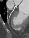
Figure 1. Coronary Artery Disease Reporting and Data System 0 in a 45-year-old man with a history of chronic chest pain. Curved multiplanar reformatted computed tomography angiographic image of the RCA, LAD artery, and LCx artery reveal normal coronary arteries with no atherosclerotic disease or stenosis. No further imaging was recommended.
CX: circumflex artery, DIST: distal, LAD: left anterior descending, LCx: left circumflex, LMCA: left main coronary artery, PDA: posterior descending artery, RCA: right coronary artery.
Figure 2
Figure 2. Coronary Artery Disease Reporting and Data System 1 in a 38-year-old man with atypical chest pain. Curved multiplanar reformatted computed tomography angiographic image show a discrete partially calcified plaque in the proximal LAD (white arrowhead) causing minimal stenosis (<25%). No further imaging was recommended.
LAD: left anterior descending.
Figure 3
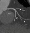
Figure 3. Coronary Artery Disease Reporting and Data System 2 in a 56-year-old man with chronic chest pain. Curved multiplanar reformatted computed tomography image of the RCA reveals a noncalcified plaque (white arrowhead) causing mild stenosis (25%–49%). No further imaging was recommended.
AM1: first acute marginal, AM2: second acute marginal, RCA: right coronary artery.
Figure 4
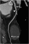
Figure 4. Coronary Artery Disease Reporting and Data System 3 in a 72-year-old woman with atypical chest pain. Curved multiplanar reformatted computed tomography angiographic image of LAD shows a partially calcified plaque (white arrowhead) in the proximal part causing moderate stenosis (50%–69%). Functional assessment was recommended. Myocardial perfusion scintigraphy (not shown) showed a stress perfusion defect in the mid anterior and anteroseptal segments, which is consistent with ischemia.
DIST: distal, LAD: left anterior descending, LMCA: left main coronary artery.
Figure 5
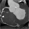
Figure 5. Coronary Artery Disease Reporting and Data System 4A in a 56-year-old woman who presented with chest pain on exertion and abnormal electrocardiogram results. Curved multiplanar reformatted computed tomography angiographic image shows a predominantly noncalcified plaque (white arrowhead) in the mid RCA causing severe luminal stenosis (70%–99%). No other significant coronary artery disease was detected. ICA was recommended. ICA results (not shown) showed severe stenosis in the RCA artery, which was treated with balloon angioplasty and stent placement.
DIST: distal, ICA: invasive coronary angiography, PDA: posterior descending artery, RCA: right coronary artery.
Figure 6
Figure 6. Coronary Artery Disease Reporting and Data System 4B in a 65-year-old man who presented with shortness of breath and chest pain. (A) Curved multiplanar reformatted computed tomography angiographic image and (B) the corresponding axial reformatted image of the LM artery shows circumferential calcified plaque (white arrowheads) causing >50% stenosis. Moderate stenosis was seen in right coronary artery and mild stenosis was seen in the left anterior descending and left circumflex (not shown). Invasive coronary angiography was recommended which revealed triple vessel disease. The patient was considered for coronary artery bypass grafting.
LM: left main coronary artery.
Figure 7
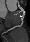
Figure 7. Coronary Artery Disease Reporting and Data System 5 in a 59-year-old man who presented with acute chest pain. Maximum intensity projection computed tomography angiographic image of the RCA shows total occlusion in the mid part (white arrowhead). Invasive coronary angiography findings (not shown) confirmed occlusion of the RCA artery.
RCA: right coronary artery.
Figure 8
Figure 8. Coronary Artery Disease Reporting and Data System N in a 40-year-old woman who presented with chronic chest pain. Curved maximum intensity projection computed tomography angiographic image of coronary arteries shows no atherosclerotic disease in the RCA, LCx, and proximal LAD. The mid LAD could not be evaluated because of a motion artifact (white arrowhead). Additional or alternative evaluation was recommended.
LAD: left anterior descending, LCx: left circumflex, RCA: right coronary artery.
Figure 9
Figure 9. Modifier N in a 56-year-old man with chest pain. (A) Curved MPR CT angiographic image of the LAD shows a non-calcified plaque (white arrowhead) causing moderate stenosis (50%–69%) in the proximal part. (B) Curved MPR CT angiographic image of LCx shows no atherosclerotic disease. (C) Axial reformatted CT angiographic image of RCA shows a non-evaluable segment in the mid part because of motion artifacts. The patient was assigned Coronary Artery Disease Reporting and Data System 3/N category. ICA was recommended. The patient underwent ICA (not shown), which confirmed moderate stenosis (70%) in the mid RCA.
CT: computed tomography, ICA: invasive coronary angiography, LAD: left anterior descending, LCx: left circumflex, MPR: multiplanar reformatted, RCA: right coronary artery.
Figure 10
Figure 10. Modifier S (stent) in a 59-year-old woman with new-onset chest pain who had a history of inferior myocardial infarction and stent placement in the RCA. Curved multiplanar reformatted computed tomography angiographic image of the RCA shows a long stent in situ with moderate in stent restenosis (50%–69%) (white arrowhead). Minimal disease was seen in the left anterior descending and left circumflex arteries (not shown). The patient was assigned Coronary Artery Disease Reporting and Data System 3/S category. Functional assessment was recommended.
RCA: right coronary artery.
Figure 11
Figure 11. Coronary Artery Disease Reporting and Data System 4A/S in a 75-year-old man with new onset chest pain and a history of myocardial infarction and stent placement in D1 branch. (A) Curved MPR CT angiographic image of the LAD shows a patent stent in D1. No evidence of in stent restenosis was seen. (B) Curved MPR CT angiographic image of the RCA shows a non-calcified plaque causing severe stenosis (70%–99%) in the proximal part (white arrowhead). ICA was recommended. ICA results (not shown) showed severe stenosis in the RCA artery, which was treated with balloon angioplasty and stent placement.
CT: computed tomography, D1: 1st diagonal, ICA: invasive coronary angiography, LAD: left anterior descending, LMCA: left main coronary artery, MPR: multiplanar reformatted, RCA: right coronary artery.
Figure 12
Figure 12. Coronary Artery Disease Reporting and Data System N/S in a 69-year-old woman with new onset chest pain and a history of myocardial infarction and stent placement in the LAD (A) Curved multiplanar reformatted CT angiographic image of the LAD shows a long patent stent in situ. No evidence of in stent restenosis seen. (B) Axial reformatted CT angiographic image of the RCA shows a non-evaluable segment in the mid part because of motion artifacts (white arrow). Left circumflex was normal. Invasive coronary angiography was recommended and revealed mild stenosis in the mid RCA (not shown).
CT: computed tomography, LAD: left anterior descending, RCA: right coronary artery.
Figure 13
Figure 13. Modifier G in an 82-year-old woman with a history of three-vessel coronary bypass graft surgery who underwent coronary CT angiography to evaluate the patency of the grafts. Curved multiplanar reformatted CT angiographic images shows patent LIMA graft to the LAD (A), patent RA graft from the ascending aorta to OM artery (B), and patent right SVG from the ascending aorta to distal RCA (C). All of the bypass grafts are patent. The patient was assigned CAD-RADS 0/G category.
CT: computed tomography, DIST: distal, LAD: left anterior descending, LIMA: left internal mammary, OM: obtuse marginal, PDA: posterior descending artery, RA: radial artery, RCA: right coronary artery, SVG: saphenous venous graft.
Figure 14
Figure 14. Modifier G in a 78-year-old man with a history of three-vessel coronary bypass graft surgery who underwent coronary CT angiography to evaluate the patency of the grafts. Curved multiplanar reformatted CT angiographic images shows patent left internal mammary to the LAD. Extensive calcifications with severe luminal stenosis is seen in the LAD proximal to the site of graft insertion. The rest of the 2 grafts (not shown) were also patent with minimum disease in one of them. The patient was assigned CAD-RADS 1/G category. The stenotic segment bypassed by graft is not taken into consideration for CAD-RADS classification.
CAD-RADS: Coronary Artery Disease Reporting and Data System, CT: computed tomography, LAD: left anterior descending.
Figure 15
Figure 15. CAD-RADS 5/G in a 86-year-old man with a history of three-vessel coronary bypass graft surgery who underwent coronary CT angiography to evaluate the patency of the grafts. Curved multiplanar reformatted CT angiographic image shows a RSVG from the ascending aorta to distal RCA. There is dense wall calcification with total luminal occlusion of the graft. All other bypass grafts were patent (not shown). The patient was assigned CAD-RADS 5/G category. Invasive coronary angiography was recommended.
CAD-RADS: Coronary Artery Disease Reporting and Data System, CT: computed tomography, RCA: right coronary artery, RSVG: reversed saphenous vein graft.
Figure 16
Figure 16. Modifier V in a 61-year-old man with chest pain. (A) Curved multiplanar reformatted CT angiographic image of RCA shows a noncalcified plaque (white arrowhead) in the distal part. The plaque shows positive remodeling and is causing moderate stenosis (50%–69%). (B) Corresponding axial reformatted CT angiographic image perpendicular to the vessel lumen (1, CT number = 380 HU) shows central low-attenuation core (2, CT number = 24 HU) and outer circumferential high-attenuation area (3, CT number = 98 HU) representing napkin-ring sign. The patient was assigned CAD-RADS: Coronary Artery Disease Reporting and Data System 3/V category.
CT: computed tomography, HU: hounsfield unit, RCA: right coronary artery.
Figure 17
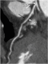
Figure 17. CAD-RADS 2 in a 48-year-old man with chest pain. Curved multiplanar reformatted computed tomography angiographic image shows noncalcified plaque (black arrow) in the proximal-LAD. It shows positive remodelling and is causing mild stenosis (25%–49%). Right coronary artery and left circumflex were normal. The patient was assigned CAD-RADS 2 category. Only one high-risk feature was present, so modifier V was not included. No further imaging was recommended.
CAD-RADS: Coronary Artery Disease Reporting and Data System, LAD: left anterior descending, LMCA: left internal mammary.
Figure 18
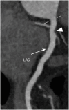
Figure 18. CAD-RADS 1 in a 45-year-old man with chest pain. Curved multiplanar reformatted computed tomography angiographic image shows noncalcified plaque (white arrowhead) causing minimal stenosis (<25%) in the proximal-LAD. The average attenuation of lesion was 7 hounsfield unit. Right coronary artery and left circumflex were normal. The patient was assigned CAD-RADS 1 category. Only one high-risk feature was present, so modifier V was not included. No further imaging was recommended.
CAD-RADS: Coronary Artery Disease Reporting and Data System, LAD: left anterior descending.
Figure 19
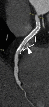
Figure 19. CAD-RADS 5/S in a 54-year-old woman. (A) Curved multiplanar reformatted computed tomography angiographic image of the LAD and (B) corresponding magnified image show a coronary artery stent in the proximal LAD with a complete gap and angulation of the stent due to fracture. The distal part of stent shows total luminal occlusion. A persistent hematoma is formed around the site of fracture (white arrowhead and dashed white line). The CAD-RADS 5/S suggests total luminal occlusion but conveys information about stent fracture.
CAD-RADS: Coronary Artery Disease Reporting and Data System, LAD: left anterior descending, LMCA: left internal mammary.
Figure 20
Figure 20. CAD-RADS 2/S in a 54-year-old man with a history of stent implantation in RCA aneurysm. Curved multiplanar reformatted computed tomography angiographic image of the RCA shows a long stent in the proximal and mid part. Mild stenosis is seen at the ostium of RCA and minimal stenosis is seen in distal part of the stent. There is evidence of endoleak (white arrowhead) into the aneurysmal sac (dashed white line). The CAD-RADS 2/S suggests mild luminal occlusion but conveys information about stent endoleak.
CAD-RADS: Coronary Artery Disease Reporting and Data System, RCA: right coronary artery.
Figure 21
Figure 21. Coronary Artery Disease Reporting and Data System 3 in a 54-year-old man with a history of chest pain. (A) Curved multiplanar reformatted computed tomography angiographic image of the LAD shows a non-calcific plaque (white arrowhead) with external remodelling in the proximal part causing moderate stenosis. (B) Iodine map (rest) shows perfusion defects (white arrowhead) in the mid ventricular anteroseptal segment, suggesting hemodynamically significant stenosis.
LAD: left anterior descending.
Figure 22
Figure 22. Coronary artery anomaly in a 45-year-old man with chest pain. (A) Axial and (B) sagittal oblique maximum intensity projection coronary computed tomography angiographic image in a 47-year-old man shows an anomalous origin of the LM from right coronary sinus. The proximal LM has an interarterial course between the ascending aorta and pulmonary trunk. No atherosclerotic disease was seen in coronary arteries. CAD-RADS does not apply to these findings, so the patient was assigned CAD-RADS category of 0.
Ao: aorta, CAD-RADS: Coronary Artery Disease Reporting and Data System, LM: left main, PA: pulmonary artery.
Figure 23
Figure 23. Coronary artery anomaly in a 49-year-old man with chest pain. (A) Sagittal and (B) short axis reformatted axial intensity projection coronary computed tomography angiographic image shows myocardial bridging of the distal LAD (white arrowheads). No atherosclerotic disease was seen in coronaries. CAD-RADS does not apply to these findings, so the patient was assigned CAD-RADS category of 0.
CAD-RADS: Coronary Artery Disease Reporting and Data System, LAD: left anterior descending, LMCA: left internal mammary.
Figure 24
Figure 24. Coronary artery aneurysm in a 58-year-old man with chest pain. Curved multiplanar reformatted angiographic image of the LAD shows fusiform aneurysm (white arrowhead) of the proximal part with eccentric calcific plaques causing minimal stenosis. The patient was assigned Coronary Artery Disease Reporting and Data System 1. No further imaging was recommended.
D1: 1st diagonal, LAD: left anterior descending, LMCA: left internal mammary.
Figure 25
Figure 25. Graft aneurysm in a 78-year-old man with new onset chest pain and history of bypass grafting. Curved multiplanar reformatted angiographic image of the saphenous venous graft to obtuse marginal shows mild diffuse disease and graft aneurysm (white arrowhead). Right saphenous vein graft to right coronary artery and left internal mammary to left anterior descending were patent. The patient was assigned Coronary Artery Disease Reporting and Data System 2/G, and no further imaging was recommended.

Similar articles

Cited by

References

    1. Leipsic J, Abbara S, Achenbach S, et al. SCCT guidelines for the interpretation and reporting of coronary CT angiography: a report of the Society of Cardiovascular Computed Tomography guidelines committee. J Cardiovasc Comput Tomogr. 2014;8:342–358. - PubMed
    1. Abbara S, Arbab-Zadeh A, Callister TQ, et al. SCCT guidelines for performance of coronary computed tomographic angiography: a report of the Society of Cardiovascular Computed Tomography guidelines committee. J Cardiovasc Comput Tomogr. 2009;3:190–204. - PubMed
    1. Halliburton SS, Abbara S, Chen MY, et al. SCCT guidelines on radiation dose and dose-optimization strategies in cardiovascular CT. J Cardiovasc Comput Tomogr. 2011;5:198–224. - PMC - PubMed
    1. Achenbach S, Delgado V, Hausleiter J, Schoenhagen P, Min JK, Leipsic JA. SCCT expert consensus document on computed tomography imaging before transcatheter aortic valve implantation (TAVI)/transcatheter aortic valve replacement (TAVR) J Cardiovasc Comput Tomogr. 2012;6:366–380. - PubMed
    1. Taylor AJ, Cerqueira M, Hodgson JM, et al. ACCF/SCCT/ACR/AHA/ASE/ASNC/ NASCI/SCAI/SCMR 2010 appropriate use criteria for cardiac computed tomography. A report of the American College of Cardiology Foundation appropriate use criteria task force, the Society of Cardiovascular Computed Tomography, the American College of Radiology, the American Heart Association, the American Society of Echocardiography, the American Society of Nuclear Cardiology, the North American Society for Cardiovascular Imaging, the Society for Cardiovascular Angiography and Interventions, and the Society for Cardiovascular Magnetic Resonance. J Cardiovasc Comput Tomogr. 2010;4:407.e1–407.e33. - PubMed