. 2022 Mar 1;135(5):jcs258186.
doi: 10.1242/jcs.258186. Epub 2021 Jun 3.

Ca2+-dependent protein acyltransferase DHHC21 controls activation of CD4+ T cells

Affiliations

Ca2+-dependent protein acyltransferase DHHC21 controls activation of CD4+ T cells

Shayahati Bieerkehazhi et al. J Cell Sci. .

Abstract

Despite the recognized significance of reversible protein lipidation (S-acylation) for T cell receptor signal transduction, the enzymatic control of this post-translational modification in T cells remains poorly understood. Here, we demonstrate that DHHC21 (also known as ZDHHC21), a member of the DHHC family of mammalian protein acyltransferases, mediates T cell receptor-induced S-acylation of proximal T cell signaling proteins. Using Zdhhc21dep mice, which express a functionally deficient version of DHHC21, we show that DHHC21 is a Ca2+/calmodulin-dependent enzyme critical for activation of naïve CD4+ T cells in response to T cell receptor stimulation. We find that disruption of the Ca2+/calmodulin-binding domain of DHHC21 does not affect thymic T cell development but prevents differentiation of peripheral CD4+ T cells into Th1, Th2 and Th17 effector T helper lineages. Our findings identify DHHC21 as an essential component of the T cell receptor signaling machinery and define a new role for protein acyltransferases in regulation of T cell-mediated immunity.

Keywords: Calmodulin; Palmitoylation; Protein acyltransferase; S-acylation; T cell receptor.


Conflict of interest statement

Competing interests The authors declare no competing or financial interests.

Figures

Fig. 1.
Fig. 1.
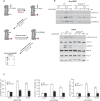
DHHC21 has a Ca2+/calmodulin-binding motif. (A) Schematic depiction of DHHC21 illustrating the approximate localization of the predicted 1-14 Ca2+/calmodulin-binding motif within the C-terminal cytoplasmic tail. The 1-14 motif is completely conserved between species. Location of the ‘dep’ mutation (ΔF233) is indicated. (B) Ca2+-dependent co-immunoprecipitation of DHHC21 and calmodulin. CD4+ T cells from WT or Zdhhcdep mice were treated for the indicated times with 10 µM thapsigargin (TG) to evoke Ca2+ release. DHHC21 was immunoprecipitated from the cell lysates and binding of calmodulin was detected by immunoblotting. Total lysates (20% of load) are depicted as input. Phosphorylated ZAP-70 and ERK1/2 are shown to confirm CD4+ T cells’ responses to TCR-stimulation. Blots are representative of three repeats.
Fig. 2.
Fig. 2.
DHHC21 mediates TCR-induced S-acylation of signaling proteins. (A) Overview of the acyl-resin assisted capture (Acyl-RAC) assay. Free cysteine thiols (-SH) are irreversibly blocked by S-methyl methanethiosulfonate (MMTS) following cell lysis. Thioester bonds between cysteine resdiues and acyl groups are then specifically cleaved by neutral hydroxylamine (HA). The newly formed free thiol groups are captured by the thiol-reactive Sepharose and S-acylated proteins are detected by immunoblotting. (B) TCR-induced protein S-acylation. CD4+ T cells from WT or Zdhhc21dep mice were stimulated with cross-linked anti-CD3–CD28 antibodies for the indicated times, and protein S-acylation (acyl-) was determined using the Acyl-RAC assay. Samples not treated with hydroxylamine (-HA) were used as a negative control. β-actin, a known S-acylated protein (Ren et al., 2013), was used as a loading control. Total lysates prior to addition of HA (5% of load) are depicted as input. Phosphorylated ZAP-70 and ERK1/2 are shown to confirm cellular responses to TG-induced changes in cytoplasmic Ca2+ levels. (C) Quantitative analysis of protein S-acylation in response to TCR stimulation with cross-linked anti-CD3–CD28 (αCD3/CD28) antibodies. Error bars represent mean±s.e.m., n=3. *P<0.05; **P<0.01; ***P<0.001 (two-tailed, unpaired Student's t-test). a.u., arbitrary units.
Fig. 3.
Fig. 3.
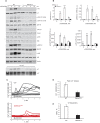
TCR signaling is impaired in Zdhhc21dep CD4+ T cells. (A) CD4+T cells from WT or Zdhhc21dep mice were stimulated with cross-linked anti-CD3–CD28 (aCD3/CD28) antibodies for indicated times and indicated phospho-proteins (p-) and total proteins were analyzed by immunoblotting. β-actin was used as a loading control. The results shown are representative of three independent experiments. (B) Quantitative analysis of phosphorylation of TCR signaling proteins in response to TCR stimulation with cross-linked anti-CD3–CD28 antibodies. Error bars represent mean±s.e.m., n=3. *P<0.05; **P<0.01; ***P<0.001 (two-tailed, unpaired Student's t-test). a.u., arbitrary units. (C) CD4+ T cells were loaded with Fura-2 Ca2+ indicator and pre-incubated with anti-CD3–CD28 antibodies. IgG antibody was added to induce cross-linking after an observation period (upward arrow). Shown are representative traces from single WT (gray) and Zdhhc21dep (red) cells. (D) Peak Ca2+ release. CD4+ T cells were treated as described in B. Data represent maximum Ca2+ values averaged from four independent experiments (∼100 WT or Zdhhc21dep cells per experiment). Data represent mean±s.e.m. *P<0.05 (two-tailed, unpaired Student's t-test). (E) Ca2+ response pattern. CD4+T cells were treated as described in B. Cells showing at least a 10% increase in Ca2+ values upon TCR stimulation were identified as responders. Data represent mean±s.e.m. percentage of responders from four independent experiments (∼100 WT or Zdhhc21dep CD4+T cells per experiment). *P<0.05 (two-tailed, unpaired Student's t-test).
Fig. 4.
Fig. 4.
DHHC21 is required for T cell activation. (A) Surface expression of CD69 on CD4+ T cells from WT or Zdhhc21dep mice. CD4+ T cells were stimulated or not (N/S) with plate-bound anti-CD3–CD28 antibodies (αCD3/CD28) for 24 h and analyzed by flow cytometry. The red section indicates the population of CD69+ cells (given as a percentage). (B) Average a percentage of CD69+ T cells from WT or Zdhhc21dep mice. CD4+ T cells were stimulated with plate-bound anti-CD3–CD28 antibodies for 24 h and analyzed by flow cytometry. The graph shows mean±s.d. from three independent experiments. **P<0.01 (two-tailed, unpaired Student's t-test). (C) IL-2 production by CD4+ T cells from WT or Zdhhc21dep mice. CD4+ T cells were stimulated with plate-bound anti-CD3/CD28 antibodies for 24 h and collected supernatants were assayed by ELISA. The graph shows mean±s.d. values, n=3, **P<0.01 (two-tailed, unpaired Student's t-test).
Fig. 5.
Fig. 5.
Thymocyte development in Zdhhc21dep mice. (A) Flow cytometry analysis of cells isolated from thymus of WT and Zdhhc21dep mice. Plots of CD4+ and CD8+ cells are gated on CD3+ cells. Plots of CD25+ and CD44+ cells are gated on CD4CD8 cells. Values are percentages for each section. (B) The absolute numbers of thymocytes from WT and Zdhhc21dep mice. Cell numbers were calculated based on the relative percentages determined by flow cytometry analysis and total cell numbers from thymus. ns, not significant, P>0.05 (two-tailed, unpaired Student's t-test). (C) Flow cytometry analysis of Treg cells isolated from thymus of WT and Zdhhc21dep mice. Plots of CD25+ and FoxP3+ cells are gated on CD4+ cells. Values are percentages for each section.
Fig. 6.
Fig. 6.
T cell development in peripheral lymphoid organs of Zdhhc21dep mice. (A) Flow cytometry analysis of cells isolated from spleen and lymph nodes of WT and Zdhhc21dep mice. Plots of CD4+ and CD8+ cells are gated on CD3+ cells. (B) Flow cytometry analysis of naïve (CD44loCD62Lhi) and effector memory (CD44hiCD62Llo) CD4+ T cells isolated from spleen and lymph nodes of WT and Zdhhc21dep mice. (C) Flow cytometry analysis of Treg cells isolated from spleens of WT and Zdhhc21dep mice. Plots of FoxP3+ cells are gated on CD4+ cells. Values are percentages for each section. (D) The absolute numbers of thymocytes from WT and Zdhhc21dep mice. Cell numbers were calculated based on the relative percentages determined by flow cytometry analysis and total cell numbers from thymus. ns, not significant, P>0.05 (two-tailed, unpaired Student's t-test). (E) Peripheral lymphoid organs in WT and Zdhhc21dep mice. The fields of view shown are 2×4 cm (lymph nodes) and 4×8 cm (spleen).
Fig. 7.
Fig. 7.
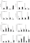
DHHC21 regulates CD4+ T activation and differentiation. Naïve CD4+ T cells from WT or Zdhhc21dep mice were stimulated with plate-bound anti-CD3–CD28 antibodies and incubated under neutral (Th0) or polarizing (Th1, Th2 and Th17) conditions for 5 days, then CD4+ T cells were re-stimulated with plate-bound anti-CD3 antibodies for 6 h. (A) Effector cytokine production measured by ELISA. The graph shows mean±s.d. values from at least three independent experiments. (B) mRNA expression of Th lineage-specific transcription factors determined by qRT-PCR normalized to 18S RNA. The graph shows mean±s.d., n=4, *P<0.05; **P<0.01; ***P<0.001 (two-tailed, unpaired Student's t-test).

References

    1. Akimzhanov, A. M. and Boehning, D. (2015). Rapid and transient palmitoylation of the tyrosine kinase Lck mediates Fas signaling. Proc. Natl. Acad. Sci. USA 112, 11876-11880. 10.1073/pnas.1509929112 - DOI - PMC - PubMed
    1. Akimzhanov, A. M., Wang, X., Sun, J. and Boehning, D. (2010). T-cell receptor complex is essential for Fas signal transduction. Proc. Natl. Acad. Sci. USA 107, 15105-15110. 10.1073/pnas.1005419107 - DOI - PMC - PubMed
    1. Au-Yeung, B. B., Shah, N. H., Shen, L. and Weiss, A. (2018). ZAP-70 in signaling, biology, and disease. Annu. Rev. Immunol. 36. 127-156. 10.1146/annurev-immunol-042617-053335 - DOI - PubMed
    1. Baekkeskov, S. and Kanaani, J. (2009). Palmitoylation cycles and regulation of protein function (Review). Mol. Membr. Biol. 26, 42-54. 10.1080/09687680802680108 - DOI - PubMed
    1. Beard, R. S., Yang, X., Meegan, J. E., Overstreet, J. W., Yang, C. G. Y., Elliott, J. A., Reynolds, J. J., Cha, B. J., Pivetti, C. D., Mitchell, D. A.et al. (2016). Palmitoyl acyltransferase DHHC21 mediates endothelial dysfunction in systemic inflammatory response syndrome. Nat. Commun. 7, 12823. 10.1038/ncomms12823 - DOI - PMC - PubMed

Publication types