. 2021 Jul;24(1):536.
doi: 10.3892/mmr.2021.12175. Epub 2021 Jun 3.

Low‑calorie sweetener D‑psicose promotes hydrogen peroxide‑mediated apoptosis in C2C12 myogenic cells favoring skeletal muscle cell injury

Affiliations

Low‑calorie sweetener D‑psicose promotes hydrogen peroxide‑mediated apoptosis in C2C12 myogenic cells favoring skeletal muscle cell injury

Zhen-Jie Wei et al. Mol Med Rep. 2021 Jul.

Abstract

Diet and exercise are the most effective approaches used to induce weight loss. D‑psicose is a low‑calorie sweetener that has been shown to reduce weight in obese individuals. However, the effect of D‑psicose on muscle cells under oxidative stress, which is produced during exercise, requires further investigation. The present study aimed to determine the effects of D‑psicose on C2C12 myogenic cells in vitro. Hydrogen peroxide (H2O2) was used to stimulate the generation of intracellular reactive oxygen species (ROS) in muscle cells to mimic exercise conditions. Cell viability was analyzed using a MTT assay and flow cytometry was used to analyze the levels of apoptosis, mitochondrial membrane potential (MMP), the generation of ROS and the cell cycle distribution following treatment. Furthermore, protein expression levels were analyzed using western blotting and cell proliferation was determined using a colony formation assay. The results of the present study revealed that D‑psicose alone exerted no toxicity on C2C12 mouse myogenic cells. However, in the presence of low‑dose (100 µM) H2O2‑induced ROS, D‑psicose induced C2C12 cell injury and significantly decreased C2C12 cell viability in a dose‑dependent manner. In addition, the levels of apoptosis and the generation of ROS increased, while the MMP decreased. MAPK family molecules were also activated in a dose‑dependent manner following treatment. Notably, the combined treatment induced G2/M phase arrest and reduced the proliferation of C2C12 cells. In conclusion, the findings of the present study suggested that D‑psicose may induce toxic effects on muscle cells in a simulated exercise situation by increasing ROS levels, activating the MAPK signaling pathway and disrupting the MMP.

Keywords: D‑psicose; MAPK signaling pathway; apoptosis; muscle cell; reactive oxygen species.


Conflict of interest statement

The authors declare that they have no competing interests.

Figures
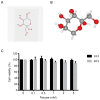
Figure 1.
Figure 1.
Safety of D-psicose in C2C12 myoblasts. (A) Two-dimensional and (B) three-dimensional chemical structure of D-psicose. (C) C2C12 cells were treated with 0, 0.1, 0.2, 1, 2 or 5 mM D-psicose for 24 or 48 h and cell viability was subsequently analyzed using a MTT assay. The results revealed that D-psicose exerted no cytotoxic effects on C2C12 myoblasts.
Figure 2.
Figure 2.
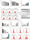
D-psicose enhances H2O2-induced C2C12 cell damage. (A) C2C12 cells were pretreated with 0–500 µM H2O2 for 2 h, after which the medium was changed. Following incubation for 24 h, a MTT assay was used to analyze cell viability. The concentration of 100 µM H2O2 was determined to be a moderate inducer of oxidative stress. C2C12 cells were pretreated with 1, 2 or 5 mM D-psicose for 3 h and then with 100 µM H2O2 for 2 h, after which the medium was changed. Following incubation for 19 h, (B) an MTT assay, (C) Annexin V/propidium iodide staining, (D) flow cytometry, (J and K) tetramethylrhodamine methyl ester perchlorate staining and (E-I) western blotting were used to analyze cell viability, cell apoptosis, MMP loss and the expression levels of apoptosis-related proteins. Data are presented as the mean ± SEM of independent experiments. #P<0.05, ##P<0.01 and ###P<0.001 vs. NT; *P<0.05, **P<0.01 and ***P<0.001 vs. H2O2 (n=3). H2O2, hydrogen peroxide; MMP, mitochondrial membrane potential; Mcl-1, MCL1 apoptosis regulator BCL2 family member; PARP-1, anti-cleaved-poly(ADP-ribose) polymerase 1; NT, No treatment.
Figure 3.
Figure 3.
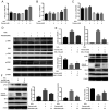
D-psicose enhances H2O2-induced apoptosis of C2C12 cells through the MAPK signaling pathway. C2C12 cells were pretreated with 1, 2 or 5 mM D-psicose for 3 h and then 100 µM H2O2 for 2 h, after which the medium was changed. Following incubation for 3 h, protein expression levels of (A) p-JNK and JNK, (B) p-P38 and P38, and (C) p-ERK and ERK were analyzed using western blotting. (D) Representative western blotting image of p-JNK, JNK, p-P38, P38, p-ERK and ERK. (E) C2C12 cells were pretreated with 5 mM D-psicose + 100 µM H2O2 or 5 mM D-psicose + 10 µM JNK inhibitor VIII + 100 µM H2O2 for 1 h. The expression levels of JNK, p-JNK, cleaved caspase-3, Bcl-2 and Bax were analyzed using western blotting. (F) C2C12 cells were pretreated with 5 mM D-psicose + 100 µM H2O2 or 5 mM D-psicose + 20 µM SB203580 + 100 µM H2O2 for 1 h. The expression levels of P38, p-P38, cleaved caspase-3, Bcl-2 and Bax were analyzed using western blotting. β-actin was used as the loading control. Data are presented as the mean ± SEM of independent experiments. ##P<0.01 and ###P<0.001 vs. NT; *P<0.05 and ***P<0.001 vs. H2O2; $P<0.05 vs. D-psicose + H2O2 (n=3). H2O2, hydrogen peroxide; p-, phosphorylated.
Figure 4.
Figure 4.
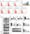
Co-treatment of D-psicose and H2O2 increases ROS generation in C2C12 cells. C2C12 cells were pretreated with 1, 2 or 5 mM D-psicose for 3 h and then 100 µM H2O2 for 2 h, after which the medium was changed. Following incubation for 30 min, ROS generation was analyzed using dihydroethidium staining and flow cytometry (A) Representative flow cytometry plots and (B) quantification of ROS generation. (C) C2C12 cells were pretreated with 5 mM D-psicose + 100 µM H2O2 or 5 mM D-psicose + 5 mM NAC + 100 µM H2O2 for 1 h. The expression levels of p-p38, p-JNK, p-ERK, cleaved caspase-3, Bcl-2 and Bax were analyzed using western blotting. (D) C2C12 cells were pretreated with 1, 2 or 5 mM D-psicose for 3 h and with 100 µM H2O2 for 2 h, after which the medium was changed. Following incubation for 3 h, the expression levels of SIRT3 and SOD2 were analyzed using western blotting. Data are presented as the mean ± SEM of independent experiments. #P<0.05 and ###P<0.001 vs. NT; *P<0.05 vs. H2O2; $P<0.05 vs. D-psicose + H2O2 (n=3). H2O2, hydrogen peroxide; ROS, reactive oxygen species; p-, phosphorylated; SIRT3, sirtuin 3; SOD2, superoxide dismutase 3; NAC, N-acetylcysteine; NT, No treatment.
Figure 5.
Figure 5.
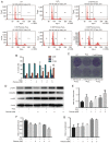
Co-treatment with D-psicose and H2O2 induces G2/M phase cell cycle arrest in C2C12 myoblasts. C2C12 cells were pretreated with 1, 2 or 5 mM D-psicose for 3 h and then with 100 µM H2O2 for 2 h, after which the medium was changed. Following incubation for 24 h, the SubG1 cell population was analyzed using propidium iodide staining and flow cytometry. (A) Representative flow cytometry plots and (B) quantification of cells in each state of the cell cycle. (C) Colony formation assays were performed using cells seeded at a density of 1×10³ cells/well into 6-well plates for 24 h, treated as described in part (A) and incubated for 5 days. (D) C2C12 cells were pretreated with 1, 2 or 5 mM D-psicose for 3 h and then with 100 μM H2O2 for 2 h, after which the medium was changed. Following incubation for 3 h, the expression levels of p-Chk1, CDC25C and p-CDC2 were analyzed using western blotting. The relative expression levels of (E) p-Chk1, (F) CDC25C and (G) p-CDC2 were quantified relative to the value of β-actin. Data are presented as the mean ± SEM of independent experiments. ##P<0.01 and ###P<0.001 vs. NT; *P<0.05 and ***P<0.001 vs. H2O2 (n=3). H2O2, hydrogen peroxide; p-, phosphorylated; Chk1, checkpoint kinase 1; CDC25C, cell division cycle 25 C; CDC2, DNA polymerase δ 1, catalytic subunit; NT, No treatment.
Figure 6.
Figure 6.
Proposed molecular mechanism through which D-psicose and H2O2 may induce apoptosis and G2/M cell cycle arrest. H2O2, hydrogen peroxide; CDC25C, cell division cycle 25 C; CDC2, DNA polymerase δ 1, catalytic subunit; Chk1, checkpoint kinase 1; H2O2, hydrogen peroxide; MMP, membrane mitochondrial potential; ROS, reactive oxygen species; NAC, N-acetylcysteine; p-, phosphorylated.

References

    1. Chu DT, Minh Nguyet NT, Dinh TC, Thai Lien NV, Nguyen KH, Nhu Ngoc VT, Tao Y, Son LH, Le DH, Nga VB, et al. An update on physical health and economic consequences of overweight and obesity. Diabetes Metab Syndr. 2018;12:1095–1100. doi: 10.1016/j.dsx.2018.05.004. - DOI - PubMed
    1. Sylvetsky AC, Rother KI. Trends in the consumption of low-calorie sweeteners. Physiol Behav. 2016;164:446–450. doi: 10.1016/j.physbeh.2016.03.030. - DOI - PMC - PubMed
    1. Swithers SE. Artificial sweeteners produce the counterintuitive effect of inducing metabolic derangements. Trends Endocrinol Metab. 2013;24:431–441. doi: 10.1016/j.tem.2013.05.005. - DOI - PMC - PubMed
    1. He W, Mu W, Jiang B, Yan X, Zhang T. Construction of a food grade recombinant bacillus subtilis based on replicative plasmids with an auxotrophic marker for biotransformation of d-Fructose to d-Allulose. J Agric Food Chem. 2016;64:3243–3250. doi: 10.1021/acs.jafc.6b00278. - DOI - PubMed
    1. Sui L, Dong Y, Watanabe Y, Yamaguchi F, Hatano N, Tsukamoto I, Izumori K, Tokuda M. The inhibitory effect and possible mechanisms of D-allose on cancer cell proliferation. Int J Oncol. 2005;27:907–912. - PubMed

MeSH terms