

Role of sex hormones in lung cancer
- PMID: 34080912
- PMCID: PMC8524770
- DOI: 10.1177/15353702211019697
Role of sex hormones in lung cancer
Abstract
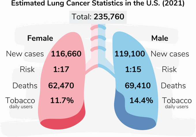
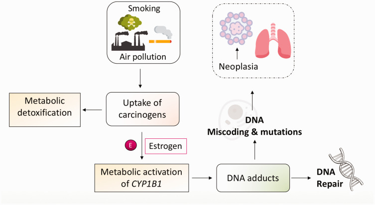
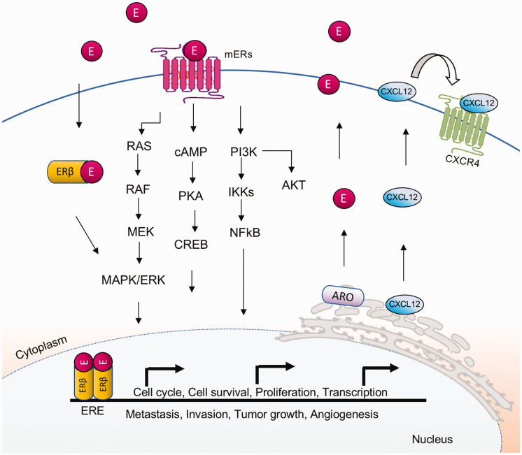
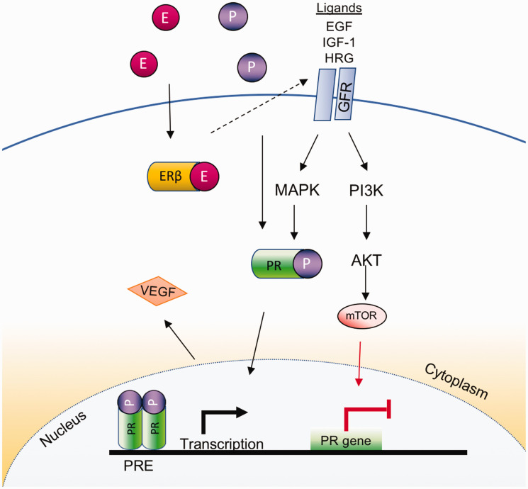
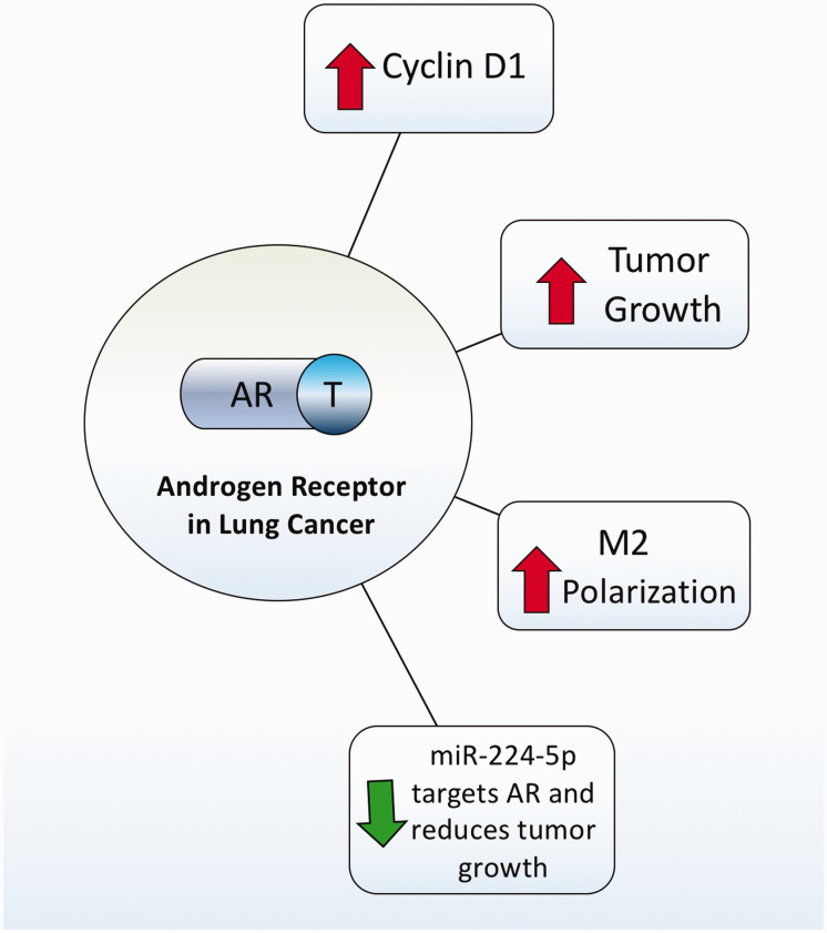
Lung cancer represents the world's leading cause of cancer deaths. Sex differences in the incidence and mortality rates for various types of lung cancers have been identified, but the biological and endocrine mechanisms implicated in these disparities have not yet been determined. While some cancers such as lung adenocarcinoma are more commonly found among women than men, others like squamous cell carcinoma display the opposite pattern or show no sex differences. Associations of tobacco product use rates, susceptibility to carcinogens, occupational exposures, and indoor and outdoor air pollution have also been linked to differential rates of lung cancer occurrence and mortality between sexes. While roles for sex hormones in other types of cancers affecting women or men have been identified and described, little is known about the influence of sex hormones in lung cancer. One potential mechanism identified to date is the synergism between estrogen and some tobacco compounds, and oncogene mutations, in inducing the expression of metabolic enzymes, leading to enhanced formation of reactive oxygen species and DNA adducts, and subsequent lung carcinogenesis. In this review, we present the literature available regarding sex differences in cancer rates, associations of male and female sex hormones with lung cancer, the influence of exogenous hormone therapy in women, and potential mechanisms mediated by male and female sex hormone receptors in lung carcinogenesis. The influence of biological sex on lung disease has recently been established, thus new research incorporating this variable will shed light on the mechanisms behind the observed disparities in lung cancer rates, and potentially lead to the development of new therapeutics to treat this devastating disease.
Keywords: Lung; cancer; carcinogenesis; estrogen receptors; sex differences; sex hormones.
Conflict of interest statement
Figures
Similar articles
-
Sex differences in lung cancer susceptibility: a review.Gend Med. 2010 Oct;7(5):381-401. doi: 10.1016/j.genm.2010.10.002. Gend Med. 2010. PMID: 21056866 Review.
-
Gender and Bladder Cancer: A Collaborative Review of Etiology, Biology, and Outcomes.Eur Urol. 2016 Feb;69(2):300-10. doi: 10.1016/j.eururo.2015.08.037. Epub 2015 Sep 4. Eur Urol. 2016. PMID: 26346676 Review.
-
The Sex Bias of Cancer.Trends Endocrinol Metab. 2020 Oct;31(10):785-799. doi: 10.1016/j.tem.2020.07.002. Epub 2020 Sep 6. Trends Endocrinol Metab. 2020. PMID: 32900596 Review.
-
Sex steroid metabolism and actions in non-small cell lung carcinoma.J Steroid Biochem Mol Biol. 2019 Oct;193:105440. doi: 10.1016/j.jsbmb.2019.105440. Epub 2019 Aug 3. J Steroid Biochem Mol Biol. 2019. PMID: 31386890 Review.
-
Associations between female lung cancer risk and sex steroid hormones: a systematic review and meta-analysis of the worldwide epidemiological evidence on endogenous and exogenous sex steroid hormones.BMC Cancer. 2021 Jun 10;21(1):690. doi: 10.1186/s12885-021-08437-9. BMC Cancer. 2021. PMID: 34112140 Free PMC article.
Cited by
-
Sexual dimorphism of colorectal cancer in humans and colorectal tumors in a murine model.Front Oncol. 2024 Aug 6;14:1398175. doi: 10.3389/fonc.2024.1398175. eCollection 2024. Front Oncol. 2024. PMID: 39165688 Free PMC article.
-
Sex Hormones and Chronic Obstructive Pulmonary Disease: A Cross-Sectional Study and Mendelian Randomization Analysis.Int J Chron Obstruct Pulmon Dis. 2024 Jul 15;19:1649-1660. doi: 10.2147/COPD.S463849. eCollection 2024. Int J Chron Obstruct Pulmon Dis. 2024. PMID: 39050738 Free PMC article.
-
Sexual dimorphism in chronic respiratory diseases.Cell Biosci. 2023 Mar 7;13(1):47. doi: 10.1186/s13578-023-00998-5. Cell Biosci. 2023. PMID: 36882807 Free PMC article. Review.
-
Association between physical activity and cancer risk among Chinese adults: a 10-year prospective study.Int J Behav Nutr Phys Act. 2022 Dec 12;19(1):150. doi: 10.1186/s12966-022-01390-1. Int J Behav Nutr Phys Act. 2022. PMID: 36510257 Free PMC article.
-
Association of Metabolic Syndrome With Risk of Lung Cancer: A Population-Based Prospective Cohort Study.Chest. 2024 Jan;165(1):213-223. doi: 10.1016/j.chest.2023.08.003. Epub 2023 Aug 10. Chest. 2024. PMID: 37572975 Free PMC article.
References
-
- Sung H, Ferlay J, Siegel RL, Laversanne M, Soerjomataram I, Jemal A, Bray F. Global cancer statistics 2020: GLOBOCAN estimates of incidence and mortality worldwide for 36 cancers in 185 countries. CA Cancer J Clin. Epub ahead of print 4 February 2021. DOI: 10.3322/caac.21660. - PubMed
-
- Siegel RL, Miller KD, Fuchs HE, Jemal A. Cancer statistics, 2021. CA A Cancer J Clin 2021; 71:7–33 - PubMed
-
- Malhotra J, Malvezzi M, Negri E, La Vecchia C, Boffetta P. Risk factors for lung cancer worldwide. Eur Respir J 2016; 48:889–902 - PubMed
Publication types
MeSH terms
Substances
Grants and funding
LinkOut - more resources
Full Text Sources
Medical