. 2021 Aug 19;32(18):1724-1736.
doi: 10.1091/mbc.E21-03-0106. Epub 2021 Jun 3.

Calcium signaling mediates a biphasic mechanoadaptive response of endothelial cells to cyclic mechanical stretch

Affiliations

Calcium signaling mediates a biphasic mechanoadaptive response of endothelial cells to cyclic mechanical stretch

Yekaterina A Miroshnikova et al. Mol Biol Cell. .

Abstract

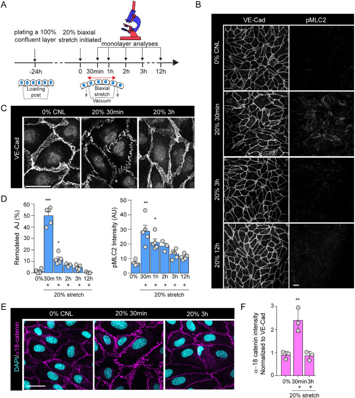
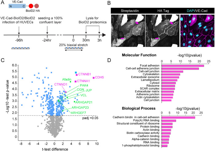
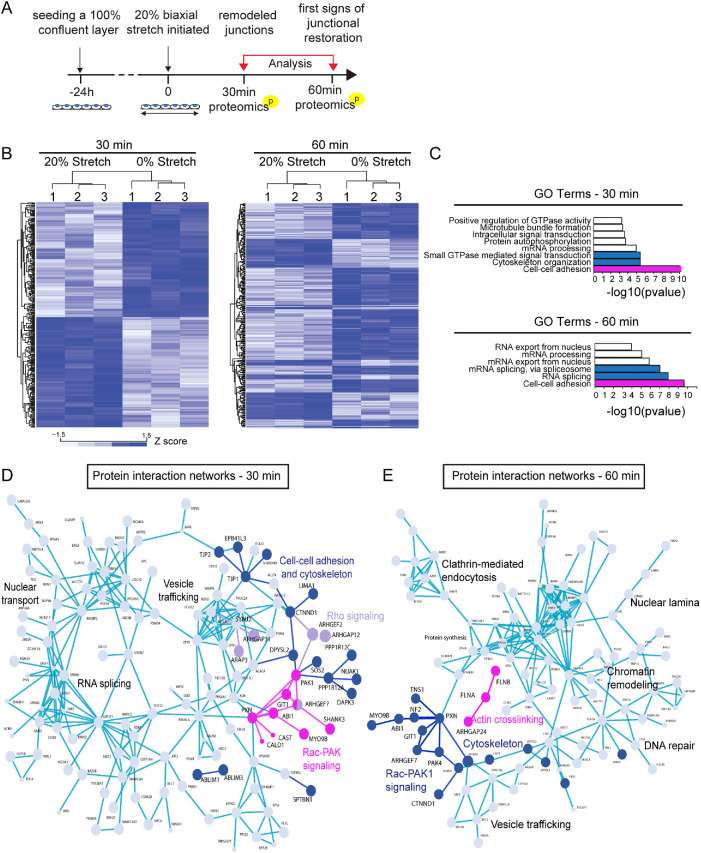
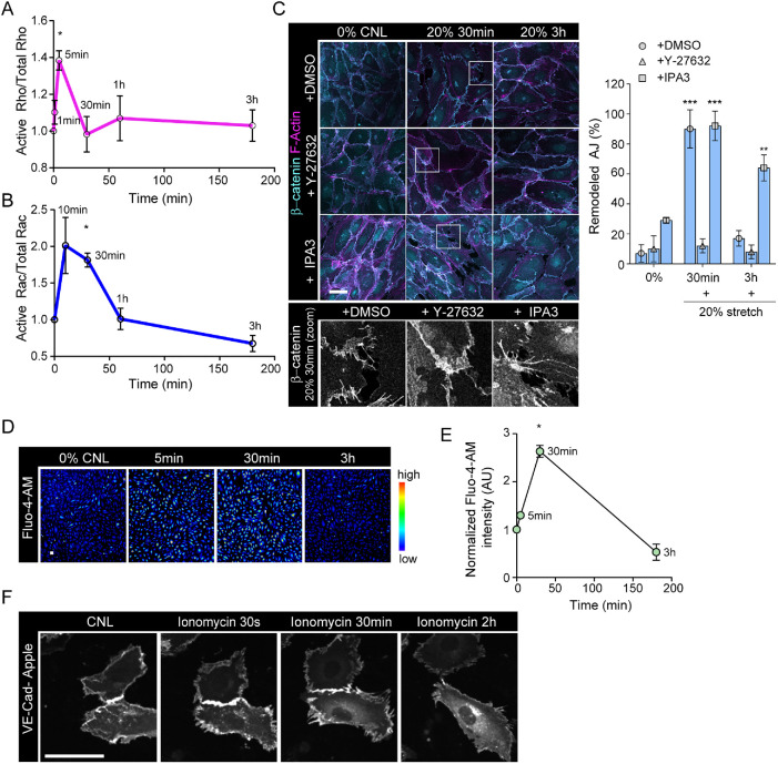
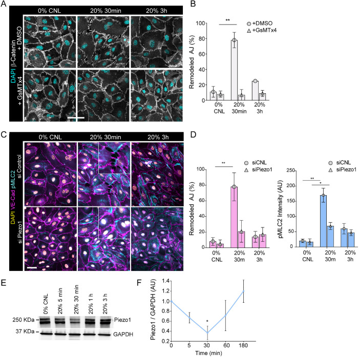
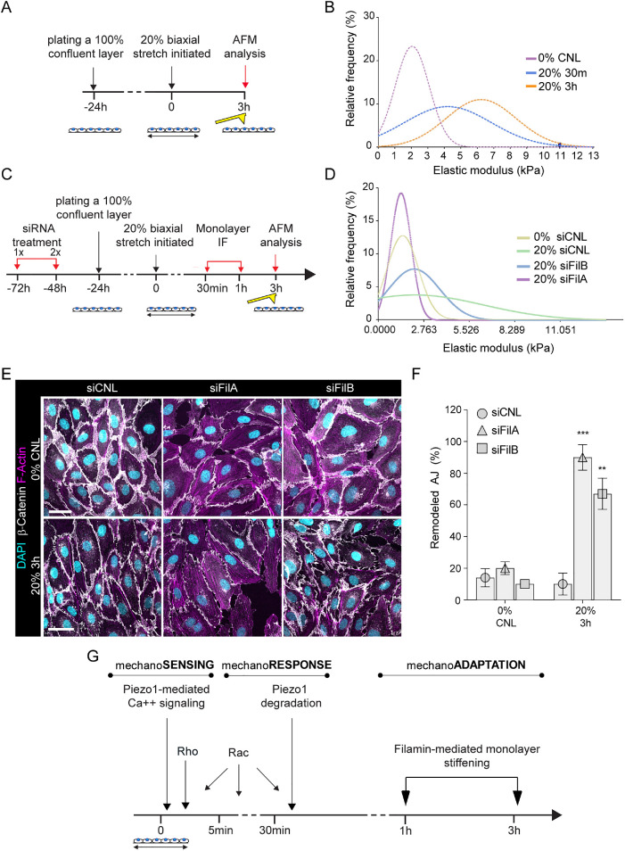
The vascular system is precisely regulated to adjust blood flow to organismal demand, thereby guaranteeing adequate perfusion under varying physiological conditions. Mechanical forces, such as cyclic circumferential stretch, are among the critical stimuli that dynamically adjust vessel distribution and diameter, but the precise mechanisms of adaptation to changing forces are unclear. We find that endothelial monolayers respond to cyclic stretch by transient remodeling of the vascular endothelial cadherin-based adherens junctions and the associated actomyosin cytoskeleton. Time-resolved proteomic profiling reveals that this remodeling is driven by calcium influx through the mechanosensitive Piezo1 channel, triggering Rho activation to increase actomyosin contraction. As the mechanical stimulus persists, calcium signaling is attenuated through transient down-regulation of Piezo1 protein. At the same time, filamins are phosphorylated to increase monolayer stiffness, allowing mechanoadaptation to restore junctional integrity despite continuing exposure to stretch. Collectively, this study identifies a biphasic response to cyclic stretch, consisting of an initial calcium-driven junctional mechanoresponse, followed by mechanoadaptation facilitated by monolayer stiffening.


Figures

FIGURE 1:
FIGURE 1:
Cyclic mechanical stretch triggers transient adherens junction remodeling and actomyosin contraction. (A) Schematic illustration of stretch experiments. HUVECs were plated at confluency on silicon elastomers 24 h before stretch application, using negative pressure to deform the elastomer substrates (20%, 100 mHz), after which monolayers were analyzed at time points indicated. (B) Representative immunofluorescence images of VE-cadherin (VE-Cad) and pMLC2-stained HUVEC monolayers exposed to stretch. Note transient emergence of zipper-patterned adhesions and increased pMLC signal at 30 min of stretch. Scale bars 30 μm. (C) Close-up images of junctional rearrangements show reversibility of junctional zippering upon stretch. Scale bars 30 μm. (D) Quantification of AJ remodeling from VE-cadherin staining (left panel) and pMLC2 intensity (right panel). Mean ± SEM; n = 5 independent experiments; ***p = 0.0006, *p = 0.0203 (VE-cadherin) and) *p = 0.0168 and *p = 0.0071 (pMLC2), ANOVA, Dunnett’s. (E) Representative immunofluorescence images of α-18-stained HUVEC monolayers exposed to stretch. Note transient increase in α-18 intensity at 30 min of stretch. Scale bars 30 μm. (F) Quantification of α-18 catenin normalized to VE-cadherin intensity. Mean ± SD; n = 3 independent experiments; **p = 0.0018, ANOVA, Dunnett’s.
FIGURE 2:
FIGURE 2:
AJ remodeling and reinforcement is associated with subtle changes in VE-cadherin interactome. (A) Schematic illustration of VE-cadherin (VE-Cad)–BioID2 pulldown and mass spectrometry to identify stretch-dependent interactome changes. (B) Representative immunofluorescence images showing localization of HA-tagged VE-cadherin–BioID2 at AJs and enrichment of streptavidin as a result of biotin binding at this site. Scale bars 30 μm. (C) Volcano plot of protein enrichment in VE-cadherin–BioID2 compared with BioID2 control. Note enrichment of VE-cadherin itself (CDH5) along with α- and β-catenin (CTNNA1, CTNNB1), indicative of successful enrichment of AJ components. Also highlighted are cytoskeletal and adhesion remodelers. Dotted line marks padj cutoff of 0.05, moderated t test/Benjamini Hochberg. (D) GO term enrichment analysis of proteins significantly enriched in VE-cadherin–BioID2 pulldowns.
FIGURE 3:
FIGURE 3:
Phosphoproteome analyses implicate activation of RhoA and Rac-PAK1 signaling and subsequent cytoskeletal remodeling upon stretch. (A) Schematic illustration of the timeline of the phosphoproteome analyses. (B) Heat map and Euclidean distance-based clustering of differentially phosphorylated proteins at 30 min (left panel) and 60 min (right panel) of stretch. At 30 min, 185 up-regulated and 193 down-regulated phosphorylations are detected, and at 60 min, 86 up-regulated and 190 down-regulated phosphorylations are detected. (C) Protein–protein interaction network analysis (STRING) of differentially phosphorylated proteins at 30 min. Various components of the Rho A (purple) and Rac-Pak (magenta) signaling pathways as well as cytoskeletal and cell–-cell adhesion regulators (dark blue) are identified. (D) GO term enrichment analyses of differentially phosphorylated proteins show enrichment of components and regulators of cell–cell adhesions both at 30 and 60 min. In addition, cytoskeletal organization and small GTPases are enriched at 30 min, whereas mRNA splicing is enriched at 60 min. (E) Protein–protein interaction network analysis of differentially phosphorylated proteins at 60 min. Various components of the Rac-Pak signaling pathway, cytoskeletal and cell–cell adhesion regulators (all in dark blue), and filamins (magenta) are identified.
FIGURE 4:
FIGURE 4:
Stretch triggers transient calcium signaling and activation of Rho and GTPases to remodel junctions. (A) Quantification of RhoA activity over time shows peak of Rho activation at 5 min of stretch (mean ± SD; n = 3 independent experiments; *p = 0.0461, ANOVA with Dunnett’s). (B) Quantification of Rac1 activity over time shows peak of Rac activation at 30 min of stretch (mean ±SD; n = 4 independent experiments; *p = 0.0163, ANOVA with Dunnett’s). (C) Representative immunofluorescence images and quantification of AJ remodeling from β-catenin and F-actin (phalloidin)-stained HUVEC monolayers exposed to stretch in the presence of either Y-27632 or IPA3. Note prevention of adhesion zippering in Y-27632-treated cells and attenuated junction restoration in IPA3-treated cells. Scale bars 30 μm (mean ± SD; n = 3 independent experiments; ***p < 0.0001,**p = 0.0015, ANOVA with Dunnett’s). (D, E) Representative images, D, and quantification, E, of calcium imaging with Fluo-4-AM shows a transient increase in intracellular calcium at 30 min of stretch (mean ± SD; n = 3 independent experiments; *p = 0.0106, ANOVA with Dunnett’s; scale bars 30 μm). (F) Representative immunofluorescence images of confluent, transiently VE-cadherin-Apple transfected HUVEC monolayers showing remodeling of VE-cadherin junctions in response to ionophore application. Scale bars 30 μm.
FIGURE 5:
FIGURE 5:
Stretch reduces Piezo1 levels to attenuate calcium signaling. (A, B) Representative immunofluorescence images, A, and quantification, B, of AJ remodeling from β-catenin stained junctions show prevention of junctional remodeling in the presence of GsMTx4 at 30 min of stretch (mean ± SD; n = 3 independent experiments; **p = 0.007, ANOVA with Dunnett’s; scale bars 30 μm). (C, D) Representative immunofluorescence images, C, and quantification, D, of pMLC2 and AJ remodeling from VE-cadherin stained HUVECs with control or Piezo1 siRNA cells show attenuation of pMLC2 elevation and junctional remodeling in Piezo1-depleted at 30 min of stretch (mean ± SD; n = 3 independent experiments; **p = 0.0017 for remodeled AJ and *p = 0.0219,**p = 0.0014 for pMLC2 intensity, ANOVA with Dunnett’s; scale bars 30 μm). (E, F) Representative Western blot, E, and quantification, F, show reduction of Piezo1 protein levels at 30 min of stretch (mean ± SD; n = 3 independent experiments; *p = 0.025, ANOVA with Dunnett’s).
FIGURE 6:
FIGURE 6:
Filamin-mediated monolayer stiffening facilitates long-term junction mechanoadaptation. (A) Schematic illustration of the atomic force microscopy experiments to quantify monolayer cortical stiffness. (B) Frequency distribution of monolayer elastic moduli show stiffening in response to stretch (n = 150–200 force curves pooled across three independent experiments; ***p < 0.0001, 30 min, 20% versus 0%, **p < 0.005, 3 h, 20% versus 0%, Kolmogorov–Smirnov test). (C) Schematic illustration of the atomic force microscopy experiments to quantify monolayer cortical stiffness in filamin-depleted cells. (D) Frequency distribution of monolayer elastic moduli shows that depletion of filamin A and to a lesser extent filamin B prevents monolayer stiffening in response to stretch (n = 200–250 force curves pooled across three independent experiments; ***p ≤ 0.0001, Kolmogorov–Smirnov test). (E, F) Representative immunofluorescence images, E, and quantification, F, of AJ remodeling from β-catenin show that depletion of filamin A or B prevents full restoration of adherens junction architecture at 3 h of stretch (mean ± SD; n = 3 independent experiments; ***p = 0009, **p = 0.0053, ANOVA with Dunnett’s; scale bars 30 μm). (G) Model of temporal dynamics of AJ remodeling and reinforcement in response to stretch.

Similar articles

Cited by

References

    1. Abriel H, Staub O (2005). Ubiquitylation of ion channels. Physiology (Bethesda) 20, 398–407. - PubMed
    1. Acharya BR, Nestor-Bergmann A, Liang X, Gupta S, Duszyc K, Gauquelin E, Gomez GA, Budnar S, Marcq P, Jensen OE, et al. (2018). A mechanosensitive RhoA pathway that protects epithelia against acute tensile stress. Dev Cell 47, 439–452 e436. - PubMed
    1. Albarran-Juarez J, Iring A, Wang S, Joseph S, Grimm M, Strilic B, Wettschureck N, Althoff TF, Offermanns S (2018). Piezo1 and Gq/G11 promote endothelial inflammation depending on flow pattern and integrin activation. J Exp Med 215, 2655–2672. - PMC - PubMed
    1. Bae C, Sachs F, Gottlieb PA (2011). The mechanosensitive ion channel Piezo1 is inhibited by the peptide GsMTx4. Biochemistry 50, 6295–6300. - PMC - PubMed
    1. Baeyens N, Schwartz MA (2016). Biomechanics of vascular mechanosensation and remodeling. Mol Biol Cell 27, 7–11. - PMC - PubMed

Publication types

MeSH terms