. 2021 Jul 8;6(13):e148855.
doi: 10.1172/jci.insight.148855.

Epitope-specific antibody responses differentiate COVID-19 outcomes and variants of concern

Affiliations

Epitope-specific antibody responses differentiate COVID-19 outcomes and variants of concern

Courtney Voss et al. JCI Insight. .

Abstract

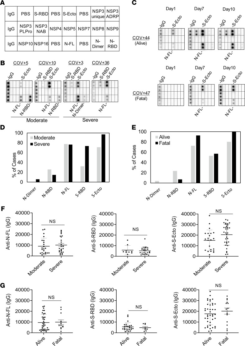
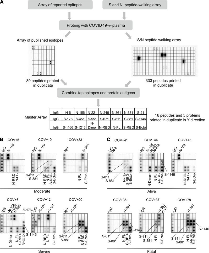
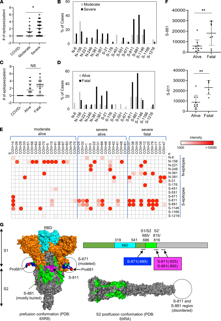
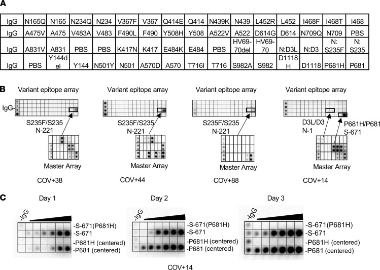
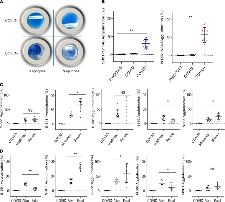
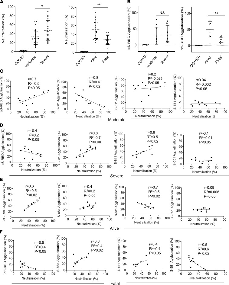
BACKGROUNDThe role of humoral immunity in COVID-19 is not fully understood, owing, in large part, to the complexity of antibodies produced in response to the SARS-CoV-2 infection. There is a pressing need for serology tests to assess patient-specific antibody response and predict clinical outcome.METHODSUsing SARS-CoV-2 proteome and peptide microarrays, we screened 146 COVID-19 patients' plasma samples to identify antigens and epitopes. This enabled us to develop a master epitope array and an epitope-specific agglutination assay to gauge antibody responses systematically and with high resolution.RESULTSWe identified linear epitopes from the spike (S) and nucleocapsid (N) proteins and showed that the epitopes enabled higher resolution antibody profiling than the S or N protein antigen. Specifically, we found that antibody responses to the S-811-825, S-881-895, and N-156-170 epitopes negatively or positively correlated with clinical severity or patient survival. Moreover, we found that the P681H and S235F mutations associated with the coronavirus variant of concern B.1.1.7 altered the specificity of the corresponding epitopes.CONCLUSIONEpitope-resolved antibody testing not only affords a high-resolution alternative to conventional immunoassays to delineate the complex humoral immunity to SARS-CoV-2 and differentiate between neutralizing and non-neutralizing antibodies, but it also may potentially be used to predict clinical outcome. The epitope peptides can be readily modified to detect antibodies against variants of concern in both the peptide array and latex agglutination formats.FUNDINGOntario Research Fund (ORF) COVID-19 Rapid Research Fund, Toronto COVID-19 Action Fund, Western University, Lawson Health Research Institute, London Health Sciences Foundation, and Academic Medical Organization of Southwestern Ontario (AMOSO) Innovation Fund.

Keywords: COVID-19; Immunoglobulins; Infectious disease.


Conflict of interest statement

Conflict of interest: CV, SE, XL, and SSCL are inventors on a US provisional patent application submitted by Western University (application number 63/118,826).

Figures

Figure 1
Figure 1. Lack of correlation between the spike or nucleocapsid antibody response and disease severity or outcome.
(A) Layout of the SARS-CoV-2 proteome array. The array included immunoglobulin G (IgG), phosphate-buffered saline (PBS), spike receptor-binding domain (S-RBD), spike ectodomain (S-ecto), nonstructural protein (NSP), ADP-ribose-1′′-monophosphatase (ADRP), papain-like protease (PLPro), nucleic acid binding (NAB), nucleocapsid full length (N-FL), nucleocapsid dimerization domain (N-dimer), and nucleocapsid RNA-binding domain (N-RBD). (B) Representative images (from n = 65 unique patient samples) of antibody responses for COVID-19 patients with moderate or severe disease determined by the proteome array. (C) Dynamic IgG antibody profiles for 2 patients with severe (but alive) or fatal disease on days 1, 7, and 10 of intensive care unit (ICU) admission. (D and E) Prevalence of antibody responses to the S or N protein/domain for the indicated patient groups determined by the proteome array (based on high-exposure images). (F and G) The intensity of antibody response to the S or N protein antigen was not correlated with disease severity (F) or outcome (G). IgG-binding signals were based on low-exposure array images. Intensity cutoff value was set at 2 SDs of the mean background signal at low exposure. Moderate, n = 31; severe, n = 34; alive, n = 51; fatal n = 14. NS, not significant from unpaired Student’s t test with Welch’s correction.
Figure 2
Figure 2. Identification of SARS-CoV-2 epitopes and epitope-resolved antibody profiling.
(A) Workflow for identifying antigenic epitopes by peptide arrays and the layout of a master array for SARS-CoV-2 antibody profiling. (B and C) Representative images of epitope-resolved antibody profiles for the different groups of COVID-19 patients (n = 65 unique patient samples).
Figure 3
Figure 3. Epitope-specific antibody responses distinguish COVID-19 patients with disparate disease severity and outcome.
(A) Antibodies from patients with severe disease (n = 34) recognized significantly more epitopes than those with moderate conditions (n = 31). (B) Distribution of epitopes in moderate versus severe cases. (C) Number of epitopes/patient in the alive (n = 51) versus fatal (n = 14) groups. (D) Distribution of epitopes in alive versus fatal cases. (E) Heatmap representation of epitope-specific antibodies detected by the master array. Note that the heatmap was based on signals detected at low exposure. (F) Fatal cases showed significantly stronger antibody responses for the S-811 (alive n = 10, fatal n = 6) and S-881 (alive n = 10, fatal n = 8) epitopes. *, P < 0.05; **, P < 0.002; NS, not significant; unpaired Student’s t test with Welch’s correction. (G) Structure models to show location of the critical epitopes on the S protein. The epitopes S-671, S-811, and S-881 are shown on the domain structure diagram of S as well as its prefusion (left) and postfusion (right) conformation. The S protein has 2 cleavage sites, S1/S2 and S2′. The S-671 epitope is located at the C-terminus of S1 and disordered in the prefusion cryo–electron microscopy structure (left panel: Protein Data Bank 6XR8). A homology model from the SWISS-MODEL repository was employed to draw an S-671 epitope model in the left panel (colored blue), without cleavage at S1/S2. The Pro681 site is shown with a red sphere. The S2′ cleavage site is located on the S-811 epitope. The S-881 epitope is buried and inaccessible in the prefusion state but is disordered in the postfusion conformation (right panel: Protein Data Bank 6XRA). The S1 region is colored orange, except for the RBD, which is in cyan. The region between the S1/S2 and S2′ cleavage sites is shown in green. The S-811 and S-881 epitopes are colored magenta in the prefusion conformation.
Figure 4
Figure 4. SARS-CoV-2 variants feature mutated epitopes not recognized by antibodies for the corresponding WT epitopes.
(A) Layout of a SARS-CoV-2 variant epitope array. (B) Examples of COVID-19 cases that showed distinct IgG responses to the mutated and WT epitopes (boxed). (C) Dilution series of P681/P681H-containing epitopes demonstrating the loss of binding for the mutant epitopes by the patient plasma.
Figure 5
Figure 5. Rapid epitope-dependent agglutination assay for SARS-CoV-2 antibodies effectively differentiates patient groups.
(A) Latex bead agglutination assay to gauge antibody responses to SARS-CoV-2. The latex beads were coated with 1 or more biotinylated S or N epitope peptides and mixed with SARS-CoV-2–negative (COVID, top) or –positive (COVID+, bottom) plasma. The presence of antibodies against the epitopes promoted the agglutination of the latex beads. Images shown were taken after 2 minutes’ incubation at room temperature. (B) Epitope-based latex agglutination assay distinguished COVID-19+ from COVID-19 or PreCOVID-19 plasma. The epitope peptides used were S-811 and S-1146 from the S and N-156 and N-361 from the N proteins. (C) Correlation of disease severity with antibody responses to the S-811, N-156, and N-361 epitopes determined by latex bead agglutination. (D) Correlation of disease outcome with antibody responses to the S-551, S-811, S-881, and N-156 epitopes determined by latex bead agglutination. P values calculated based on unpaired 1-tailed Student’s t test with Welch’s correction (no assumption of equal SD) (n = 20 for B; n = 10 for C and D). *P < 0.05, **P < 0.002. Error bars represent the SD.
Figure 6
Figure 6. Antibody specificity predicts neutralization efficiency and disease outcome.
(A) Correlation of neutralization efficiency with clinical severity (left) or outcome (right). *, P < 0.05; **, P < 0.01. (B) Correlation of S-RBD antibody response measured by latex agglutination with COVID-19 severity (left) or outcome (right). **, P < 0.01. Error bars represent SD. (CF) Pearson’s (r) correlation between epitope-dependent agglutination and neutralization. The coefficient of determination (R2) was calculated based on linear regression analysis. Confidence interval: 95%. The P values were calculated using a 2-tailed t distribution with n – 2 degrees of freedom (n = 10). P values are shown on each graph.

References

    1. Ravi N, et al. Diagnostics for SARS-CoV-2 detection: a comprehensive review of the FDA-EUA COVID-19 testing landscape. Biosens Bioelectron. 2020;165:112454. - PMC - PubMed
    1. Long QX, et al. Clinical and immunological assessment of asymptomatic SARS-CoV-2 infections. Nat Med. 2020;26(8):1200–1204. doi: 10.1038/s41591-020-0965-6. - DOI - PubMed
    1. Sakurai A, et al. Natural history of asymptomatic SARS-CoV-2 infection. N Engl J Med. 2020;383(9):885–886. doi: 10.1056/NEJMc2013020. - DOI - PMC - PubMed
    1. Keeley AJ, et al. Asymptomatic SARS-CoV-2 infection: the tip or the iceberg? Thorax. 2020;75(8):621–622. doi: 10.1136/thoraxjnl-2020-215337. - DOI - PubMed
    1. Lisboa Bastos M, et al. Diagnostic accuracy of serological tests for Covid-19: systematic review and meta-analysis. BMJ. 2020;370:m2516. - PMC - PubMed

MeSH terms