Assessment of Lung Reaeration at 2 Levels of Positive End-expiratory Pressure in Patients With Early and Late COVID-19-related Acute Respiratory Distress Syndrome
- PMID: 34081643
- PMCID: PMC8386391
- DOI: 10.1097/RTI.0000000000000600
Assessment of Lung Reaeration at 2 Levels of Positive End-expiratory Pressure in Patients With Early and Late COVID-19-related Acute Respiratory Distress Syndrome
Abstract
Purpose: Patients with novel coronavirus disease (COVID-19) frequently develop acute respiratory distress syndrome (ARDS) and need invasive ventilation. The potential to reaerate consolidated lung tissue in COVID-19-related ARDS is heavily debated. This study assessed the potential to reaerate lung consolidations in patients with COVID-19-related ARDS under invasive ventilation.
Materials and methods: This was a retrospective analysis of patients with COVID-19-related ARDS who underwent chest computed tomography (CT) at low positive end-expiratory pressure (PEEP) and after a recruitment maneuver at high PEEP of 20 cm H2O. Lung reaeration, volume, and weight were calculated using both CT scans. CT scans were performed after intubation and start of ventilation (early CT), or after several days of intensive care unit admission (late CT).
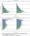
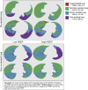
Results: Twenty-eight patients were analyzed. The median percentages of reaerated and nonaerated lung tissue were 19% [interquartile range, IQR: 10 to 33] and 11% [IQR: 4 to 15] for patients with early and late CT scans, respectively (P=0.049). End-expiratory lung volume showed a median increase of 663 mL [IQR: 483 to 865] and 574 mL [IQR: 292 to 670] after recruitment for patients with early and late CT scans, respectively (P=0.43). The median decrease in lung weight attributed to nonaerated lung tissue was 229 g [IQR: 165 to 376] and 171 g [IQR: 81 to 229] after recruitment for patients with early and late CT scans, respectively (P=0.16).
Conclusions: The majority of patients with COVID-19-related ARDS undergoing invasive ventilation had substantial reaeration of lung consolidations after recruitment and ventilation at high PEEP. Higher PEEP can be considered in patients with reaerated lung consolidations when accompanied by improvement in compliance and gas exchange.
Copyright © 2021 Wolters Kluwer Health, Inc. All rights reserved.
Conflict of interest statement
The authors declare no conflicts of interest.
Figures


References
-
- Soni N, Williams P. Positive pressure ventilation: what is the real cost? Br J Anaesth. 2008;101:446–457. - PubMed
-
- Goligher EC, Kavanagh BP, Rubenfeld GD, et al. . Oxygenation response to positive end-expiratory pressure predicts mortality in acute respiratory distress syndrome: a secondary analysis of the LOVS and express trials. Am J Respir Crit Care Med. 2014;190:70–76. - PubMed
-
- Cressoni M, Chiumello D, Algieri I, et al. . Opening pressures and atelectrauma in acute respiratory distress syndrome. Intensive Care Med. 2017;43:603–611. - PubMed
-
- Rocco PRM, Santos C Dos, Pelosid P, et al. . Pathophysiology of ventilator-associated lung injury. Curr Opin Anaesthesiol. 2012;25:123–130. - PubMed
MeSH terms
LinkOut - more resources
Full Text Sources
Medical

