. 2021 Aug 3;33(8):1565-1576.e5.
doi: 10.1016/j.cmet.2021.05.013. Epub 2021 May 18.

SARS-CoV-2 infects human pancreatic β cells and elicits β cell impairment

Affiliations

SARS-CoV-2 infects human pancreatic β cells and elicits β cell impairment

Chien-Ting Wu et al. Cell Metab. .

Abstract

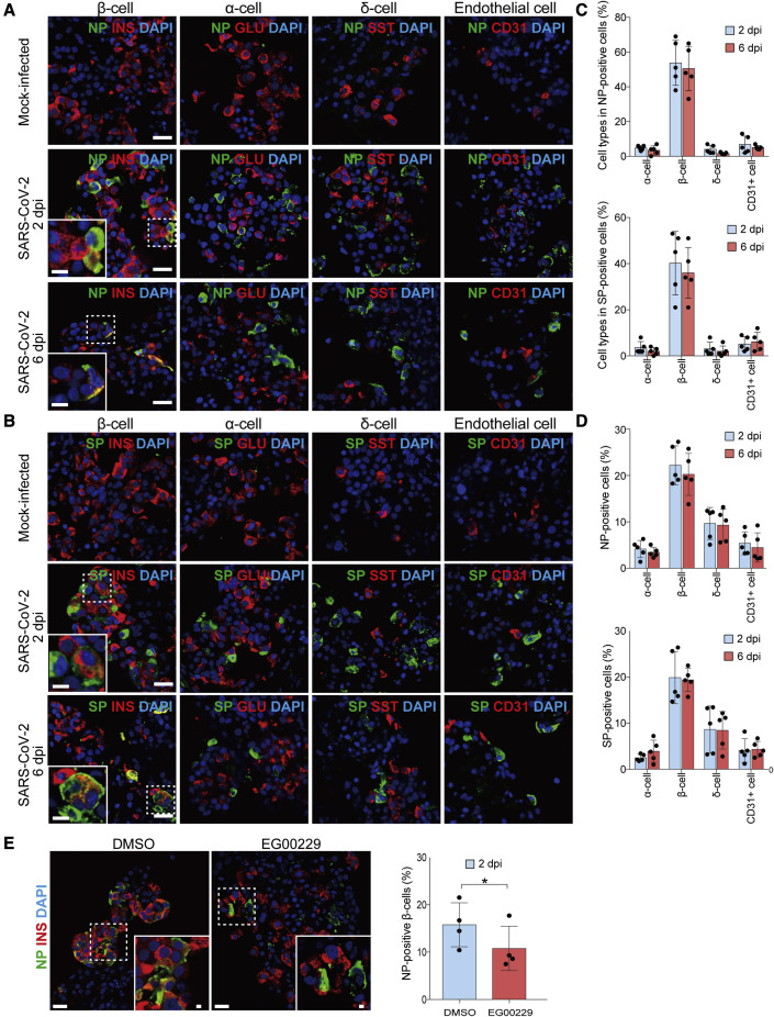
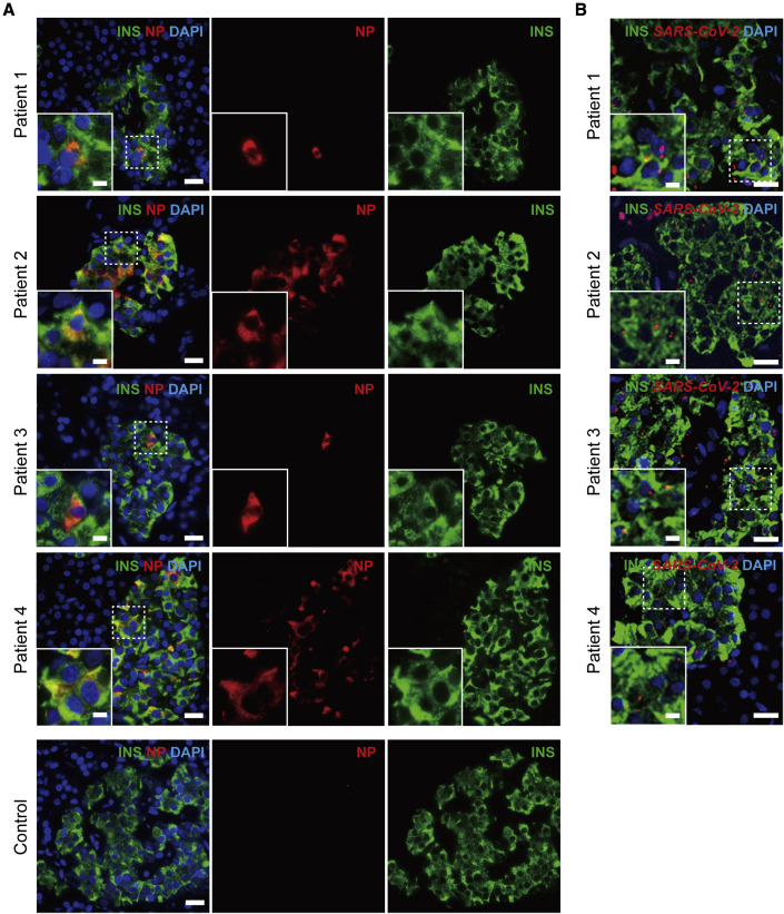
Emerging evidence points toward an intricate relationship between the pandemic of coronavirus disease 2019 (COVID-19) and diabetes. While preexisting diabetes is associated with severe COVID-19, it is unclear whether COVID-19 severity is a cause or consequence of diabetes. To mechanistically link COVID-19 to diabetes, we tested whether insulin-producing pancreatic β cells can be infected by SARS-CoV-2 and cause β cell depletion. We found that the SARS-CoV-2 receptor, ACE2, and related entry factors (TMPRSS2, NRP1, and TRFC) are expressed in β cells, with selectively high expression of NRP1. We discovered that SARS-CoV-2 infects human pancreatic β cells in patients who succumbed to COVID-19 and selectively infects human islet β cells in vitro. We demonstrated that SARS-CoV-2 infection attenuates pancreatic insulin levels and secretion and induces β cell apoptosis, each rescued by NRP1 inhibition. Phosphoproteomic pathway analysis of infected islets indicates apoptotic β cell signaling, similar to that observed in type 1 diabetes (T1D). In summary, our study shows SARS-CoV-2 can directly induce β cell killing.

Keywords: ACE2; COVID-19; SARS-CoV-2; SARS-CoV-2 spike protein; apoptosis; insulin; neuropilin 1; pancreatic beta cell; phosphoproteomics; type 1 diabetes.


Conflict of interest statement

Declaration of interests The authors declare no competing interests.

Figures

None
Graphical abstract
Figure 1
Figure 1
SARS-CoV-2-associated receptors are expressed in pancreatic β cells (A) Representative double immunofluorescence staining of ACE2, TMPRSS2, NRP1, and TFRC with the β cell marker, insulin (INS), and α cell marker, glucagon (GLU), in the normal human pancreas, donor 1. See Table 1. (B) Quantification of ACE2, TMPRSS2, NRP1, and TFRC in β cells (INS +) and α cells (GLU +) from a normal pancreas. No statistically significant changes in ACE2 and TMPRSS2 expression were detected between β and α cells. NRP1 and TFRC expression was statistically significantly higher in β cells compared with α cells. Rabbit anti-NRP1 (Abcam, ab81321, 1:200) and mouse anti-TFRC (Thermo Fisher, # 13-6800, 1:200) were used for the experiments shown here. Error bars represent mean ± SD (~10–15 islets from the pancreas of 5 non-COVID-19 donors; see Table 1). ∗∗p < 0.001, one-way ANOVA with Tukey’s post-test. Each dot represents one donor. Scale bars, 5 μm (A) and 2 μm (insets). See also Figures S1 and S2 and Table 1.
Figure 2
Figure 2
SARS-CoV-2 preferentially infects β cells of human pancreatic islets ex vivo (A–D) Mock-treated or SARS-CoV-2-infected human pancreatic islets were stained after 2 or 6 dpi. (A) Representative double immunofluorescence staining of SARS-CoV-2 nucleocapsid protein (NP) in combination with β cell marker, insulin (INS); ɑ cell marker, glucagon (GLU); δ cell marker, somatostatin (SST); and endothelial cell marker (CD31). (B) Representative double immunofluorescence staining of SARS-CoV-2 spike protein (SP) in combination with a similar combination of markers as (A). The nuclei were stained using DAPI (blue) as a counterstain. (C) Quantified percentages of SARS-CoV-2 NP and SP within α, β, δ, and endothelial cells of pancreatic islets. Around 40% to 60% NP and SP staining, respectively, are present within β cells. (D) Quantified percentages of SARS-CoV-2 NP- and SP-positive α, β, δ, and endothelial cells. (C and D) Error bars represent mean ± SD (~500–1,000 cells were quantified from healthy isolated human islets from donors 1–5; see Table 2). (E) Representative double immunofluorescence staining of SARS-CoV-2 NP in combination with insulin after pre-treating islets with dimethyl sulfoxide (DMSO) or 100 μM EG00229 for 1 h before infection with SARS-CoV-2. Islets were fixed at 2 dpi and stained for SARS-CoV-2 NP and β cell marker, insulin (INS). Quantification of the percentages of β cells containing NP-positive β cells (right). Error bars represent mean ± SD (~500–1,000 cells were quantified from healthy isolated human islets from donors 10–13; see Table 2). p < 0.05, two-tailed Student’s t test. Each dot represents one donor. Scale bars, 5 μm (A, B, and E) and 2 μm (insets). See also Table 2.
Figure 3
Figure 3
SARS-CoV-2 infects pancreatic β cells of patients with COVID-19 (A) Representative double immunofluorescence staining of pancreatic islets from patients with COVID-19 and healthy controls using antibodies against SARS-CoV-2 NP and INS. (B) Representative multiplexed images of in situ hybridization against the SARS-CoV-2 spike mRNA, in combination with immunofluorescence staining of insulin (INS). SARS-CoV-2 spike mRNA expression (red dots) was detected within pancreatic β cells. The nuclei were stained using DAPI (blue) as a counterstain. Scale bars, 5 μm (A and B) and 2 μm (insets). See also Figure S3 and Table 3.
Figure 4
Figure 4
SARS-CoV-2 infection interferes with insulin content/secretion and induces β cell apoptosis (A–F) Pancreatic islet functionality was analyzed by insulin content, glucose-stimulated insulin secretion (GSIS), and TUNEL staining ex vivo. (A) Insulin content is decreased in SARS-CoV-2-infected islets compared with mock-treated islets. (B) GSIS is decreased in SARS-CoV-2-infected islets compared with mock-treated islets. (A and B) Error bars represent mean ± SD (data were collected from 7 healthy isolated human islets, donors 2–8; see Table 2). p < 0.05, two-tailed Student’s t test. (C) Representative staining of β cell apoptosis by in situ TUNEL and DAPI staining in β cells (INS) of mock- or SARS-CoV-2-treated human islets. DNase-treated sections were used as a positive control in the TUNEL assay. (D and F) Quantification of the percentages of islets containing TUNEL-positive β cells. Error bars represent mean ± SD (~500–1,000 cells were quantified from each of 3–5 separate healthy isolated human islets, donors 1–5 [D] and 7–9 [F]; see Table 2). (E) Representative staining of β cell apoptosis by in situ TUNEL and DAPI staining in β cells (INS) of mock-treated versus SARS-CoV-2-SP-treated human islets. p < 0.05, ∗∗p < 0.01, two-tailed Student’s t test. Scale bars, 5 μm (C and E). See also Figures S3–S5 and Tables 2, S1, S2, and S3.

Comment in

References

    1. Arda H.E., Li L., Tsai J., Torre E.A., Rosli Y., Peiris H., Spitale R.C., Dai C., Gu X., Qu K., et al. Age-dependent pancreatic gene regulation reveals mechanisms governing human beta cell function. Cell Metab. 2016;23:909–920. - PMC - PubMed
    1. Baron M., Veres A., Wolock S.L., Faust A.L., Gaujoux R., Vetere A., Ryu J.H., Wagner B.K., Shen-Orr S.S., Klein A.M., et al. A single-cell transcriptomic map of the human and mouse pancreas reveals inter- and intra-cell population structure. Cell Syst. 2016;3:346–360.e4. - PMC - PubMed
    1. Blodgett D.M., Nowosielska A., Afik S., Pechhold S., Cura A.J., Kennedy N.J., Kim S., Kucukural A., Davis R.J., Kent S.C., et al. Novel observations from next-generation RNA sequencing of highly purified human adult and fetal islet cell subsets. Diabetes. 2015;64:3172–3181. - PMC - PubMed
    1. Boddu S.K., Aurangabadkar G., Kuchay M.S. New onset diabetes, type 1 diabetes and COVID-19. Diabetes Metab. Syndr. 2020;14:2211–2217. - PMC - PubMed
    1. Cantuti-Castelvetri L., Ojha R., Pedro L.D., Djannatian M., Franz J., Kuivanen S., van der Meer F., Kallio K., Kaya T., Anastasina M., et al. Neuropilin-1 facilitates SARS-CoV-2 cell entry and infectivity. Science. 2020;370:856–860. - PMC - PubMed

Publication types

MeSH terms