. 2021 Aug:44:102022.
doi: 10.1016/j.redox.2021.102022. Epub 2021 May 26.

Protease-activated receptor 2 induces ROS-mediated inflammation through Akt-mediated NF-κB and FoxO6 modulation during skin photoaging

Affiliations

Protease-activated receptor 2 induces ROS-mediated inflammation through Akt-mediated NF-κB and FoxO6 modulation during skin photoaging

EunJin Bang et al. Redox Biol. 2021 Aug.

Abstract

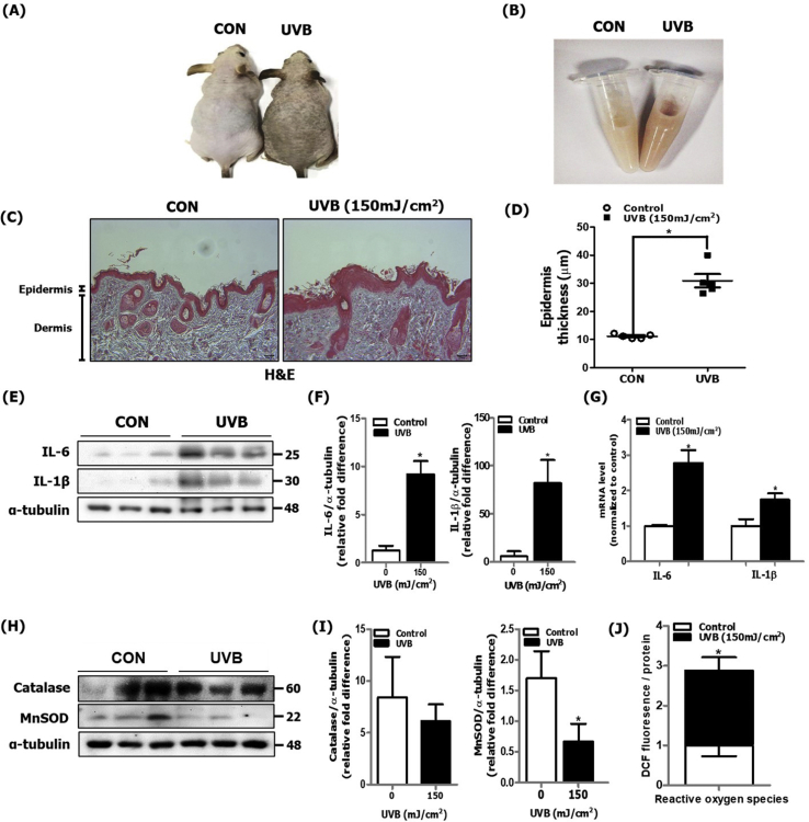
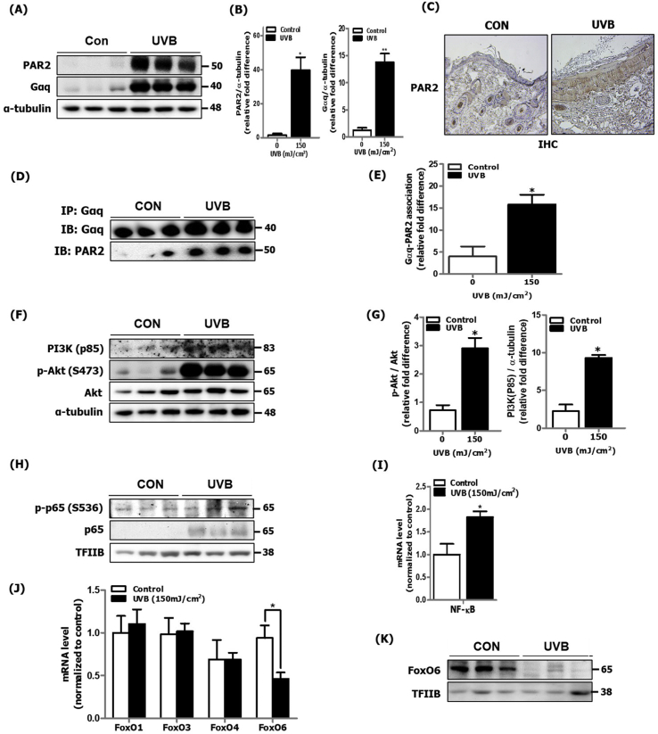
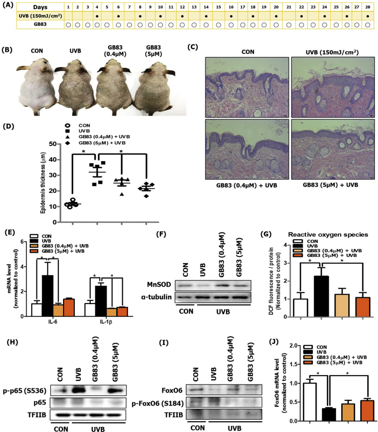
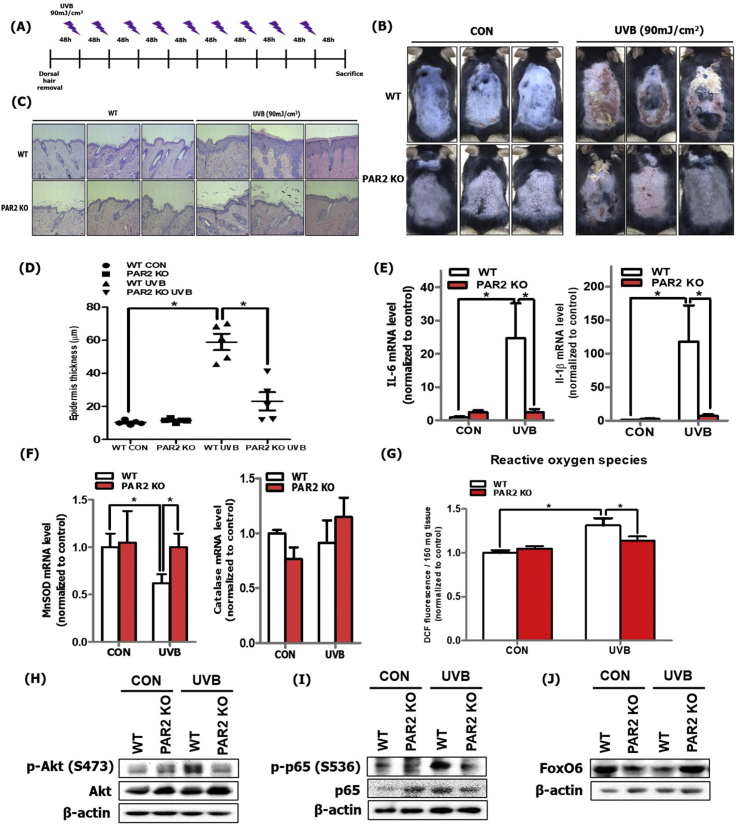
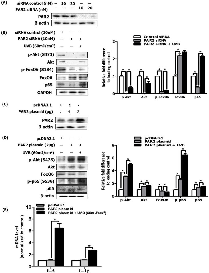
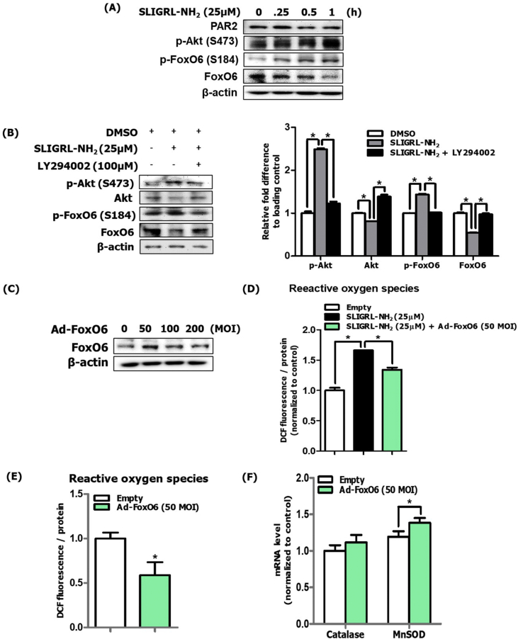
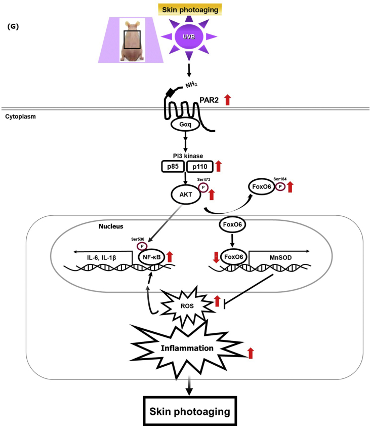
Long-term exposure to ultraviolet irradiation to skin leads to deleterious intracellular effects, including reactive oxygen species (ROS) production and inflammatory responses, causing accelerated skin aging. Previous studies have demonstrated that increased expression and activation of protease-activated receptor 2 (PAR2) and Akt is observed in keratinocyte proliferation, suggesting their potential regulatory role in skin photoaging. However, the specific underlying molecular mechanism of PAR2 and the Akt/NF-κB/FoxO6-mediated signaling pathway is not clearly defined. In this study, we first used the UVB-irradiated photoaged skin of hairless mice and observed an increase in PAR2 and Gαq expression and PI3-kinase/Akt, NF-κB, and suppressed FoxO6. Consequently, increased levels of proinflammatory cytokines and decreased levels of antioxidant MnSOD was observed. Next, to investigate PAR2-specific roles in inflammation and oxidative stress, we used photoaged hairless mice topically applied with PAR2 antagonist GB83 and photoaged PAR2 knockout mice. PAR2 inhibition and deletion significantly suppressed inflammatory and oxidative stress levels, which were associated with decreased IL-6 and IL-1β levels and increased MnSOD levels, respectively. Furthermore, NF-κB phosphorylation and decreased FoxO6 was reduced by PAR2 inhibition and deletion in vivo. To confirm the in vivo results, we conducted PAR2 knockdown and overexpression in UVB-irradiated HaCaT cells. In PAR2 knockdown cells by si-PAR2 treatment, it suppressed Akt/NF-κB and increased FoxO6, whereas PAR2 overexpression reversed these effects and subsequently modulated proinflammatory target genes. Collectively, our data define that PAR2 induces oxidative stress and inflammation through Akt-mediated phosphorylation of NF-κB (Ser536) and FoxO6 (Ser184), which could be a critical upstream regulatory mechanism in ROS-mediated inflammatory response.

Keywords: FoxO6; Inflammation; NF-κB; Protease-activated receptor 2; ROS; Skin photoaging.


Conflict of interest statement

There are no conflicts of interest to declare.

Figures

Fig. 1
Fig. 1
Increased inflammatory response and oxidative stress in UVB-irradiated dorsal skin of hairless mice. After HRM2 mice were repeatedly exposed to UVB radiation (150 mJ/cm2) for 28 days, skin tissue was excised and homogenized to separate the nuclear and cytosolic fractions (N = 5 per group). (A) A photograph of the dorsal skin of control and UVB-irradiated mice was taken for phenotype analysis. (B) A photograph of skin tissue homogenates was taken to visualize discoloration. (C) H&E-stained histological image of skin section of control and UVB-irradiated mice was captured and (D) epidermis thickness was quantified using Motic Image Plus 2.0 software (N = 5 per group). (E) The protein expression levels of IL-6 and IL-1β were measured using western blotting and (F) quantified using ImageJ software. α-tubulin was the loading control of the cytosolic fractions. (G) mRNA levels of IL-6 and IL-1β were quantified using qPCR (N = 5 per group). (H) The protein levels of catalase and MnSOD were measured using western blotting and (I) quantified using ImageJ software. α-tubulin was the loading control of the cytosolic fractions. (J) ROS production was determined by measuring the DCF fluorescence level in the skin cytosolic fraction (N = 5 per group). All data are represented as the mean ± SEM, and significance was determined using an unpaired t-test; *P < 0.05 vs. control.
Fig. 2
Fig. 2
Increased PAR2 expression and Akt/NF-κB/FoxO6 modulation in UVB-irradiated dorsal skin of hairless mice. UVB-irradiated mice skin was homogenized for mRNA and protein extraction (N = 5 per group). (A) The protein expression level of PAR2 and the Gαq subunit was detected in cytosol using western blotting and (B) quantified using ImageJ software (N = 3 per group). α-tubulin was the loading control of the cytosolic fractions. (C) PAR2 localization was examined in skin sections using immunohistochemistry. (D) The level of physical association between PAR2 and Gαq was examined using immunoprecipitation and (E) quantified using ImageJ software (N = 3 per group). (F) PI3K and Akt phosphorylation was detected in the cytosol using western blotting and (G) quantified using ImageJ software (N = 3 per group). α-tubulin was the loading control of the cytosolic fractions. (H) The activation level of p65 was measured by detecting p65 phosphorylation in the nucleus using western blotting and (I) quantified mRNA level using qRT-PCR (N = 5 per group). TFIIB was the loading control of the nuclear fraction. (J) The mRNA expression levels of FoxO isoforms were investigated using qRT-PCR (N = 5 per group). (K) The protein expression level of FoxO6 was detected using western blotting. TFIIB was the loading control of the nuclear fraction. All data are represented as the mean ± SEM and significance was determined using an unpaired t-test; *P < 0.05 vs. control.
Fig. 3
Fig. 3
PAR2 inhibition decreased oxidative stress and inflammation in UVB-irradiated dorsal skin of hairless mice. (A) Approved experimental procedure for topical application of GB83 during skin photoaging (N = 5 per group). (B) A photograph of the dorsal skin of control, UVB-treated, and GB83 with UVB-treated hairless mice was taken for phenotype analysis. (C) H&E-stained histological image of a dorsal skin section from the skin of control, UVB-treated, and GB83 with UVB-treated hairless mice was captured. (D) The thickness of the epidermis was quantified using Motic Image Plus 2.0 software (N = 5 per group). (E) The mRNA expression levels of IL-6 and IL-1β were measured using qRT-PCR (N = 5 per group). (F) The protein expression level of MnSOD was measured using western blotting (N = 3 per group). α-tubulin was the loading control of the cytosolic fractions. (G) The ROS production level was measured using the DCF fluorescence level in skin cytosol fraction (N = 5 per group). (H) The protein expression levels of p-p65 and p65 and (I) p-FoxO6 and FoxO6 were measured using western blotting (N = 3 per group). TFIIB was the loading control of the nuclear fraction. (J) The mRNA expression level of FoxO6 was measured using qRT-PCR (N = 5 per group). All data are represented as the mean ± SEM and significance was determined using an one-factor analysis of variance (ANOVA); *P < 0.05.
Fig. 4
Fig. 4
PAR2 KO mice showed reduced oxidative stress and inflammation during skin photoaging. (A) The experimental procedure for induction of UVB-irradiated skin photoaging using C57BL/6-strained WT and PAR2 KO mice (N = 5 per group). (B) Photographs of dorsal skin of control and UVB-irradiated mice of WT and PAR2 KO mice were taken for phenotype analysis. (B) H&E-stained histological image of a skin section of the control and UVB-irradiated WT and PAR2 KO mice was captured and (C) epidermal thickness was measured and (D) quantified using Motic Image Plus 2.0 software (N = 5 per group). (E) The mRNA levels of IL-6 and IL-1β and (F) MnSOD and catalase were quantified using qRT-PCR (N = 5 per group). (G) ROS production was determined by measuring DCF fluorescence level in the skin cytosol fraction (N = 5 per group). (H) The protein expression levels of phosphorylated Akt, total-Akt (I) phosphorylated p65, p65, and (J) total FoxO6 were detected using western blotting (N = 5 per group). β-actin was the loading control. All data are represented as the mean ± SEM, and significance was determined using an one-factor ANOVA; *P < 0.05.
Fig. 5
Fig. 5
PAR2 induced oxidative stress and the inflammatory response through the Akt/NF-κB/FoxO6 signaling pathway in HaCaT cells. (A) After PAR2 knockdown using siRNA, PAR2 protein level was analyzed via western blotting and observed PAR2 was efficiently knocked-down at 10 nM. β-actin was the loading control of the cytosolic fractions. (B) After treatment with 10 nM siRNA under UVB-irradiated condition, cell lysates were analyzed via western blotting for phosphorylated Akt, total-Akt, phosphorylated FoxO6, total-FoxO6, and p65 and were quantified using ImageJ software (N = 3). GAPDH was the loading control of the whole lysis. (C) The protein expression levels of PAR2 were measured in the HaCaT cell line expressing pcDNA3, a PAR2 plasmid, using transfection method. β-actin was the loading control of the cytosolic fractions. (D) The protein expression levels for phosphorylated Akt, total-Akt, FoxO6, phosphorylated p65 and p65 were measured in the UVB-irradiated HaCaT cells overexpressing PAR2 plasmid and were quantified using ImageJ software (N = 3). β-actin was the loading control of the whole lysis. The mRNA levels of (E) IL-6 and IL-1β were quantified under the same experimental condition. All data are represented as the mean ± SEM (N = 3 per group) and significance was determined using an one-factor ANOVA; *P < 0.05.
Fig. 6
Fig. 6
FoxO6 suppressed PAR2-induced ROS production in HaCaT cells. (A) Protein levels of PAR2, p-Akt, p-FoxO6, and FoxO6 in SLIGRL-NH2 treated cells. β-actin was the loading control of the whole lysis. (B) Suppression of these phosphorylated Akt and FoxO6 proteins was observed in LY294002-pretreated cells and these were quantified using ImageJ software (N = 3). (C) HaCaT cells were treated with FoxO6 adenovirus dose-dependently, and 50 MOI was the effective treatment dose. β-actin was the loading control of the whole lysis. (D) Suppression of PAR2-induced ROS levels by FoxO6 adenovirus (50 MOI) (E) Suppression of ROS levels by FoxO6 adenovirus (50 MOI). ROS levels in HaCaT cells were measured using DCF fluorescence levels (F) mRNA levels of antioxidant catalase and MnSOD genes were quantified. All data are represented as the mean ± SEM (N = 3 per group) and significance was determined using an unpaired t-test and one-factor ANOVA; *P < 0.05. (G) Graphical description of PAR2 inducing ROS-mediated inflammation through Akt-mediated NF-κB and FoxO6 modulation during skin photoaging.
Fig. 6
Fig. 6
FoxO6 suppressed PAR2-induced ROS production in HaCaT cells. (A) Protein levels of PAR2, p-Akt, p-FoxO6, and FoxO6 in SLIGRL-NH2 treated cells. β-actin was the loading control of the whole lysis. (B) Suppression of these phosphorylated Akt and FoxO6 proteins was observed in LY294002-pretreated cells and these were quantified using ImageJ software (N = 3). (C) HaCaT cells were treated with FoxO6 adenovirus dose-dependently, and 50 MOI was the effective treatment dose. β-actin was the loading control of the whole lysis. (D) Suppression of PAR2-induced ROS levels by FoxO6 adenovirus (50 MOI) (E) Suppression of ROS levels by FoxO6 adenovirus (50 MOI). ROS levels in HaCaT cells were measured using DCF fluorescence levels (F) mRNA levels of antioxidant catalase and MnSOD genes were quantified. All data are represented as the mean ± SEM (N = 3 per group) and significance was determined using an unpaired t-test and one-factor ANOVA; *P < 0.05. (G) Graphical description of PAR2 inducing ROS-mediated inflammation through Akt-mediated NF-κB and FoxO6 modulation during skin photoaging.

References

    1. Fisher G.J., Kang S., Varani J. Mechanisms of photoaging and chronological skin aging. Arch. Dermatol. Nov 2002;138(11):1462–1470. doi: 10.1001/archderm.138.11.1462. - DOI - PubMed
    1. Pandel R., Poljšak B., Godic A., Dahmane R. Skin photoaging and the role of antioxidants in its prevention. ISRN Dermatol. Sep 12 2013;2013:930164. doi: 10.1155/2013/930164. - DOI - PMC - PubMed
    1. Oh J.E., Kim M.S., Jeon W.K. A nuclear factor kappa B-derived inhibitor tripeptide inhibits UVB-induced photoaging process. J. Dermatol. Sci. Dec 2014;76(3):196–205. doi: 10.1016/j.jdermsci.2014.10.002. - DOI - PubMed
    1. Mittal M., Siddiqui M.R., Tran K., Reddy S.P., Malik A.B. Reactive oxygen species in inflammation and tissue injury. Antioxidants Redox Signal. Mar 1 2014;20(7):1126–1167. doi: 10.1089/ars.2012.5149. - DOI - PMC - PubMed
    1. Coughlin S.R. Thrombin signalling and protease-activated receptors. Nature. Sep 14 2000;407(6801):258–264. doi: 10.1038/35025229. - DOI - PubMed

Publication types