. 2021 Jun 3;16(1):43.
doi: 10.1186/s13020-021-00451-0.

Electroacupuncture reduces cold stress-induced pain through microglial inactivation and transient receptor potential V1 in mice

Affiliations

Electroacupuncture reduces cold stress-induced pain through microglial inactivation and transient receptor potential V1 in mice

Hsien-Yin Liao et al. Chin Med. .

Abstract

Background: The treatment, and efficacy thereof, is considered to be inadequate with specificity to alleviation of Fibromyalgia and its associated pain. Fibromyalgia patients suffer from chronic and persistent widespread pain and generalized tenderness. Transient receptor potential V1 (TRPV1), which is reported as a Ca2+ permeable ion channel that can be activated by inflammation, is reported to be involved in the development of fibromyalgia pain.

Methods: The current study explored the TRPV1 channel functions as a noxious sensory input in mice cold stress model. It remains unknown whether electroacupuncture (EA) attenuates fibromyalgia pain or affects the TRPV1 pathway.

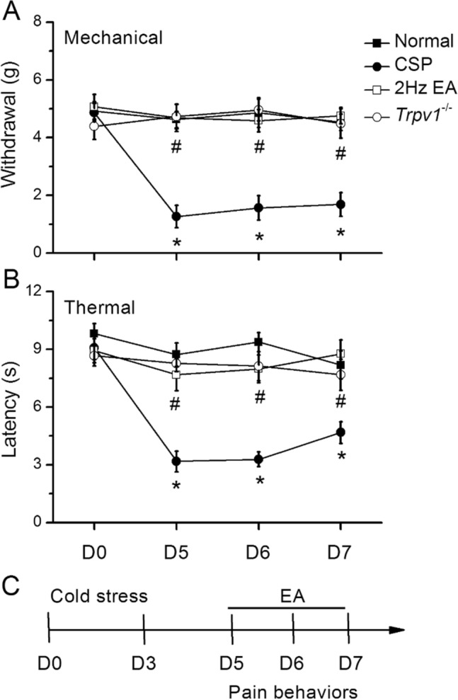
Results: We show that cold stress increases mechanical and thermal pain (day 7: mechanical: 1.69 ± 0.41 g; thermal: 4.68 ± 0.56 s), and that EA and Trpv1 deletion counter this increase. EA and Trpv1 deletion reduced the cold stress-induced increase in inflammatory mediators and TRPV1-related molecules in the hypothalamus, periaqueductal gray (PAG), and cerebellum of mice.

Conclusions: Our results imply that EA has an analgesic effect associated with TRPV1 downregulation. We provide novel evidence that these inflammatory mediators can modulate the TRPV1 signaling pathway and suggest new potential therapeutic targets for fibromyalgia pain.

Keywords: Cerebellum; Electroacupuncture; Fibromyalgia; Hypothalamus; Microglia; TRPV1.


Conflict of interest statement

There are no financial or other relationships that might lead to a conflict of interest for all authors.

Figures

Fig. 1
Fig. 1
Mechanical withdrawal, thermal latency, and experimental flow in normal, CSP, EA, and Trpv1−/− mice. A Mechanical threshold from the von Frey tests. B Thermal latency from the Hargreaves’ test. C Experimental flow in normal, CSP, EA, and Trpv1−/− mice. *Indicates statistical significance when compared with the normal group. #Indicates statistical significance when compared with the CSP groups. n = 10 in all groups
Fig. 2
Fig. 2
Inflammatory mediators plasma concentrations in mice. A IL-1α, IL-1β, IL-2, and IL-5, B IL-6, IL-9, IL-10, and IL-12, C IL-13, IL-17 A, TNF-α, and IFN-γ. **Indicates statistical significance when compared with the normal group. #Indicates statistical significance when compared with the CSP group. IL Interleukin, IFN Interferon, TNF Tumor necrosis factor. n = 6 in all groups
Fig. 3
Fig. 3
Levels of TRPV1 and related molecules in the mice hypothalamus. The western blot bands contain four lanes of protein expression corresponding to the Normal, CSP, EA, and Trpv1−/− groups. A TRPV1, B HMGB1, C S100B, D TLR4, E RAGE, F pPI3K, G pAkt, H pmTOR, I pERK, J pp38, K pJNK, and L pNFκB protein levels. *Indicates statistical significance when compared with the normal group. #Indicates statistical significance when compared with the CSP group. n = 6 in all groups
Fig. 4
Fig. 4
Levels of TRPV1 and related molecules in the mice PAG. The western blot bands contain four lanes of protein expression corresponding to the Normal, CSP, EA, and Trpv1−/− groups. A TRPV1, B HMGB1, C S100B, D TLR4, E RAGE, F pPI3K, G pAkt, H pmTOR, I pERK, J pp38, K pJNK, and L pNFκB protein levels. *Indicates statistical significance when compared with the normal group. #Indicates statistical significance when compared with the CSP group. n = 6 in all groups
Fig. 5
Fig. 5
Levels of TRPV1 and related molecules in the mice cerebellum CVI. The western blot bands contain four lanes of protein expression corresponding to the Normal, CSP, EA, and Trpv1−/− groups. A TRPV1, B HMGB1, C S100B, D TLR4, E RAGE, F pPI3K, G pAkt, H pmTOR, I pERK, J pp38, K pJNK, and L pNFκB protein levels. *Indicates statistical significance when compared with the normal group. #Indicates statistical significance when compared with the CSP group. n = 6 in all groups
Fig. 6
Fig. 6
Levels of TRPV1 and related molecules in the mice cerebellum CVII. The western blot bands contain four lanes of protein expression corresponding to the Normal, CSP, EA, and Trpv1−/− groups. A TRPV1, B HMGB1, C S100B, D TLR4, E RAGE, F pPI3K, G pAkt, H pmTOR, I pERK, J pp38, K pJNK, and L pNFκB protein levels. *Indicates statistical significance when compared with the normal group. #Indicates statistical significance when compared with the CSP group. n = 6 in all groups
Fig. 7
Fig. 7
Immunofluorescence staining of TRPV1, Iba1, and double staining protein expression in the mice hypothalamus. A TRPV1, B Iba1, and C TRPV1/Iba1 double staining, immuno-positive (green, red, or yellow) signals in the mice hypothalamus region. Scale bar: 100 μm. n = 4 in all groups
Fig. 8
Fig. 8
Immunofluorescence staining of TRPV1, Iba1, and double staining protein expression in the mice ventrolateral PAG. A TRPV1, B Iba1, and C TRPV1/Iba1 double staining, immuno-positive (green, red, or yellow) signals in the mice vlPAG region. Scale bar: 100 μm. n = 4 in all groups
Fig. 9
Fig. 9
Immunofluorescence staining of TRPV1, Iba1, and double staining protein expression in the mice cerebellar lobule VI. A TRPV1, B Iba1, and C TRPV1/Iba1 double staining, immuno-positive (green, red, or yellow) signals in the mice cerebellar lobule VI region. Scale bar: 100 μm. n = 4 in all groups
Fig. 10
Fig. 10
Immunofluorescence staining of TRPV1, Iba1, and double staining protein expression in the mice cerebellar lobule VII. A TRPV1, B Iba1, and C TRPV1/Iba1 double staining, immuno-positive (green, red, or yellow) signals in the mice cerebellar lobule VII region. Scale bar: 100 μm. n = 4 in all groups
Fig. 11
Fig. 11
TRPV1 and related molecular pathways in the mouse brain

Similar articles

Cited by

References

    1. Wolfe F, Walitt B, Perrot S, Rasker JJ, Hauser W. Fibromyalgia diagnosis and biased assessment: sex, prevalence and bias. PLoS ONE. 2018;13(9):e0203755. doi: 10.1371/journal.pone.0203755. - DOI - PMC - PubMed
    1. Sluka KA, Clauw DJ. Neurobiology of fibromyalgia and chronic widespread pain. Neuroscience. 2016;338:114–29. doi: 10.1016/j.neuroscience.2016.06.006. - DOI - PMC - PubMed
    1. Yen LT, Hsieh CL, Hsu HC, Lin YW. Targeting ASIC3 for relieving mice fibromyalgia pain: roles of electroacupuncture, opioid, and adenosine. Sci Rep. 2017;7:46663. doi: 10.1038/srep46663. - DOI - PMC - PubMed
    1. Hung CH, Lee CH, Tsai MH, Chen CH, Lin HF, Hsu CY, Lai CL, Chen CC. Activation of acid-sensing ion channel 3 by lysophosphatidylcholine 16:0 mediates psychological stress-induced fibromyalgia-like pain. Ann Rheum Dis. 2020;79(12):1644–56. doi: 10.1136/annrheumdis-2020-218329. - DOI - PMC - PubMed
    1. Lin YW, Chou AIW, Su H, Su KP. Transient receptor potential V1 (TRPV1) modulates the therapeutic effects for comorbidity of pain and depression: the common molecular implication for electroacupuncture and omega-3 polyunsaturated fatty acids. Brain Behav Immun. 2020;89:604–14. doi: 10.1016/j.bbi.2020.06.033. - DOI - PubMed

LinkOut - more resources