Review
. 2021 Jun 3;41(1):16.
doi: 10.1186/s41232-021-00166-7.

VEGFR1-tyrosine kinase signaling in pulmonary fibrosis

Affiliations
Review

VEGFR1-tyrosine kinase signaling in pulmonary fibrosis

Hideki Amano et al. Inflamm Regen. .

Abstract

Vascular endothelial growth factor (VEGF) is not only an important factor for angiogenesis but also lung development and homeostasis. VEGF-A binds three tyrosine kinase (TK) receptors VEGFR1-3. Idiopathic pulmonary fibrosis (IPF) is one of the poor prognoses of lung diseases. The relationship of VEGF and IPF remains to be clarified. Treatment with nintedanib used for the treatment of IPF reduced fibroblast proliferation, inhibited TK receptors, platelet-derived growth factor receptor (PDGFR), fibroblast growth factor receptor (FGFR), and VEGFR. Because the effect of that treatment is still not satisfactory, the emergence of new therapeutic agents is needed. This review describes the enhancement of pulmonary fibrosis by VEGFR1-TK signal and suggests that the blocking of the VEGFR1-TK signal may be useful for the treatment of pulmonary fibrosis.

Keywords: CXCR4; Pulmonary fibrosis; SDF-1; VEGFR1+ cells; VEGFR1-TK.


Conflict of interest statement

The authors declare that they have no competing interests.

Figures

Fig. 1
Fig. 1
The protein level of VEGF receptors in the lung after BLM treatment. a The protein level of VEGF-A, VEGFR1, and VEGFR2 were evaluated by western blot analysis. bd Statistical analysis of VEGF-A (d), VEGFR1 (e), and VEGFR2 (f) was determined and normalized to β-actin. Data are expressed as the mean ± SD (n = 3 mice per group) [11]
Fig. 2
Fig. 2
BLM-induced pulmonary fibrosis formation was suppressed in TKKO mice. a Hematoxylin-eosin staining of the lung in WT mice and TKKO mice on day 21 after BLM treatment. The red arrow indicates the fibrotic area. Bar = 200 μm. b The fibrotic area in the lung on day 21. Data are expressed as the mean ± SD (n = 8-12 mice per group). **p < 0.01 versus WT mice. c Ashcroft score WT mice and TKKO mice on day 21. Data are expressed as the mean ± SD (n = 14-17 mice per group). ****p < 0.0001 versus WT mice [11]
Fig. 3
Fig. 3
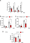
VEGFR1 TK signaling induces the expression of fibrosis-stimulating factors. a TNF-α levels in the lung after BLM treatment. Data are expressed as the mean ± SD (n = 4 mice per group) *p < 0.05 versus WT mice. b-d Expression of S100A4 (b), type I collagen (c), and TGF-β (d) in the lung on day 21 after BLM treatment. Data are expressed as the mean ± SD (n = 4-5 mice per group). *p < 0.05 versus WT mice [11]
Fig. 4
Fig. 4
VEGFR1-TK signaling induces pulmonary fibrosis by VEGFR1+ cells accumulation through the expression of SDF-1/CXCR7/CXCR4 axis. The expression of SDF-1 (a), CXCR7 (b), and CXCR4 (c) in the lung on days 0, 7, 14, and 21 after BLM treatment. Data are expressed as the mean ± SD (n = 3 mice per group). *p < 0.05 versus WT mice. (c) Representative immunohistochemical staining of VEGFR1 in the lung in WT mice and TKKO mice on day 21 after BLM treatment. Bar = 50 μm (d). The number of VEGFR1+ cells in the lung WT mice and TKKO mice on day 21. Data are expressed as the mean ± SD (n = 16 mice per group). ***p < 0.001 versus WT mice. (e) Scheme of VEGFR1-TK signaling on pulmonary fibrosis formation after BLM treatment [11]

Similar articles

Cited by

References

    1. Aiello M, Bertorelli G, Bochchino M, Chetta A, Fiore-Donati A, Fois A, et al. The earlier, the better: impact of early diagnosis on clinical outcome in idiopathic pulmonary fibrosis. Pulm Pharmacol Ther. 2017;44:7–15. doi: 10.1016/j.pupt.2017.02.005. - DOI - PubMed
    1. Wynn TA, Barron L. Macrophages: master regulators of inflammation and fibrosis. Semin Liver Dis. 2010;30(03):245–257. doi: 10.1055/s-0030-1255354. - DOI - PMC - PubMed
    1. Oishi Y, Manabe I. Macrophages in inflammation, repair and regeneration. Int Immunol. 2018;30:511–529. - PubMed
    1. Folkman J. Angiogenesis in cancer, vascular, rheumatoid and other disease. Nat Med. 1995;1(1):27–31. doi: 10.1038/nm0195-27. - DOI - PubMed
    1. Lederer DJ, Martinez FJ. Idiopathic pulmonary fibrosis. N Engl J Med. 2018;378(19):1811–1823. doi: 10.1056/NEJMra1705751. - DOI - PubMed

LinkOut - more resources