. 2021 Jun 1;71(3):256-266.
doi: 10.30802/AALAS-CM-20-000078. Epub 2021 Jun 3.

Fibrous Osteodystrophy, Chronic Renal Disease, and Uterine Adenocarcinoma in Aged Gray Mouse Lemurs (Microcebus murinus)

Affiliations

Fibrous Osteodystrophy, Chronic Renal Disease, and Uterine Adenocarcinoma in Aged Gray Mouse Lemurs (Microcebus murinus)

Kerriann M Casey et al. Comp Med. .

Abstract

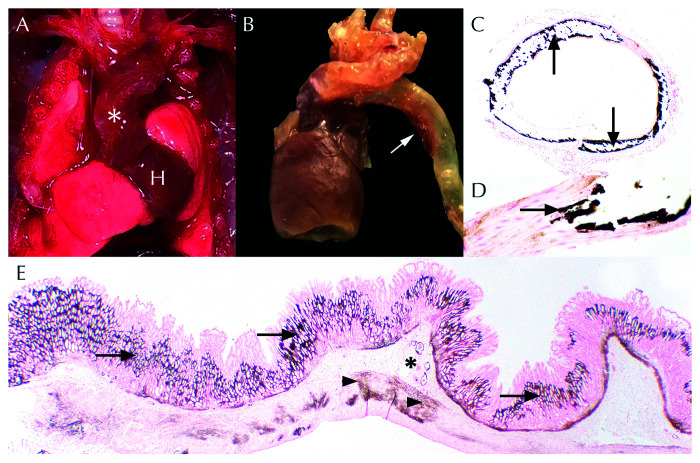
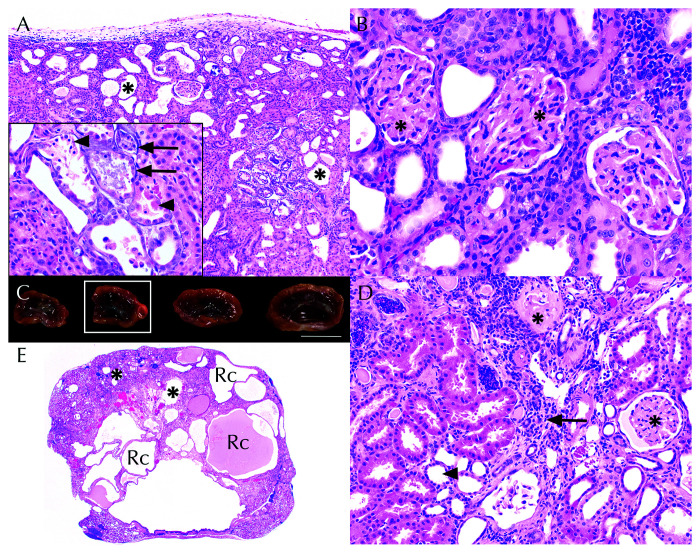
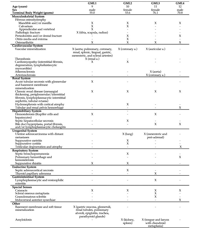
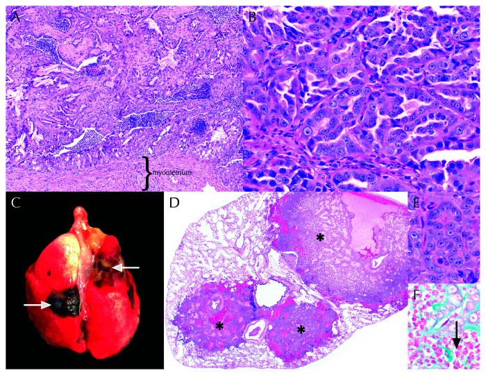
The gray mouse lemur (Microcebus murinus, GML) is a nocturnal, arboreal, prosimian primate that is native to Madagascar. Captive breeding colonies of GMLs have been established primarily for noninvasive studies on questions related to circadian rhythms and metabolism. GMLs are increasingly considered to be a strong translational model for neurocognitive aging due to overlapping histopathologic features shared with aged humans. However, little information is available describing the clinical presentations, naturally occurring diseases, and histopathology of aged GMLs. In our colony, a 9 y-old, male, GML was euthanized after sudden onset of weakness, lethargy, and tibial fracture. Evaluation of this animal revealed widespread fibrous osteodystrophy (FOD) of the mandible, maxilla, cranium, appendicular, and vertebral bones. FOD and systemic metastatic mineralization were attributed to underlying chronic renal disease. Findings in this GML prompted periodic colony-wide serum biochemical screenings for azotemia and electrolyte abnormalities. Subsequently, 3 additional GMLs (2 females and 1 male) were euthanized due to varying clinical and serum biochemical presentations. Common to all 4 animals were FOD, chronic renal disease, uterine adenocarcinoma (females only), cataracts, and osteoarthritis. This case study highlights the concurrent clinical and histopathologic abnormalities that are relevant to use of GMLs in the expanding field of aging research.


Figures

Figure 1.
Figure 1.
Appendicular fractures in gray mouse lemur 1 (GML1). (A) Closed, oblique, middiaphyseal fracture of the right tibia (asterisk) and diffuse “moth eaten” lucenies of the right tibia and tarsus. (B) Postmortem medial deviation of the right tibia (arrow) relative to the stifle and tarsus. (C) Postmortem lateral deviation of the right middiaphyseal radius and ulna (arrow). (D) Postmortem radiographs revealed comminuted fractures of the radius and ulna (asterisk) in addition to “moth eaten” lucencies of the distal humerus, radius, and ulna.
Figure 2.
Figure 2.
Systemic metastatic mineralization in gray mouse lemur 1 (GML1). (A) In-vivo thickening and tortuosity (white asterisk) of the aorta and brachiocephalic trunk as it exits the heart (H). (B) Ex-vivo dilation and opacity (white arrow) of the aortic arch and descending aorta. (C) Transverse sections of the aorta exhibited circumferential deposition of dark brown mineral (black arrows). Von Kossa, 4×. (D) Higher magnification of aorta with replacement of tunica media smooth muscle by dark brown mineral (black arrow). Von Kossa, 40× . (E) Widespread gastric mineralization (dark brown) seen throughout the lamina propria (arrows), submucosal vessels (asterisk), and smooth muscle (arrowheads). Von Kossa, 1.25×.
Figure 3.
Figure 3.
Fibrous osteodystrophy (FOD) in gray mouse lemurs (GMLs) (A) Transverse section of the sacroiliac joint (S = sacrum; I = ilium) of GML1 exhibits diffuse expansion and replacement of bone by highly cellular fibrous connective tissue. Sc = spinal cord. H and E, 1.25×. (B) Cortical and medullary bone is replaced by fibrosis (arrows) that surrounds poorly mineralized islands of woven bone (asterisk). High numbers of osteoclasts (arrowheads) are scattered throughout. H and E, 20 ×. (C) Transverse section of the oral cavity in GML2 revealed widespread replacement of the maxillary (asterisk) and mandibular (arrowhead) alveolar bone by fibrous connective tissue and immature woven bone. (D) Spicules of woven bone (asterisk) were trichrome-positive. Trichrome, 20×. MxM = maxillary molar; MdM = mandibular molar; Bc = buccal cavity. H and E, 1.25×.
Figure 4.
Figure 4.
Chronic renal disease in gray mouse lemurs (GMLs) (A) In GML1, renal tubular dilation (asterisks) was compounded by widespread basement membrane mineralization (inset, arrows) and acute tubular necrosis (inset, arrowheads). H and E, 10×; inset, 40×. (B) Increased glomerular mesangial matrix (asterisks) obscured capillary loop profiles. H and E, 40× (C) Undulating cortical surfaces, size variability, and renal pelvis dilation in gray mouse lemur 2 (GML2). Scale bar = 1 cm. (D). Residual cortical tissue comprised interstitial fibrosis and lymphoplasmacytic interstitial nephritis (arrow), tubular dilation (arrowhead), and variable glomerular mesangial matrix thickening (asterisks). H and E, 20×. (E). Renal histology of kidney from Figure 4 C (white box) demonstrating marked cortical thinning, renal cysts (Rc), and tubular dilation (asterisks). H and E, 1.25×.
Figure 5.
Figure 5.
Histologic lesions within gray mouse lemurs (GMLs). X = feature present; - = feature absent.
Figure 6.
Figure 6.
Metastatic uterine adenocarcinoma in female gray mouse lemurs (GMLs) (A) Uterine adenocarcinoma effaced the endometrium and filled the uterine lumen of GML2. H and E, 10×. (B) Neoplastic cells in GML2 and GML3 comprised polygonal epithelial cells arranged in tubular formations. H and E, 40×. (C) Tan to black pulmonary nodules (arrows) were visible on the pleural surface and extended within the parenchyma of GML2. D. Nodules in Figure 6 C corresponded to multifocal metastatic uterine adenocarcinoma (asterisks). H and E, 1.25×. (E) Metastatic neoplastic cells were cytologically indistinguishable from the primary uterine tumors of GML2 and GML3. (Figure 6 B). H and E, 40 ×. (F) Suppurative bronchopneumonia with gram-negative rods (arrow) was also present within the lung tissue of GML2. Gram stain, 100×.

References

    1. Alleaume C, Mrini ME, Laloy E, Marchal J, Aujard F, Chahory S. 2017. Scleral and corneal xanthomatous inflammation in a gray mouse lemur (Microcebus murinus). Vet Ophthalmol 20:177–180. 10.1111/vop.12374. - DOI - PubMed
    1. Barthold SW, Griffey SM, Percy DH. 2016. Mouse, p 1–118. Chapter 1. In: Barthold SW, Griffey SM, Percy DH, editors. Pathology of laboratory rodents and rabbits, 4th ed. Ames (IA): John Wiley and Sons.
    1. Baxter VK, Shaw GC, Sotuyo NP, Carlson CS, Olson EJ, Zink MC, Mankowski JL, Adams RJ, Hutchinson EK, Metcalf Pate KA. 2013. Serum albumin and body weight as biomarkers for the antemortem identification of bone and gastrointestinal disease in the common marmoset. PLoS One 8:1–10. 10.1371/journal.pone.0082747. - DOI - PMC - PubMed
    1. Beltran WA, Vanore M, Ollivet F, Nemoz-Bertholet F, Aujard F, Clerc B, Chahory S. 2007. Ocular findings in two colonies of gray mouse lemurs (Microcebus murinus). Vet Ophthalmol 10:43–49. 10.1111/j.1463-5224.2007.00491.x. - DOI - PubMed
    1. Bertrand A, Pasquier A, Petiet A, Wiggins C, Kraska A, Joseph-Mathurin N, Aujard F, Mestre-Francés N, Dhenain M. 2013. Micro-MRI study of cerebral aging: ex vivo detection of hippocampal subfield reorganization, microhemorrhages and amyloid plaques in mouse lemur primates. PLoS One 8:1–8. 10.1371/journal.pone.0056593. - DOI - PMC - PubMed

Publication types