

Rhegmatogenous retinal detachment with giant retinal tear in a child with Marfan's syndrome: a rare ocular emergency
- PMID: 34083189
- PMCID: PMC8174519
- DOI: 10.1136/bcr-2020-241354
Rhegmatogenous retinal detachment with giant retinal tear in a child with Marfan's syndrome: a rare ocular emergency
Abstract
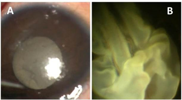
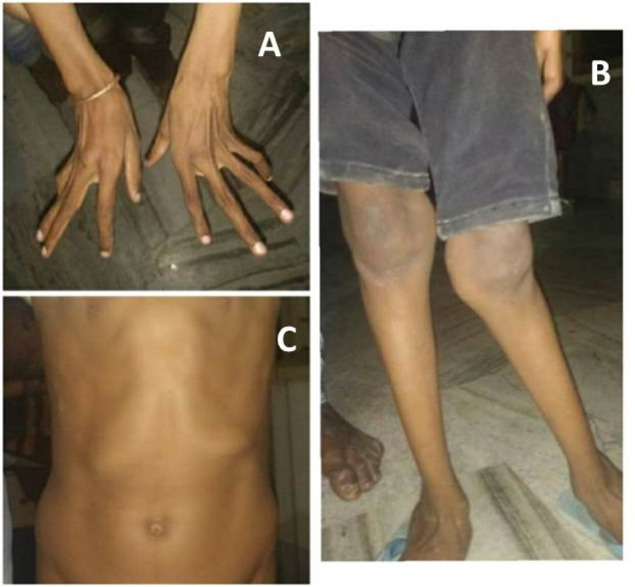
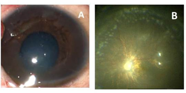
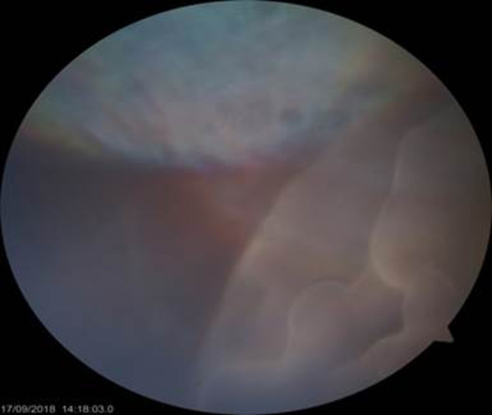
A 7-year-old boy with Marfanoid habitus presented with sudden and painless decrease in the vision of the right eye. Ocular examination revealed rhegmatogenous retinal detachment with 360° giant retinal tear in the right eye and small peripheral retinal breaks with lattice degeneration in the left eye. The patient underwent a 23-gauge pars plana vitrectomy with scleral buckling in the right eye and laser around the breaks in the left eye. At 1-week follow-up visit, the child presented with similar complaints in the left eye as were seen in the right eye. This was later managed effectively with 23-gauge pars plana vitrectomy only. So, with our case report, we would like to highlight the need for aggressive screening in children who are diagnosed with Marfan's syndrome and the need for prophylactic treatment in the unaffected eye.
Keywords: congenital disorders; ophthalmology; retina.
© BMJ Publishing Group Limited 2021. No commercial re-use. See rights and permissions. Published by BMJ.
Conflict of interest statement
Competing interests: None declared.
Figures
Similar articles
-
Retinal detachment in Marfan's syndrome. Characteristics and surgical results.Retina. 1997;17(5):390-6. doi: 10.1097/00006982-199709000-00006. Retina. 1997. PMID: 9355186
-
Incidence of retinal detachment and visual outcome in eyes presenting with posterior vitreous separation and dense fundus-obscuring vitreous hemorrhage.Ophthalmology. 2001 Dec;108(12):2273-8. doi: 10.1016/s0161-6420(01)00822-3. Ophthalmology. 2001. PMID: 11733270
-
Retinal detachment surgery in Marfan's syndrome.Retina. 1998;18(5):405-9. doi: 10.1097/00006982-199805000-00003. Retina. 1998. PMID: 9801033
-
Pars plana vitrectomy combined with scleral buckle versus pars plana vitrectomy for giant retinal tear.Cochrane Database Syst Rev. 2019 Dec 16;12(12):CD012646. doi: 10.1002/14651858.CD012646.pub2. Cochrane Database Syst Rev. 2019. PMID: 31840810 Free PMC article.
-
Pars plana vitrectomy versus scleral buckling for repairing simple rhegmatogenous retinal detachments.Cochrane Database Syst Rev. 2019 Mar 8;3(3):CD009562. doi: 10.1002/14651858.CD009562.pub2. Cochrane Database Syst Rev. 2019. PMID: 30848830 Free PMC article.
Cited by
-
Genetics and Clinical Findings Associated with Early-Onset Myopia and Retinal Detachment in Saudi Arabia.Genes (Basel). 2025 Jul 21;16(7):848. doi: 10.3390/genes16070848. Genes (Basel). 2025. PMID: 40725504 Free PMC article. Review.
-
Rhegmatogenous Retinal Detachment with Giant Retinal Tear: Case Series and Literature Review.J Clin Med. 2024 Aug 9;13(16):4690. doi: 10.3390/jcm13164690. J Clin Med. 2024. PMID: 39200832 Free PMC article.
-
Genotype-phenotype Correlations of Ocular Posterior Segment Abnormalities in Marfan Syndrome.Ophthalmol Sci. 2024 Apr 6;4(5):100526. doi: 10.1016/j.xops.2024.100526. eCollection 2024 Sep-Oct. Ophthalmol Sci. 2024. PMID: 38840780 Free PMC article.
-
What Should We Pay More Attention to Marfan Syndrome Expecting Ectopia Lentis: Incidence and Risk Factors of Retinal Manifestations.J Pers Med. 2023 Feb 24;13(3):398. doi: 10.3390/jpm13030398. J Pers Med. 2023. PMID: 36983580 Free PMC article.
References
Publication types
MeSH terms
LinkOut - more resources
Full Text Sources
Medical
Miscellaneous