. 2021 Aug 1;27(15):4422-4434.
doi: 10.1158/1078-0432.CCR-21-1549. Epub 2021 Jun 3.

Aging and CNS Myeloid Cell Depletion Attenuate Breast Cancer Brain Metastasis

Affiliations

Aging and CNS Myeloid Cell Depletion Attenuate Breast Cancer Brain Metastasis

Alex Man Lai Wu et al. Clin Cancer Res. .

Abstract

Purpose: Breast cancer diagnosed in young patients is often aggressive. Because primary breast tumors from young and older patients have similar mutational patterns, we hypothesized that the young host microenvironment promotes more aggressive metastatic disease.

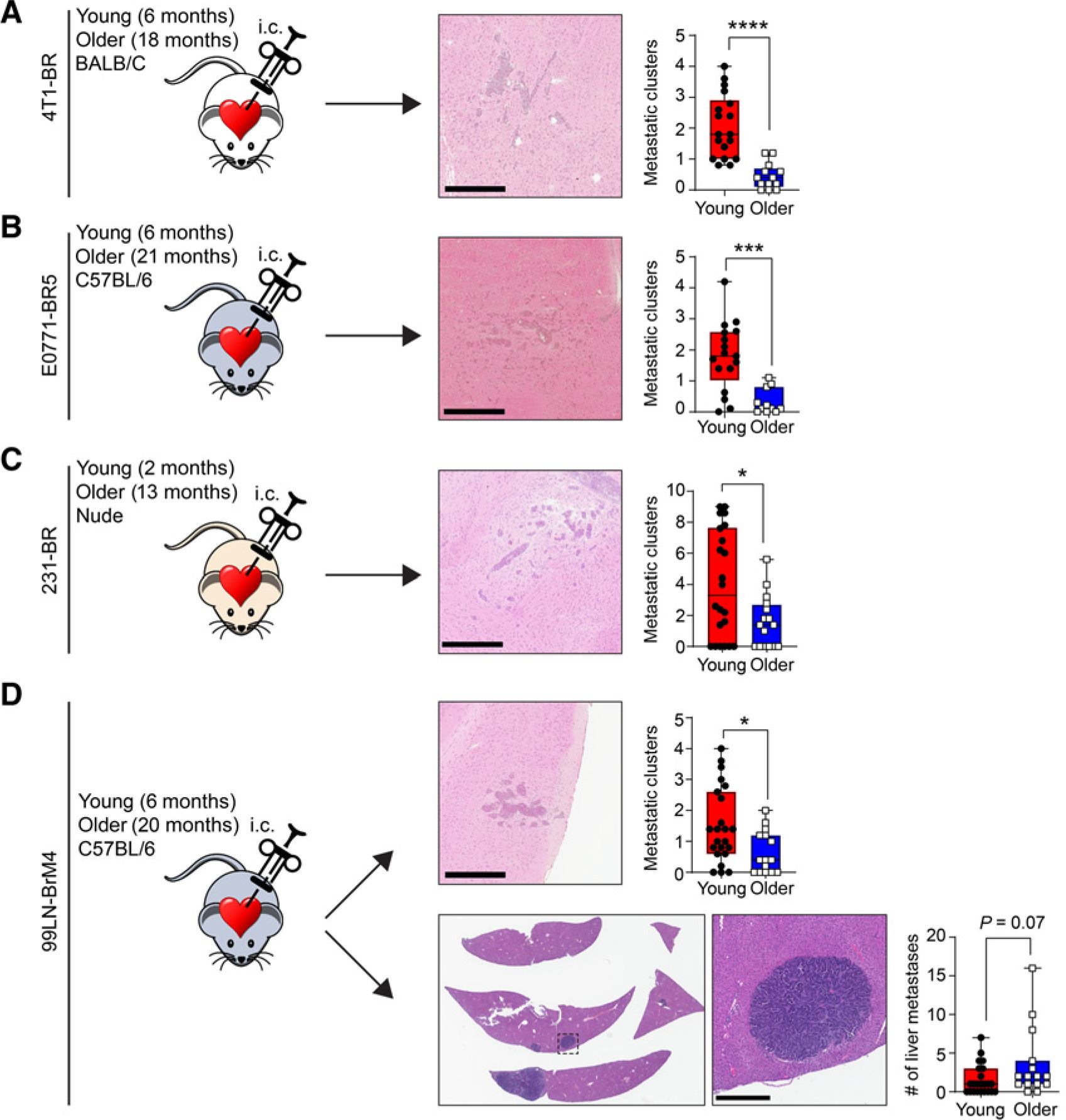
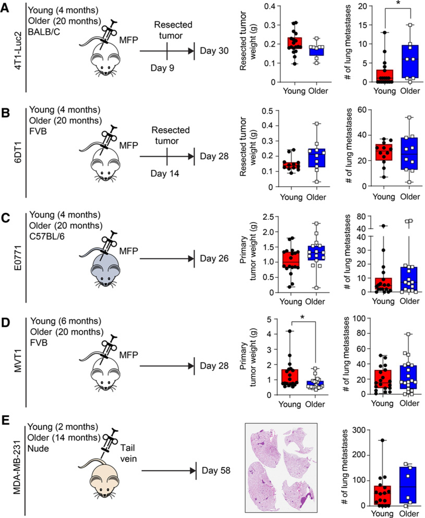
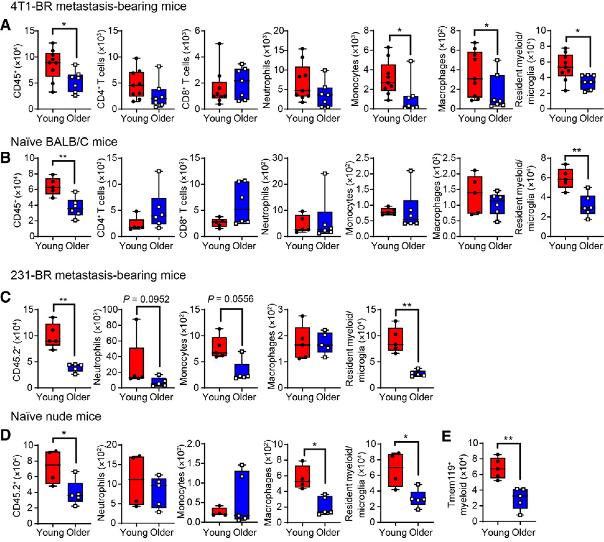
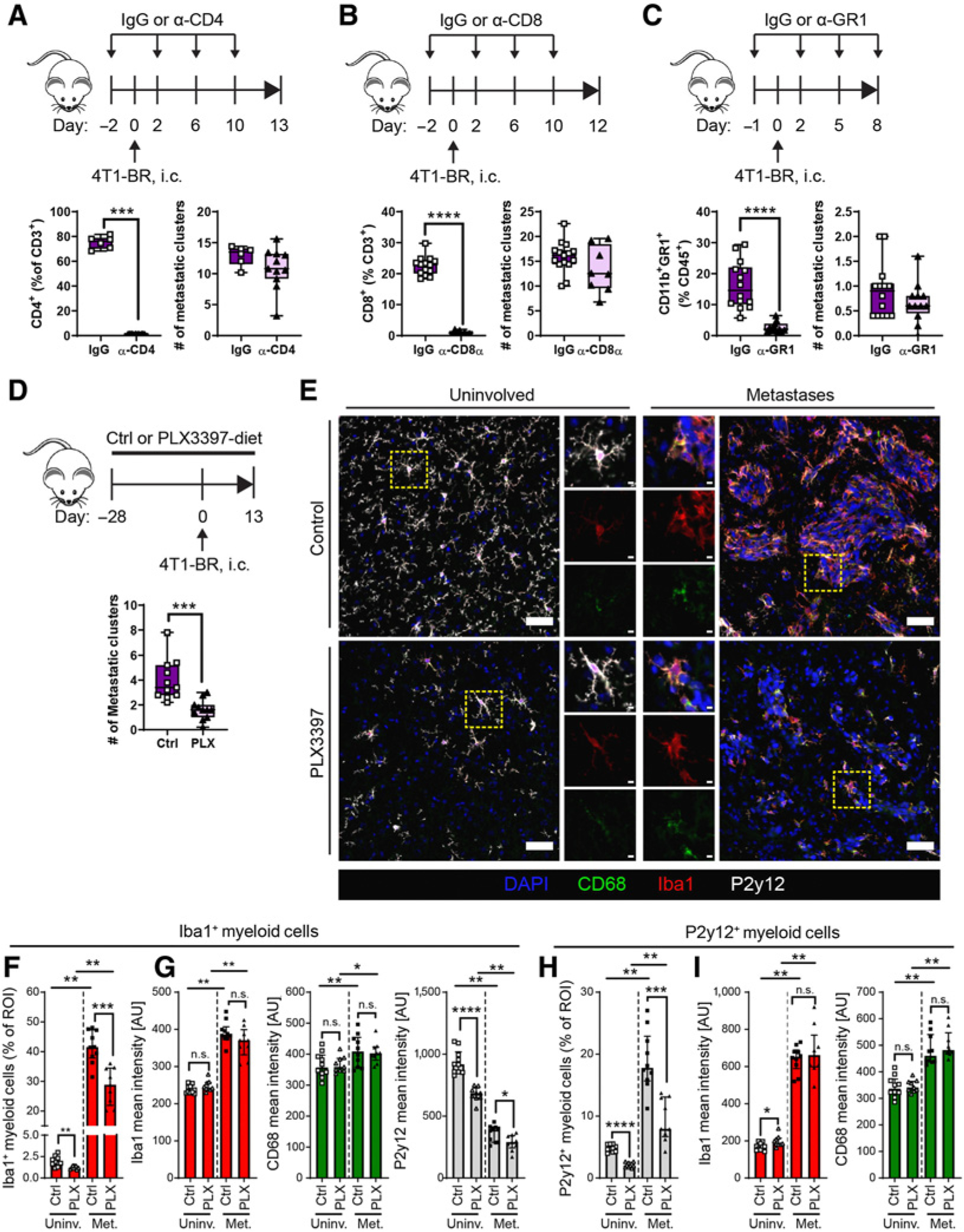
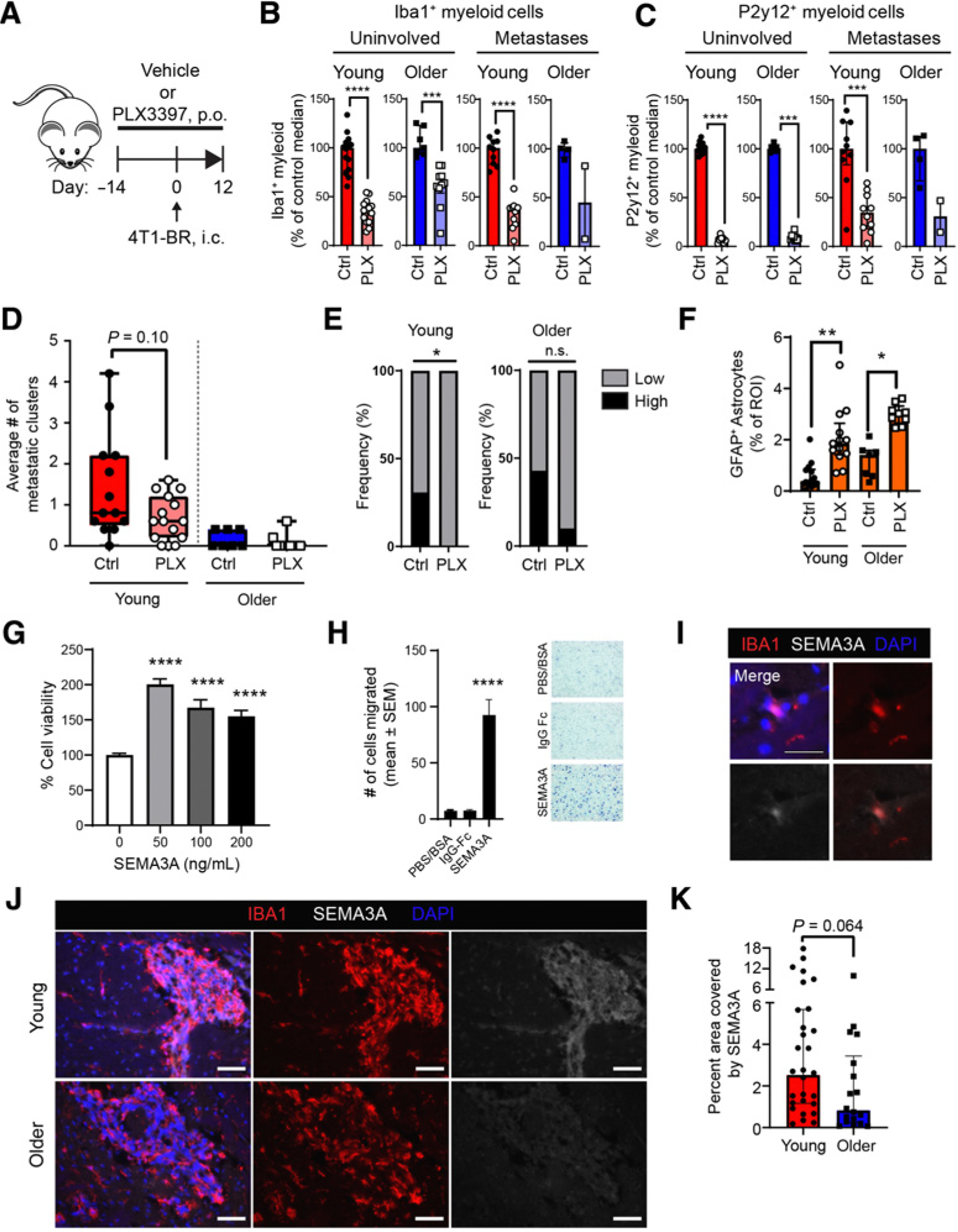
Experimental design: Triple-negative or luminal B breast cancer cell lines were injected into young and older mice side-by-side to quantify lung, liver, and brain metastases. Young and older mouse brains, metastatic and naïve, were analyzed by flow cytometry. Immune populations were depleted using antibodies or a colony-stimulating factor-1 receptor (CSF-1R) inhibitor, and brain metastasis assays were conducted. Effects on myeloid populations, astrogliosis, and the neuroinflammatory response were determined.

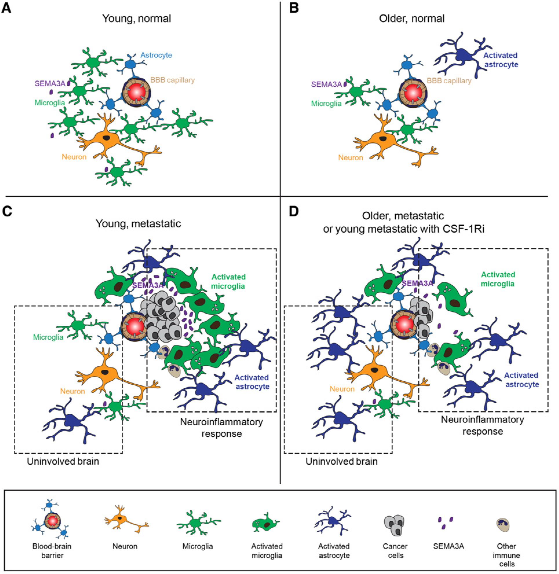
Results: Brain metastases were 2- to 4-fold higher in young as compared with older mouse hosts in four models of triple-negative or luminal B breast cancer; no age effect was observed on liver or lung metastases. Aged brains, naïve or metastatic, contained fewer resident CNS myeloid cells. Use of a CSF-1R inhibitor to deplete myeloid cells, including both microglia and infiltrating macrophages, preferentially reduced brain metastasis burden in young mice. Downstream effects of CSF-1R inhibition in young mice resembled that of an aged brain in terms of myeloid numbers, induction of astrogliosis, and Semaphorin 3A secretion within the neuroinflammatory response.

Conclusions: Host microenvironmental factors contribute to the aggressiveness of triple-negative and luminal B breast cancer brain metastasis. CSF-1R inhibitors may hold promise for young brain metastasis patients.


Figures

Figure 1.
Figure 1.
A young age promotes breast cancer metastasis in the brain. Breast cancer cells were injected intracardially into young and older mice and metastatic tumor burden was quantified. Metastatic clusters, a group of four or more closely spaced metastatic deposits, or one large lesion > 300 μm in both dimensions, was used as the histological endpoint. A, Brain metastatic tumor burden in young (n = 18) and older (n = 13) BALB/C mice 13 days after inoculation with 4T1-BR cells. B, Brain metastatic tumor burden in young (n= 17) and older (n= 11) C57BL/6 mice 13 days after inoculation with E0771-BR5 cells. C, Brain metastatic tumor burden in young (n = 22) and older (n = 16) nude mice 30 days after inoculation with MDA-MB-231-BR (231-BR) cells. D, Metastatic tumor burden in the brain (top) and liver (bottom) of young (n = 23) and older (n = 15) C57BL/6 mice 29 to 35 days after inoculation with 99LN-BrM4 cells. Metastatic tumor burden reported is the average number of metastatic clusters per brain tissue section, or number of liver lesions in one representative tissue section. H&E tissue section scale bar, 500 μm. Each data point is one mouse. One independent experiment per model (*, P < 0.05; **, P < 0.01; ***, P < 0.001; ****, P < 0.0001, Mann–Whitney test).
Figure 2.
Figure 2.
No effect of age on breast cancer metastasis in lung. A, Resected tumor weight and number of lung metastases in young (n = 18) and older (n = 8) BALB/C mice inoculated with 4T1-Luc2 cells into MFP. B, Resected tumor weight and number of lung metastases inyoung (n = 12) and older (n = 10) FVB mice inoculated with 6DT1 cells into MFP. C, Primary tumor weight and number of lung metastases in young (n = 18) and older (n= 15) C57BL/6 mice inoculated with parental E0771 cells into MFP. D, Primary tumor weight and number of lung metastases in young (n = 19) and older (n = 19) FVB mice inoculated with MVT1 cells into MFP. E, Number of lung metastases inyoung (n = 16) and older (n = 6) nude mice injected via tail-veinwith MDA-MB-231 cells. Each data point is one mouse. One independent experiment per model (*, P < 0.05, Mann–Whitney test).
Figure 3.
Figure 3.
Effect of age and metastatic status on brain immune composition. A and B, Number of brain immune cells in young (5–6 months) and older (18–20 months) BALB/C mice with (A) or without (B) 4T1-BR brain metastases. Results shown in A are combined data from two independent experiments, terminated at day 13 and 14 after injection with cancer cells and in B, are one independent experiment. A and B, Immune subsets were defined as follows: CD4+ T cells (CD3+CD4+), CD8+ T cells (CD3+CD8α+), neutrophils (CD11b+Ly6G+), monocytes (CD11b+Ly6C+Ly6GF4/80), macrophages(CD11b+Ly6C+Ly6GF4/80+), and resident myeloid/microglia (CD45LoCD11b+CD39+CD3Ly6CLy6G). C and D, Number of brain immune cells in young (2–3 months) and older (15 months) athymic nude mice with (C) or without (D) 231-BRbrain metastases. C,Mice were euthanized at day28 after intracardial injection with cancer cells. C and D, Immune subsets were defined as follows: neutrophils (CD11b+Ly6G+), monocytes (CD11b+Ly6C+Ly6GF4/80), macrophages (CD11b+Ly6C+Ly6GF4/80+), and resident myeloid/microglia (CD45.2+CD11b+Thy1.2Ly6CLy6G). E, Number of Tmem119+ myeloid cells (CD45+CD11b+Tmem119+) in young (2 months) and older (15–17 months) athymic nude mice. Each data point is one mouse. One independent experiment (*, P < 0.05; **, P < 0.01, Mann–Whitney test).
Figure 4.
Figure 4.
Depletion of immune subsets reveals a contributory role for CNS myeloid cells in brain metastasis. A–C, Frequency of immune subsets in terminal blood draws and number of 4T1-BR brain metastatic clusters quantified from BALB/C mice (2 months) injected with anti-CD4 (A), anti-CD8α (B), or anti-GR1 (C) antibodies or isotype IgG controls. Each data point is one mouse (n = 6–15 per experimental group). One independent experiment per depletion strategy (*, P < 0.05; **, P < 0.01, Mann–Whitney test). D–H, BALB/C mice (2 months) were given control diet (Ctrl, n = 11) or diet supplemented with 300 mg/kg PLX3397, a CSF-1R inhibitor (PLX, n = 11) for 28 days prior to injection of 4T1-BR cancer cells (day 0) and continuously thereafter until study termination at day 13. D, Schematic representation of the experiment and average number of metastatic clusters per brain tissue section. Each data point is one mouse. One independent experiment (***, P < 0.001, Mann–Whitney test). E, Representative immunofluorescence images of uninvolved cortical brain tissue and brain metastases in control and PLX3397-treated mice. Images of uninvolved tissue and brain metastases are from the same animal. Scale bar, 50 μm. Inset scale bar, 10 μm. F, Quantification of percent of region-of-interest (ROI) covered by Iba1. G, Staining intensity [arbitrary units (AU)] for Iba1, CD68, and P2y12 within the Iba1+ regions. H, Quantification of percent of ROI covered by P2y12. I, Staining intensity for Iba1 and CD68 within the P2y12+ regions. F–I, The Wilcoxon matched-pairs test was used to compare differences in percent coverage and staining intensity between uninvolved (Uninv.) brain regions and brain metastases (Met.) within each treatment group (*, P < 0.05; **, P < 0.01, significant difference from uninvolved brain). The Mann–Whitney test was used to compare control versus PLX3397 within each tissue compartment (*, P < 0.05; **, P < 0.01; ***, P < 0.001; ****, P < 0.0001, n.s., not significant). ROI for uninvolved brain is the whole field whereas ROI for metastases is area covered by brain metastases.
Figure 5.
Figure 5.
Effect of resident CNS myeloid cell depletion on brain metastasis development in young and older mice. Young (4.5 months) and older (18 months) BALB/C mice were given daily oral gavages of PLX3397 (PLX; 100 mg/kg) or vehicle control (Ctrl) and injected with 4T1-BR cancer cells. Mice were euthanized at day 12 after injection of cancer cells. Young-Ctrl (n = 13), Young-PLX (n = 15), Older-Ctrl (n = 7), Older-PLX (n = 10), one independent experiment. A, Schematic representation of the experiment. B and C, Percent reduction in Iba1+ myeloid (B) and P2y12+ microglia (C) in PLX3397-treated mice relative to control mice within the same tissue compartment (***, P < 0.001, Mann–Whitney test). Too few aged mice developed brain metastases therefore no statistical test could be performed. D, Average number of metastatic clusters per brain tissue section. Each data point is one mouse. The Mann–Whitney test was used to test statistical difference between treatment arms within one age group. E, Frequency of mice within each age group defined as having high and low brain metastatic tumor burden using age-specific cut-offs (*, P < 0.05, Fischer exact test). F, Percent of ROI in uninvolved brain regions covered with GFAP+ astrocytes (*, P < 0.05; **, P < 0.0001 Kruskal–Wallis test, followed by Dunn post hoc pairwise comparison). ROI for uninvolved brain is the whole field. G, Cellular viability of 4T1-BR cells treated with increasing concentrations of recombinant mouse SEMA3A Fc chimera protein were assessed at 48 hours (mean ± SEM, n = 8–16 wells). Representative of two independent experiments (****, P < 0.0001 compared with untreated control, one-way ANOVA, Dunnett post hoc comparison). H, Results from Boyden chamber motility assay used to test the migration of 4T1-BR cells towards recombinant SEMA3A Fc chimera protein (100 μg/mL), control IgG Fc, and diluent PBS/BSA (0.1%). Pooled results from three independent experiments (n = 6–22 wells). I, Representative immunofluorescent image of an Iba1þ myeloid cell expressing SEMA3A. J, Representative immunofluorescent images of SEMA3A in 4T1-BR brain metastases of young (n = 6) and older (n = 5) mice. Scale bar = 50 μm. K, Quantitation of SEMA3A expression in 4T1-BR brain metastases. Each data point is a metastatic cluster. Statistical test, Mann–Whitney test.
Figure 6.
Figure 6.
Schema of brain metastasis in young and older mice. A, The young normal brain microenvironment features capillaries enclosed by the blood–brain barrier, neurons, astrocytes, myeloid cells (primarily microglia), and other cells. B, With age, the normal brain has fewer myeloid cells/microglia, less Semaphorin 3a (Sema3a) production, and more activated astrocytes, defined by GFAP expression. C, The young metastatic brain contains a relatively high metastatic burden and shows alterations in two areas: in uninvolved (noncancer containing) regions, activated astrocytes are observed and may be inhibitory to initiation of colonization. Prominent alterations are observed in the neuroinflammatory response that forms around a metastatic lesion. These include high numbers of neuroinflammatory microglia and high levels of Sema3a, which may potentiate a developing metastatic lesion. D, With age, the metastatic brain contains fewer metastases and decreased myeloid cells overall. In the uninvolved brain, greater GFAP+ activated astrocytes occur. In the neuroinflammatory response, less Sema3a is produced. Many of these same features (fewer microglia, activated astrocytes) are also observed in young metastatic brains treated with a CSF-1R inhibitor, suggesting that CSF-1R inhibition phenocopies aspects of aging.

Similar articles

Cited by

References

    1. Anders CK, Hsu DS, Broadwater G, Acharya CR, Foekens JA, Zhang Y, et al. Young age at diagnosis correlates with worse prognosis and defines a subset of breast cancers with shared patterns of gene expression. J Clin Oncol 2008;26:3324–30. - PubMed
    1. Partridge AH, Hughes ME, Warner ET, Ottesen RA, Wong YN, Edge SB, et al. Subtype-dependent relationship between young age at diagnosis and breast cancer survival. J Clin Oncol 2016;34:3308–14. - PubMed
    1. van der Hage JA, Mieog JSD, van de Velde CJH, Putter H, Bartelink H, van de Vijver MJ. Impact of established prognostic factors and molecular subtype in very young breast cancer patients: pooled analysis of four EORTC randomized controlled trials. Breast Cancer Res 2011;13:R68. - PMC - PubMed
    1. Cancello G, Maisonneuve P, Rotmensz N, Viale G, Mastropasqua MG, Pruneri G, et al. Prognosis and adjuvant treatment effects in selected breast cancer subtypes of very young women (<35 years) with operable breast cancer. Ann Oncol 2010;21:1974–81. - PubMed
    1. Anders CK, Fan C, Parker JS, Carey LA, Blackwell KL, Klauber-DeMore N, et al. Breast carcinomas arising at a young age: unique biology or a surrogate for aggressive intrinsic subtypes? J Clin Oncol 2011;29:E18–20. - PMC - PubMed

Publication types

Substances