. 2021 Jun;9(6):e002484.
doi: 10.1136/jitc-2021-002484.

Direct and indirect immune effects of CMP-001, a virus-like particle containing a TLR9 agonist

Affiliations

Direct and indirect immune effects of CMP-001, a virus-like particle containing a TLR9 agonist

Shakoora A Sabree et al. J Immunother Cancer. 2021 Jun.

Abstract

Background: CMP-001, also known as vidutolimod, is a virus-like particle containing a TLR9 agonist that is showing promise in early clinical trials. Our group previously demonstrated that the immunostimulatory effects of CMP-001 are dependent on an anti-Qβ antibody response which results in opsonization of CMP-001 and uptake by plasmacytoid dendritic cells (pDCs) that then produce interferon (IFN)-α. IFN-α then leads to an antitumor T-cell response that is responsible for the in vivo efficacy of CMP-001. Here, we explore mechanisms by which the initial effects of CMP-001 on pDCs activate other cells that can contribute to development of an antitumor T-cell response.

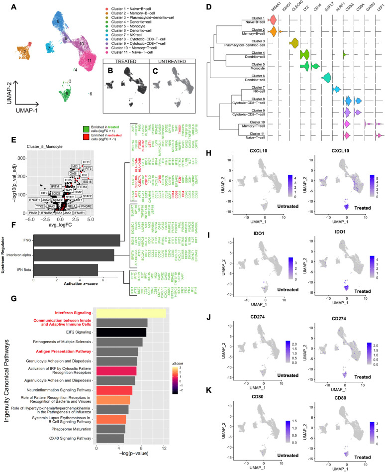
Methods: Uptake of CMP-001 by various peripheral blood mononuclear cell (PBMC) populations and response to anti-Qβ-coated CMP-001 were evaluated by flow cytometry and single-cell RNA sequencing. Purified monocytes were treated with anti-Qβ-coated CMP-001 or recombinant IFN-α to evaluate direct and secondary effects of anti-Qβ-coated CMP-001 on monocytes.

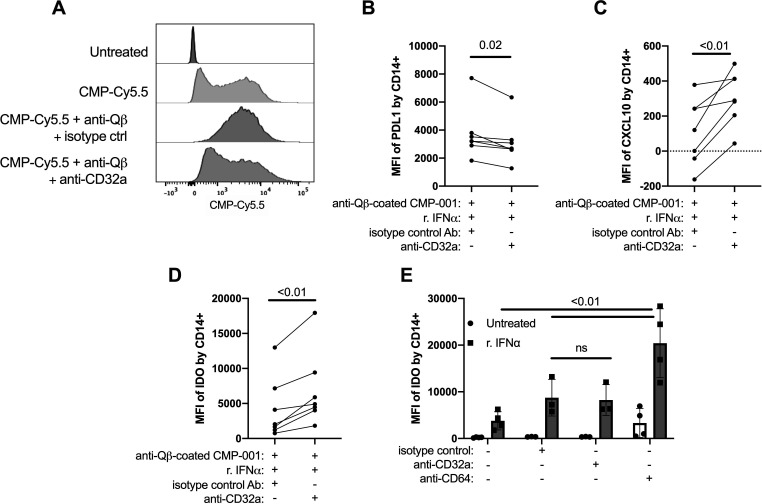
Results: Monocytes had the highest per cell uptake of anti-Qβ-coated CMP-001 with lower levels of uptake by pDCs and other cell types. Treatment of PBMCs with anti-Qβ-coated CMP-001 induced upregulation of IFN-responsive genes including CXCL10, PDL1, and indoleamine-2,3-dioxygenase (IDO) expression by monocytes. Most of the impact of anti-Qβ-coated CMP-001 on monocytes was indirect and mediated by IFN-α, but uptake of anti-Qβ-coated CMP-001 altered the monocytic response to IFN-α and resulted in enhanced expression of PDL1, IDO, and CD80 and suppressed expression of CXCL10. These changes included an enhanced ability to induce autologous CD4 T-cell proliferation.

Conclusions: Anti-Qβ-coated CMP-001 induces IFN-α production by pDCs which has secondary effects on a variety of cells including monocytes. Uptake of anti-Qβ-coated CMP-001 by monocytes alters their response to IFN-α, resulting in enhanced expression of PDL1, IDO and CD80 and suppressed expression of CXCL10. Despite aspects of an immunosuppressive phenotype, these monocytes demonstrated increased ability to augment autologous CD4 T-cell proliferation. These findings shed light on the complexity of the mechanism of action of anti-Qβ-coated CMP-001 and provide insight into pathways that may be targeted to further enhance the efficacy of this novel approach to immunotherapy.

Keywords: immunotherapy; tumor microenvironment.


Conflict of interest statement

Competing interests: SEB holds stock options in Checkmate Pharmaceuticals. GJW received research funding from Checkmate Pharmaceuticals. All of the other authors declare no competing interests.

Figures

Figure 1
Figure 1
Uptake of fluorescently labeled CMP-001 by various immune cell subsets. Unfractionated human peripheral blood mononuclear cells (A) or monocytes polarized into classical M1 (B) and M2 (C) macrophages were treated with Cy5.5-labeled CMP-001 plus or minus anti-Qβ for 1 hour. Cells were stained for unique surface markers and analyzed via flow cytometry. Data are representative of two to three individual experiments. NK, natural killer.
Figure 2
Figure 2
scRNA-seq of PBMCs treated with anti-Qβ-coated CMP-001 demonstrates upregulation of genes in monocytes that can impact on T-cell function, including those in the IFN pathway. scRNA-seq was performed on unfractionated normal donor PBMCs treated with and without anti-Qβ-coated CMP-001. Data from one of two similar experiments are shown. (A) Clustering of merged datasets. (B) Clustering of treated cells. (C) Clustering of untreated cells. (D) Violin plots of classic cell-type surface markers. (E) Volcano plot of differentially expressed genes in monocytes (cluster 5) following anti-Qβ-coated CMP-001 treatment. For most genes, treatment resulted in positive logFCs. (F) Ingenuity pathway analysis of potential upstream regulators of differentially expressed genes in monocytes following anti-Qβ-coated CMP-001 treatment. (G) Ingenuity pathway analysis of upregulated signaling pathways in monocytes following anti-Qβ-coated CMP-001 treatment. UMAP plots of CXCL10 (H), IDO1 (IDO) (I), CD274 (PDL1) (J), and CD80 (K) untreated (left) and treated with anti-Qβ-coated CMP-001 (right). IRF, interferon regulatory factor; IFN, interferon; PBMC, peripheral blood mononuclear cell; scRNA-seq, single-cell RNA sequencing; UMAP, uniform manifold approximation and projection; IDO, indoleamine-2,3-dioxygenase.
Figure 3
Figure 3
Anti-Qβ-coated CMP-001-mediated induction of CXCL10 is dependent on monocytes and pDC-derived type I IFN. Various cell populations were treated with CMP-001 with and without anti-Qβ for 24 hours and supernatant evaluated for CXCL10, IFN-α, and IFN-β. (A) CXCL10 production in response to anti-Qβ-coated CMP-001 by PBMCs with and without monocyte depletion. (B) CXCL10 production by monocytes combined with pDCs in response to anti-Qβ-coated CMP-001. (C) CXCL10 production in response to anti-Qβ-coated CMP-001 by monocytes plus pDCs when cells were placed on the same (–) or opposite (+) sides of a transwell. (D) Production of IFN-α by pDCs treated with anti-Qβ-coated CMP-001. (E) Production of IFN-β by pDCs treated with anti-Qβ-coated CMP-001. (F) Impact of antibody blocking of IFN-α on CXCL10 production by PBMCs treated with anti-Qβ-coated CMP-001. (G) Impact of antibody blocking of IFNAR2 on CXCL10 production by PBMCs treated with anti-Qβ-coated CMP-001. Graphs depict means±SEM. Statistical significance was determined using two-way ANOVA with multiple comparisons test (A), one-way ANOVA with multiple comparisons test (B, C), and paired t-test (D–G) with alpha=0.05 (n=3–6). ANOVA, analysis of variance; IFN, interferon; ns, not significant; PBMC, peripheral blood mononuclear cell; pDC, plasmacytoid dendritic cell.
Figure 4
Figure 4
Anti-Qβ-coated CMP-001 enhanced expression of IDO and PDL1 by monocytes is dependent on type I IFN. Unfractionated PBMCs from healthy donors were treated with CMP-001 with and without anti-Qβ, and expression of cytoplasmic IDO and surface PDL1 by monocytes was determined by flow cytometry. (A, D) Expression of IDO and PDL1 induced by CMP-001 with and without anti-Qβ. (B, E) Impact of antibody blocking of IFNAR2 on expression of IDO and PDL1 induced by anti-Qβ-coated CMP-001. (C, F) Expression of IDO and PDL1 induced by recombinant IFN-α. Graphs illustrate expression (MFI) of IDO and PDL1 on Zombie Aqua negative CD14+ cells. Statistical significance was determined using one-way analysis of variance with multiple comparisons test (A, D) and paired t-test (B, C, E, F) with alpha=0.05 (n=6). IFN, interferon; MFI, median fluorescence intensity; ns, not significant; PBMC, peripheral blood mononuclear cell; IDO, indoleamine-2,3-dioxygenase.
Figure 5
Figure 5
Phagocytosis by monocytes of antibody-coated particles alters their response to IFN-α. Monocytes from healthy donors were treated with recombinant IFN-α plus or minus anti-Qβ-coated CMP-001, anti-Qβ-coated methylated CMP-001 (mCMP-001), G10 (soluble TLR9 agonist) or IgG +protein L beads for 18–20 hours. Expression of cytoplasmic IDO and CXCL10 and surface PDL1 was determined by flow cytometry. (A, E, I) Impact of recombinant IFN-α on IDO, PDL1, and CXCL10 expression by monocytes. (B, F, J) Impact of anti-Qβ-coated CMP-001 on IDO, PDL1, and CXCL10 expression by monocytes in response to IFN-α. (C, G, K) Impact of anti-Qβ-coated CMP-001, anti-Qβ-coated mCMP-001, or G10 on IDO, PDL1, and CXCL10 expression by monocytes in response to IFN-α. (D, H, L) Impact of IgG+ protein L beads on IDO, PDL1, and CXCL10 expression by monocytes in response to IFN-α. Statistical significance was determined using one-way analysis of variance with multiple comparisons test and paired t-test with alpha=0.05 (n=6–10). IFN, interferon; IDO, indoleamine-2,3-dioxygenase; MFI, median fluorescence intensity; ns, not significant.
Figure 6
Figure 6
Impact of CD32a blockade on uptake of anti-Qβ-coated CMP-001 by monocytes and concomitant response to IFN-α. (A) Monocytes from healthy donors were pretreated anti-CD32a or isotype control for 15 min followed by treatment with 10 μg/mL Cy5.5-labeled CMP-001 plus or minus anti-Qβ. Data are representative of two independent experiments. (B–D) Monocytes from healthy donors were pretreated with anti-CD32a or isotype control for 15 min followed by treatment with recombinant IFN-α and anti-Qβ-coated CMP-001 for 18–20 hours. Expression of PDL1, CXCL10, and IDO were determined by flow cytometry. (E) Monocytes from healthy donors were treated with 10 μg/mL anti-CD32a, anti-CD64, or isotype control (in the absence of particles) for 18–20 hours. Expression of IDO was determined by flow cytometry. Statistical significance was determined using paired t-test (B–D) and two-way analysis of variance (E) with alpha=0.05 (n=3–7). IFN, interferon; IDO, indoleamine-2,3-dioxygenase; MFI, median fluorescence intensity; ns, not significant.
Figure 7
Figure 7
Monocytes treated with anti-Qβ-coated CMP-001 and IFN-α upregulate CD80 expression and induce autologous CD4 T-cell proliferation. (A) Monocytes from healthy donors were treated with recombinant IFN-α plus or minus anti-Qβ-coated CMP-001 for 18–20 hours. CD80 surface expression was determined by flow cytometry. (B) Monocytes from healthy donors were treated with recombinant IFN-α plus or minus anti-Qβ-coated CMP-001 for 18–20 hours. The next day, autologous CFSE-labeled T cells were added to treated monocytes at a ratio of 1:10 (monocyte:T cell), and percent of CFSE-diluted CD3+ T cells was determined after 4 days as a measure of T-cell proliferation. (C) Purified CFSE-labeled T cells were treated directly with recombinant IFN-α plus or minus anti-Qβ-coated CMP-001 in the absence of monocytes, and percent of CFSE-diluted CD3+ T cells was determined after 4 days as a measure of T-cell proliferation. (D) Representative histograms of CFSE-diluted CD3+ T cells. (E, F) Phenotypical characterization of proliferated CD3+ T cells into CD4 and CD8 subsets. Statistical significance was determined using one-way analysis of variance with multiple comparisons test with alpha=0.05 (n=3–11). IFN, interferon; MFI, median fluorescence intensity; ns, not significant.

Similar articles

Cited by

References

    1. Hammerich L, Bhardwaj N, Kohrt HE, et al. . In situ vaccination for the treatment of cancer. Immunotherapy 2016;8:315–30. 10.2217/imt.15.120 - DOI - PubMed
    1. Cheng Y, Lemke-Miltner CD, Wongpattaraworakul W, et al. . In situ immunization of a TLR9 agonist virus-like particle enhances anti-PD1 therapy. J Immunother Cancer 2020;8:e000940. 10.1136/jitc-2020-000940 - DOI - PMC - PubMed
    1. Lemke-Miltner CD, Blackwell SE, Yin C, et al. . Antibody opsonization of a TLR9 Agonist-Containing virus-like particle enhances in situ immunization. J Immunol 2020;204:1386–94. 10.4049/jimmunol.1900742 - DOI - PMC - PubMed
    1. Miller AM, Lemke-Miltner CD, Blackwell S, et al. . Intraperitoneal CMP-001: a novel immunotherapy for treating peritoneal carcinomatosis of gastrointestinal and pancreaticobiliary cancer. Ann Surg Oncol 2021;28:1187–97. 10.1245/s10434-020-08591-7 - DOI - PMC - PubMed
    1. Weiner GJ. Cpg oligodeoxynucleotide-based therapy of lymphoid malignancies. Adv Drug Deliv Rev 2009;61:263–7. 10.1016/j.addr.2008.12.006 - DOI - PubMed

Publication types

MeSH terms