. 2021 Jun 4;372(6546):1068-1073.
doi: 10.1126/science.abg1774.

Reciprocal repulsions instruct the precise assembly of parallel hippocampal networks

Affiliations

Reciprocal repulsions instruct the precise assembly of parallel hippocampal networks

Daniel T Pederick et al. Science. .

Abstract

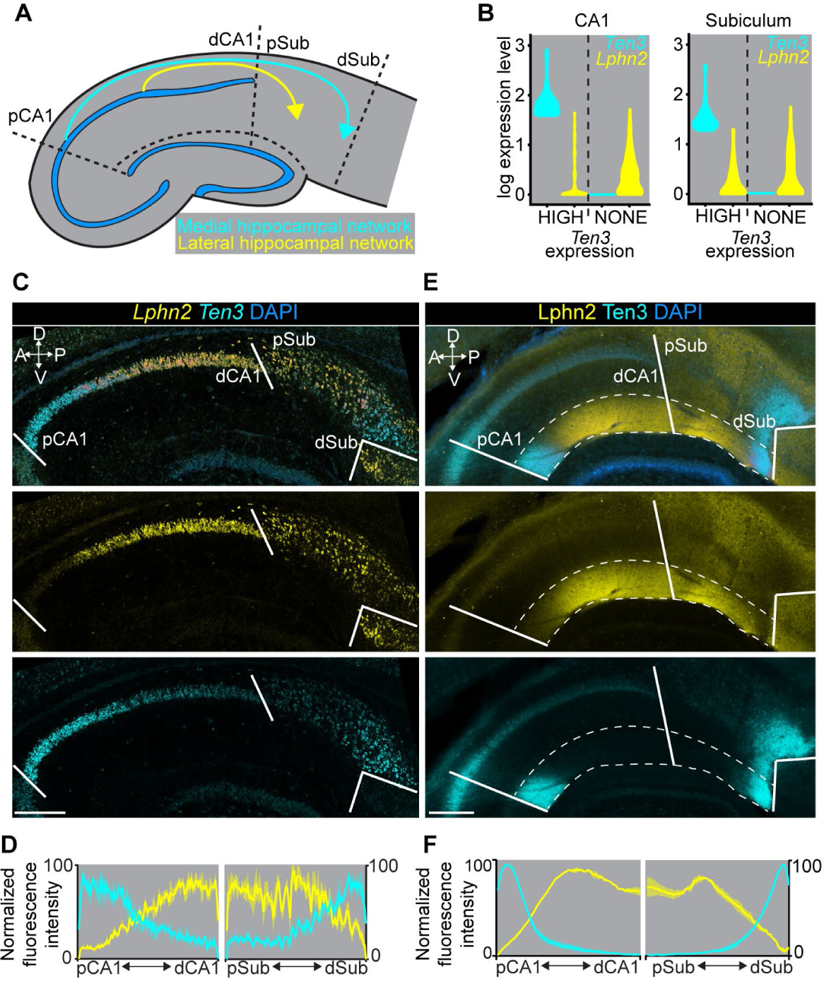
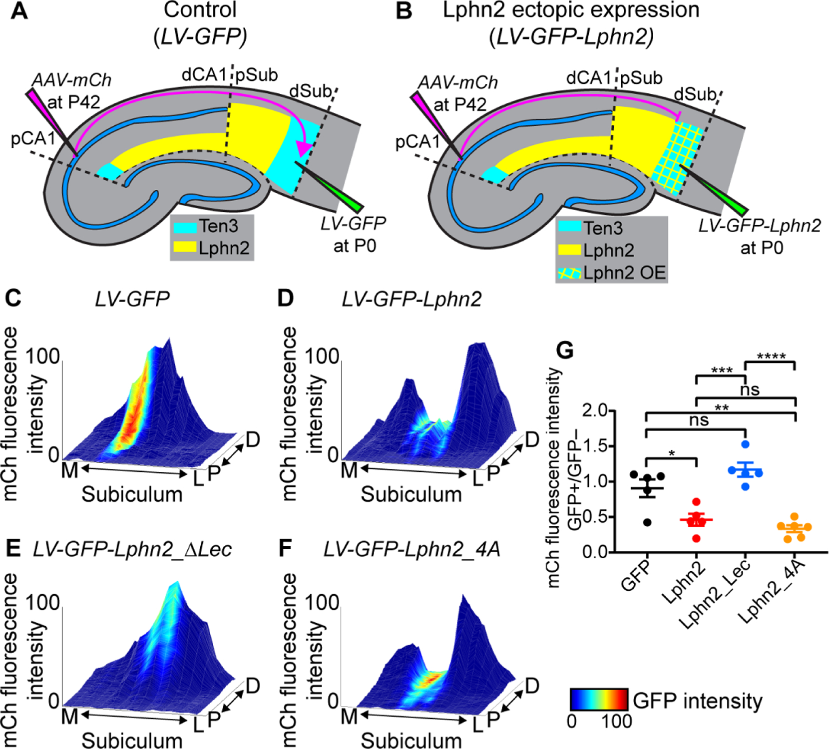
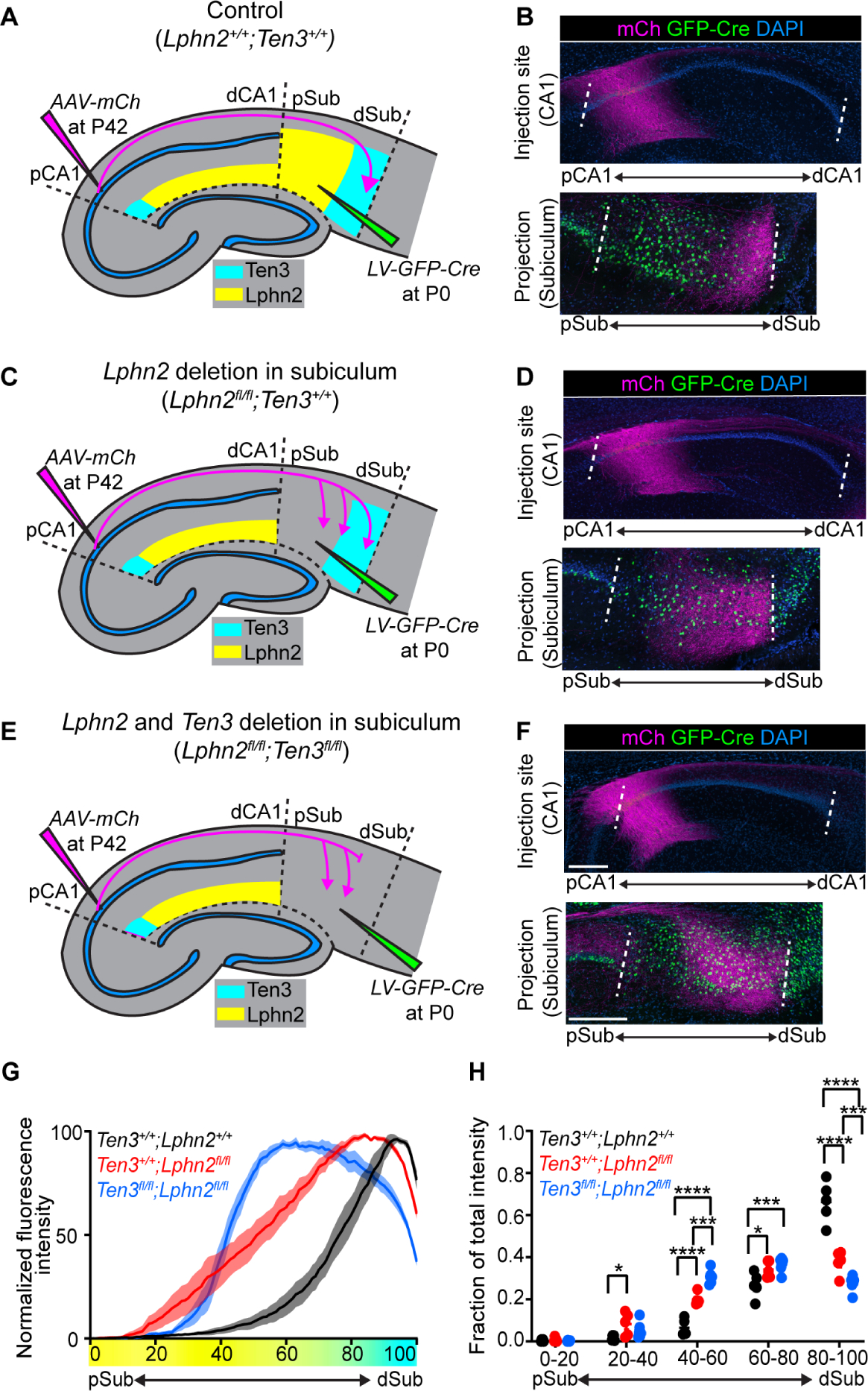
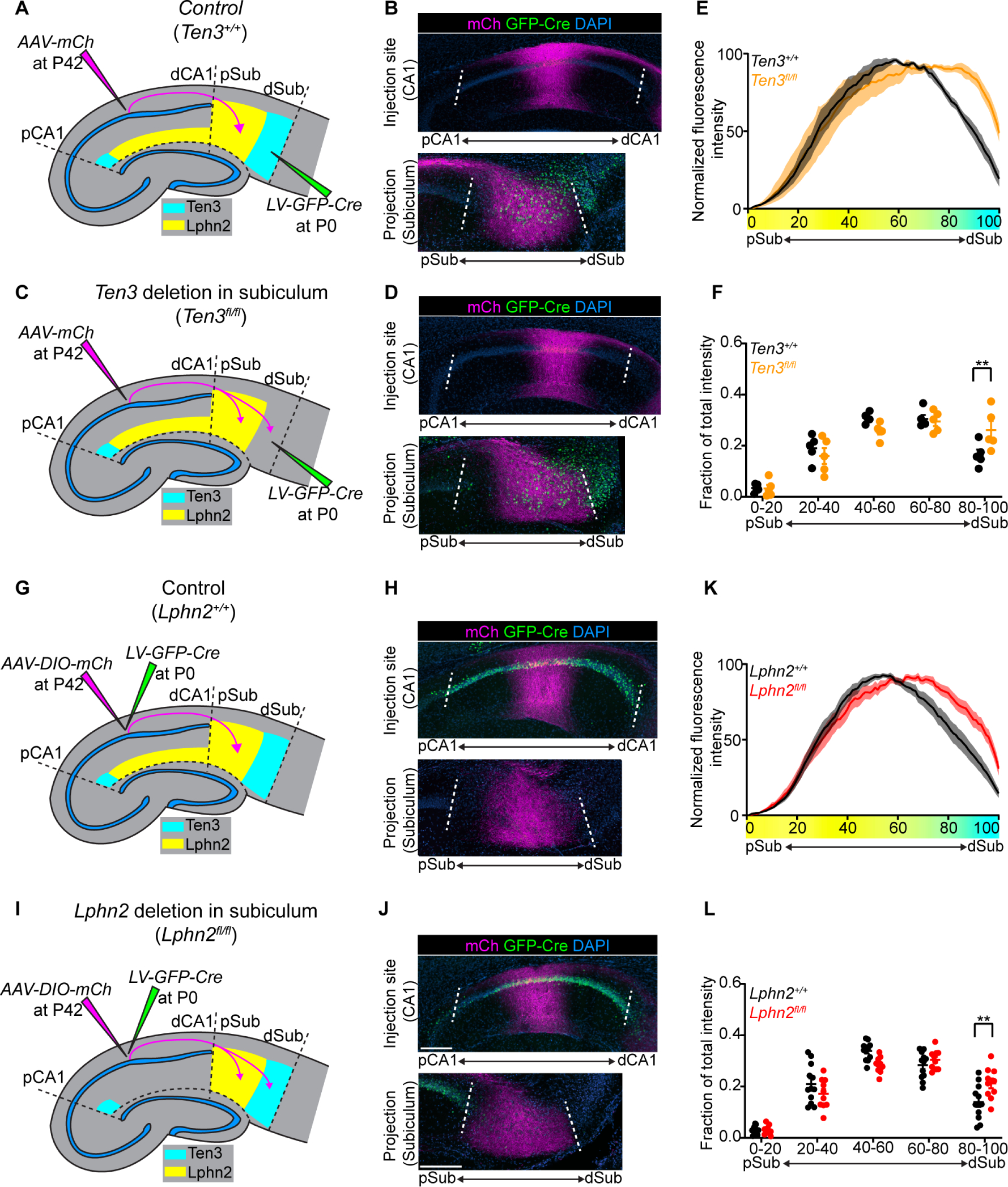
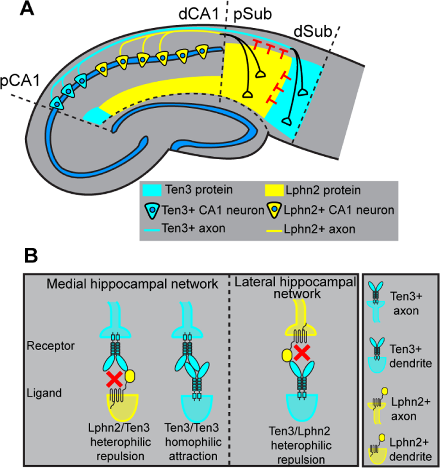
Mammalian medial and lateral hippocampal networks preferentially process spatial- and object-related information, respectively. However, the mechanisms underlying the assembly of such parallel networks during development remain largely unknown. Our study shows that, in mice, complementary expression of cell surface molecules teneurin-3 (Ten3) and latrophilin-2 (Lphn2) in the medial and lateral hippocampal networks, respectively, guides the precise assembly of CA1-to-subiculum connections in both networks. In the medial network, Ten3-expressing (Ten3+) CA1 axons are repelled by target-derived Lphn2, revealing that Lphn2- and Ten3-mediated heterophilic repulsion and Ten3-mediated homophilic attraction cooperate to control precise target selection of CA1 axons. In the lateral network, Lphn2-expressing (Lphn2+) CA1 axons are confined to Lphn2+ targets via repulsion from Ten3+ targets. Our findings demonstrate that assembly of parallel hippocampal networks follows a "Ten3→Ten3, Lphn2→Lphn2" rule instructed by reciprocal repulsions.


Conflict of interest statement

Competing interests: The authors declare no competing interests.

Figures

Fig. 1.
Fig. 1.. Complementary expression patterns of Lphn2 and Ten3 in the hippocampal network.
(A) Summary of connection patterns of medial (cyan) and lateral (yellow) hippocampal networks. pCA1 and dCA1, proximal and distal CA1; pSub and dSub, proximal and distal subiculum. (B) Violin plots highlighting Lphn2 and Ten3 expression in Ten3-HIGH and Ten3-NONE cells in CA1 and subiculum. The unit of expression level is ln [1+ (reads per 10000 transcripts)]. (C) Double in situ hybridization for Lphn2 (middle) and Ten3 (bottom) mRNA on a sagittal section of P8 mouse brain. Solid lines represent boundaries between CA1 and subiculum as labeled in the overlay (top). (D) Quantification of Lphn2 and Ten3 mRNA across the proximal–distal axis of CA1 and subiculum cell body layers (n = 3 mice). Mean ± SEM. (E) Double immunostaining for Lphn2 (middle; anti-GFP antibody) and Ten3 (bottom) on a sagittal section of P8 Lphn2-mVenus knock-in mouse (16) brain. Solid lines represent boundaries between CA1 and subiculum as labeled in the overlay (top). Region between dashed lines is the molecular layer. (F) Quantification of Lphn2 and Ten3 protein across the proximal–distal axis of molecular layers of CA1 and subiculum (n = 3 mice). Mean ± SEM. Scale bars, 200 μm. Axis labels in this and all subsequent figures: A, anterior; P, posterior; D, dorsal; V, ventral.
Fig. 2.
Fig. 2.. Ten3+ proximal CA1 axons avoid distal subiculum ectopically expressing Lphn2 in a Lphn2/Teneurin interaction–dependent and Lphn2/FLRT interaction–independent manner.
(A, B) Experimental design and summary of results. LV, lentivirus; AAV-mCh, adeno-associated virus expressing membrane-bound mCherry as an anterograde tracer. (C–F) Representative mountain-plots showing normalized GFP fluorescence intensity as color (LV expression) and normalized mCh fluorescence intensity as height (proximal CA1 axon projections) in subiculum. P, proximal; D, distal; M, medial; L, lateral. (G) Ratio of mCh fluorescent intensity of GFP+ versus GFP– regions. LV-GFP (n = 5 mice), LV-GFP-P2A-Lphn2 (wild-type Lphn2; n = 5 mice), LV-GFP-P2A-Lphn2_ΔLec (Lphn2 that does not bind Teneurins; n = 5 mice) and LV-GFP-P2A-Lphn2_4A (Lphn2 that does not bind FLRTs; n = 6 mice). Mean ± SEM; one-way ANOVA with Tukey’s multiple comparisons test. ****P ≤ 0.0001; *** P ≤ 0.001; ** P ≤ 0.01; * P ≤ 0.05; ns, not significant.
Fig. 3.
Fig. 3.. Lphn2/Ten3-mediated repulsion and Ten3/Ten3-mediated attraction cooperate to guide proximal CA1→distal subiculum target selection.
(A, C, E) Experimental design and summary of results for control (A), Lphn2 conditional knockout in subiculum (C), and Lphn2 and Ten3 double conditional knockout in subiculum (E). (B, D, F) Representative images of AAV-mCh (magenta) injections in proximal CA1 (top) and corresponding projections of proximal CA1 axons overlapping with LV-GFP-Cre (green) injection sites in subiculum (bottom). Data in (B), (D), and (F) correspond to experimental conditions in (A), (C), and (E), respectively. (G) Normalized mean fluorescence intensity traces of subiculum projections from proximal CA1 in GFP-Cre+ sections for Lphn2+/+;Ten3+/+ (n = 5 mice), Lphn2fl/fl;Ten3+/+ (n = 5 mice) and Lphn2fl/fl;Ten3fl/fl (n = 6 mice). Mean ± SEM. Color bar under x-axis represents Lphn2 (yellow) and Ten3 (cyan) expression in subiculum as quantified in Fig. 1F. (H) Fraction of total axon intensity for the same data as (G) across 20 percent intervals. Mean ± SEM, two-way ANOVA with Sidak’s multiple comparisons test. ****P ≤ 0.0001; *** P ≤ 0.001; * P ≤ 0.05. Scale bar, 200 μm. Injection site locations in CA1 are shown in fig. S11.
Fig. 4.
Fig. 4.. Lphn2+ mid-CA1 axons avoid Ten3+ distal subiculum.
(A, C) Experimental design and summary of results for tracing mid-CA1 axons in control (A) and Ten3 cKO in subiculum (C). (B, D) Representative images of AAV-mCh (magenta) injections in mid-CA1 (top) and corresponding projections overlapping with LV-GFP-Cre (green) injection sites in subiculum (bottom). Data in (B) and (D) correspond to experimental conditions in (A) and (C), respectively. (E) Normalized mean fluorescence intensity traces of subiculum projections from mid-CA1 in GFP-Cre+ sections for Ten3+/+ (n = 5 mice) and Ten3fl/fl (n = 5 mice). Mean ± SEM. Color bar under x-axis represents Lphn2 (yellow) and Ten3 (cyan) expression in subiculum as quantified in Fig. 1F. (F) Fraction of total axon intensity (same data as E) across 20 percent intervals. Mean ± SEM; two-way ANOVA with Sidak’s multiple comparisons test, ** P ≤ 0.01. (G, I) Experimental design and summary of results for tracing control (G) and Lphn2-null (I) mid-CA1 axon projections to subiculum. (H, J) Representative images of AAV-DIO-mCh (magenta; mCh expression in a Cre-dependent manner) injections in mid-CA1 (top) and corresponding projections in subiculum (bottom). Data in (H) and (J) correspond to experimental conditions in (G) and (I), respectively. (K) Normalized mean fluorescence intensity traces of subiculum projections from Lphn2+/+ (n = 12 mice) and Lphn2fl/fl (n = 10 mice) mid-CA1 axons. Mean ± SEM. Color bar under x-axis represents Lphn2 (yellow) and Ten3 (cyan) expression in subiculum as quantified in Fig. 1F. (L) Fraction of total axon intensity (same data as K) across 20 percent intervals. Mean ± SEM; two-way ANOVA with Sidak’s multiple comparisons test; ** P ≤ 0.01. Scale bars, 200 μm. Injection site locations in CA1 are shown in fig. S11.
Fig. 5.
Fig. 5.. Lphn2 and Ten3 instruct target selection of hippocampal axons through reciprocal repulsions.
(A) Ten3+ CA1 axons target Ten3+ subiculum via repulsion from Lphn2 and attraction to Ten3 in subiculum. Lphn2+ CA1 axons target Lphn2+ subiculum via repulsion from Ten3 in subiculum. (B) High-magnification view of ligand–receptor interactions that instruct target selection of Ten3+ (left) and Lphn2+ (right) axons. Red crosses symbolize repulsion.

Comment in

References

    1. O’Keefe J, Dostrovsky J, Brain Res. 34, 171–175 (1971). - PubMed
    1. Scoville WB, Milner B, Neuropsychiatry Clin J. Neurosci. 12, 103–113 (2000). - PubMed
    1. Squire LR, Stark CEL, Clark RE, Annu. Rev. Neurosci 27, 279–306 (2004). - PubMed
    1. Hafting T, Fyhn M, Molden S, Moser M-B, Moser EI, Nature 436, 801–806 (2005). - PubMed
    1. Igarashi KM, Ito HT, Moser EI, Moser M-B, FEBS Lett. 588, 2470–2476 (2014). - PubMed

Publication types

MeSH terms