. 2021 Jun 3;11(1):11793.
doi: 10.1038/s41598-021-91191-6.

Automatic and unbiased segmentation and quantification of myofibers in skeletal muscle

Affiliations

Automatic and unbiased segmentation and quantification of myofibers in skeletal muscle

Ariel Waisman et al. Sci Rep. .

Abstract

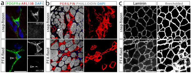
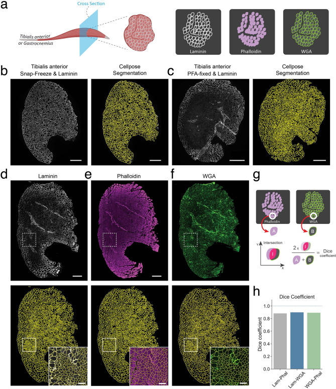
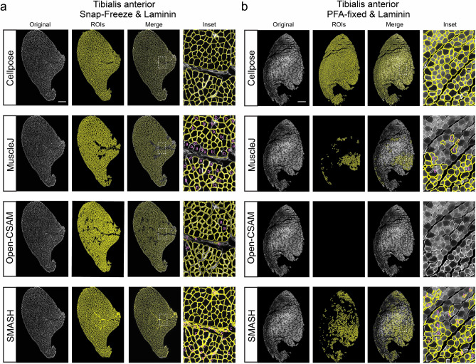
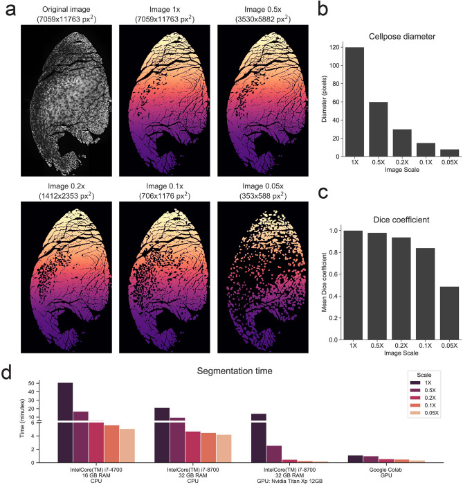
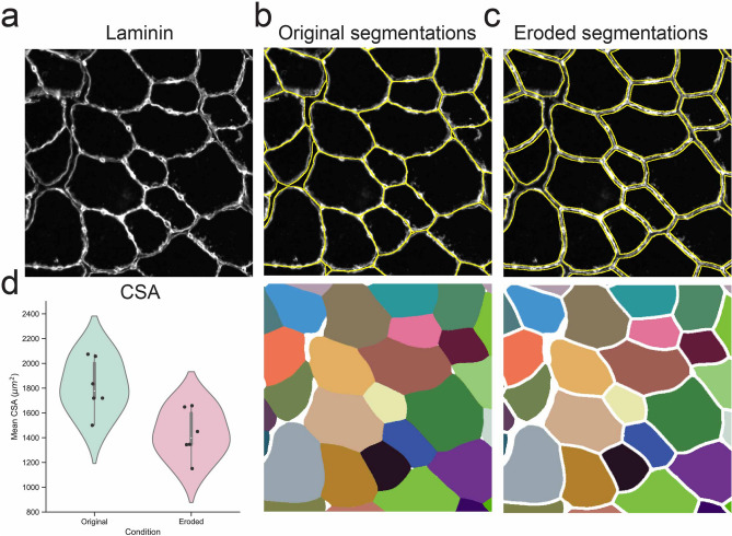
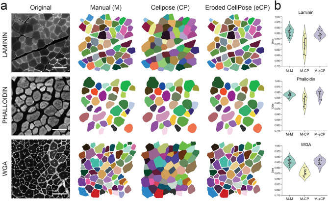
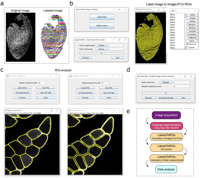
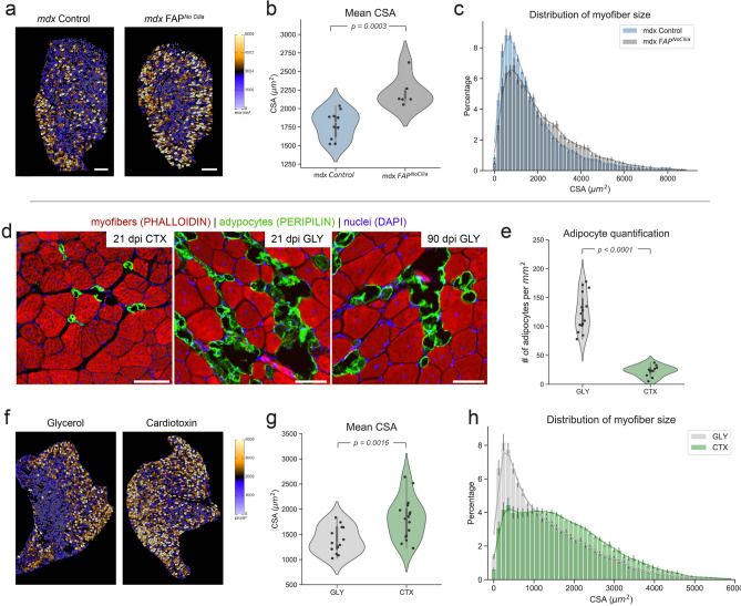
Skeletal muscle has the remarkable ability to regenerate. However, with age and disease muscle strength and function decline. Myofiber size, which is affected by injury and disease, is a critical measurement to assess muscle health. Here, we test and apply Cellpose, a recently developed deep learning algorithm, to automatically segment myofibers within murine skeletal muscle. We first show that tissue fixation is necessary to preserve cellular structures such as primary cilia, small cellular antennae, and adipocyte lipid droplets. However, fixation generates heterogeneous myofiber labeling, which impedes intensity-based segmentation. We demonstrate that Cellpose efficiently delineates thousands of individual myofibers outlined by a variety of markers, even within fixed tissue with highly uneven myofiber staining. We created a novel ImageJ plugin (LabelsToRois) that allows processing of multiple Cellpose segmentation images in batch. The plugin also contains a semi-automatic erosion function to correct for the area bias introduced by the different stainings, thereby identifying myofibers as accurately as human experts. We successfully applied our segmentation pipeline to uncover myofiber regeneration differences between two different muscle injury models, cardiotoxin and glycerol. Thus, Cellpose combined with LabelsToRois allows for fast, unbiased, and reproducible myofiber quantification for a variety of staining and fixation conditions.


Conflict of interest statement

The authors declare no competing interests.

Figures

Figure 1
Figure 1
Fixation of muscle tissues is required to preserve certain cellular structures. (a) Cilia, marked by ARL13B (red), can only be detected on FAPs, labeled by PDGFRα (green), in PFA-fixed (bottom) but not snap-frozen (top) tibialis anterior muscle sections. Scale bar is 10 μm. (b) The morphology of PERILIPIN-expressing adipocytes (red) is severely compromised without prior fixation. Scale bar is 50 μm. (c) Laminin (gray) perfectly outlines myofibers in snap-frozen tissue (top) enabling simple myofiber segmentation using ImageJ thresholding. However, fixation impacts Laminin immunoreactivity preventing myofiber segmentation. Scale bar is 250 μm.
Figure 2
Figure 2
Myofiber segmentation using Cellpose. (a) Schematic of the experimental design. (b) Cross section of a snap-frozen tibialis anterior muscle stained for Laminin. Right, original image. Left, Cellpose segmentation results. Scale bar, 500 μm. (c) Cross section of a PFA-fixed tibialis anterior muscle stained for Laminin. Right, original image. Left, Cellpose segmentation results. Scale bar, 500 μm. (d-f) Cross section of a PFA-fixed tibialis anterior muscle stained for Laminin (d), Phalloidin (e) and WGA (f). Top, original images. Bottom, result of Cellpose segmentation. Inset, detail of the specified region. Scale bar within inset, 500 μm. (g) Diagram depicting the calculation of the Dice coefficient. For each segmented object in the two label images, the Dice coefficient is the result of two times the area of the intersection divided by the sum of the areas of both objects. (h) Mean Dice coefficient for all myofibers in the comparison between the indicated stains.
Figure 3
Figure 3
Segmentation comparison between Cellpose and different software. Snap-frozen (a) and PFA-fixed (b) TA sections were stained for Laminin and segmented by Cellpose, MuscleJ, Open-CSAM and SMASH as indicated. Original image on the left and segmentation results labeled as ROIs. Inserts on right represent magnified areas as indicated by boxes in the merged image. Segmentation errors are labeled as missed fibers (a), mis-segmented fibers (b) and false-positive fibers (c). Scale bar, 500 μm.
Figure 4
Figure 4
Benchmarking of Cellpose algorithm across different image sizes and hardware specifications. (a) Top-left, original full-sized PFA-fixed TA cross-section stained for Laminin. The remaining images correspond to the Cellpose-generated label images when full-sized or downscaled versions of the original image were fed to the algorithm. (b) Myofiber diameter fed to the Cellpose algorithm for the full-sized or the downscaled images. Approximate myofiber diameter for the original image was manually determined. (c) Mean Dice coefficient for the comparison of the original 1 × label image with itself or with the downscaled label images (see “Methods”). (d) Comparison of Cellpose segmentation time for the different image sized across different hardware platforms.
Figure 5
Figure 5
Erosion of Cellpose segmentations is needed for accurate CSA measurements. (a) Example of a snap-frozen Laminin stained TA cross-section. (b) Cellpose segmentation of the image in (a) showing the overlaid segmentations in yellow (top) or the Cellpose-generated label image (bottom). (c) Segmentation results after the Cellpose label image was eroded by 4 pixels. (d) Mean CSA for all myofiber of Laminin stained TA muscle cross-sections of 6 different mice with or without a 4-pixel label erosion.
Figure 6
Figure 6
Eroded Cellpose segmentations are as accurate as human experts in delineating myofibers. (a) Left, example of cross-sectional TA images marked by Laminin, Phalloidin or WGA and, right, their corresponding labeled images manually generated by a human expert (M), and the original (CP) or the eroded Cellpose segmentation (eCP). (b) A set of four images was analyzed for each of the three indicated stains. Three human experts manually segmented each of the images, while those same images were also segmented by Cellpose. Cellpose labels were additionally eroded by a fixed number of pixels according to visual inspection. The Dice coefficient was used to compare the different label images corresponding to the same original image. The violin plot shows the mean Dice coefficient for each human pair for a specific image (M–M), the comparison of human manual segmentations to their associated Cellpose segmentations (M–CP), or for human manual segmentations to their associated Cellpose eroded segmentations (M–eCP).
Figure 7
Figure 7
Development of LabelsToROIs plugin to analyze label images in FIJI/ImageJ. As indicated in (a), the plugin takes as input an original image and a labeled image generated using Cellpose or another software. (b) The plugin allows to automatically extract the segmentations in the label images and generate the corresponding FIJI/ImageJ regions of interest (ROIs). (c) Once generated, the ROIs can be eroded by a fixed number of pixels until the segmentations correctly delineate the objects of interest. The ROI files can be saved, and different measurements can be selected to generate table measurements. (d) The LabelsToROIs plugin also allows for simultaneous processing of multiple images, enabling the generation of ROIs, ROI erosion and table measurements in one step, while automatically saving all the files for future inspection. (e) A diagram of the proposed pipeline for the automatic analysis of skeletal muscle biopsies.
Figure 8
Figure 8
Cellpose and LabelsToROIs enable the quantification of CSA across different disease and injury settings. Top Re-quantification of data from Kopinke et al., 2017. (a) TA images from 10- to 12-month-old mdx control and mdx FAPNoCilia mice stained for Laminin were re-analyzed using Cellpose. LabelsToROIs was used to erode the segmentations according to visual inspection (5 pixels). Myofiber segmentations were color-coded based on their CSA using the FIJI/ImageJ ROI color coder. Scale bar, 500 μm. (b) Mean CSA of mdx control (n=10) and mdx FAPNoCilia mice (n=6). Statistical differences were assessed by a Student T test. (c) Distribution of myofiber CSA for mdx control and mdx FAPNoCilia mice. Results are presented as means standard error of the mean (SEM) for each bin and each genotype. Bottom CSA comparisons between injury types. (d) Few adipocytes, marked by PERILPIN (green), have formed 21 days post muscle injury caused by cardiotoxin (CTX). In contrast, glycerol-induced injury (GLY) causes massive fat infiltration that persists up to 3 months. Myofibers are labeled with Phalloidin (red) and nuclei with DAPI (blue). (e) Number of adipocytes per mm2 of injured area 21 days post GLY (n=15) or CTX (n=11) injury. Statistical differences were assessed by a Student T test. (f) Cellpose and LabelsToROIs were used to segment and process TA images stained for Laminin corresponding to GLY and CTX treated mice. Myofiber segmentations were color-coded based on their CSA using the FIJI/ImageJ ROI color coder. (g) Quantification of the mean CSA of myofibers 21 days post GLY (n=12) or CTX (n=18) injury. Statistical differences were assessed by a Student T test. (h) Distribution of myofiber size for each injury. Results are presented as means standard error of the mean (SEM) for each bin and each treatment.

References

    1. Lepper C, Partridge TA, Fan CM. An absolute requirement for pax7-positive satellite cells in acute injury-induced skeletal muscle regeneration. Development. 2011;138:3639–3646. doi: 10.1242/dev.067595. - DOI - PMC - PubMed
    1. Murphy MM, Lawson JA, Mathew SJ, Hutcheson DA, Kardon G. Satellite cells, connective tissue fibroblasts and their interactions are crucial for muscle regeneration. Development. 2011 doi: 10.1242/dev.064162. - DOI - PMC - PubMed
    1. Relaix F, Zammit PS. Satellite cells are essential for skeletal muscle regeneration: The cell on the edge returns centre stage. Development. 2012 doi: 10.1242/dev.069088. - DOI - PubMed
    1. Sambasivan R, et al. Pax7-expressing satellite cells are indispensable for adult skeletal muscle regeneration. Development. 2011;138:3647–3656. doi: 10.1242/dev.067587. - DOI - PubMed
    1. Joe AW, et al. Muscle injury activates resident fibro/adipogenic progenitors that facilitate myogenesis. Nat. Cell Biol. 2010;12:2015. doi: 10.1038/ncb2015. - DOI - PMC - PubMed

Publication types

LinkOut - more resources