. 2021 Jun 3;4(1):682.
doi: 10.1038/s42003-021-02220-z.

SARS-CoV-2 proteins and anti-COVID-19 drugs induce lytic reactivation of an oncogenic virus

Affiliations

SARS-CoV-2 proteins and anti-COVID-19 drugs induce lytic reactivation of an oncogenic virus

Jungang Chen et al. Commun Biol. .

Abstract

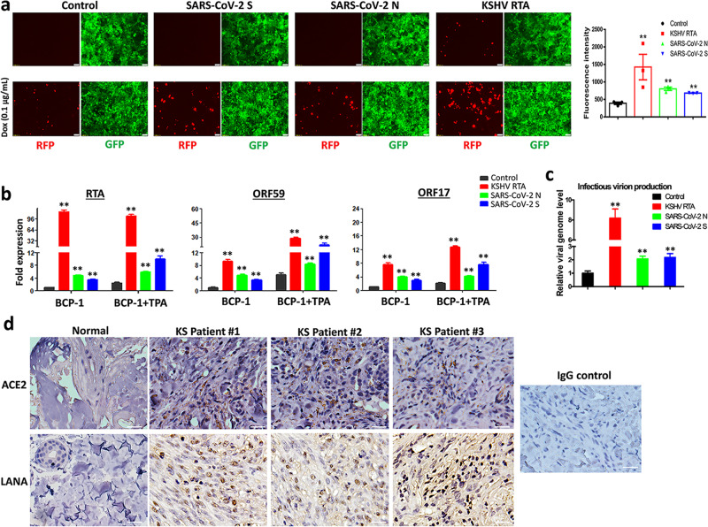
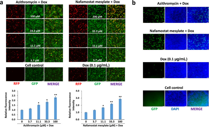
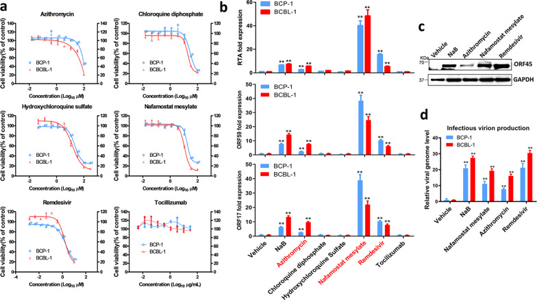
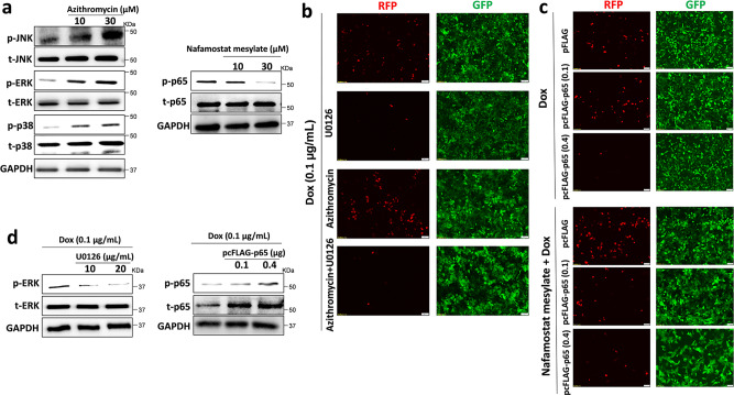
An outbreak of the novel coronavirus SARS-CoV-2, the causative agent of Coronavirus Disease-2019 (COVID-19), a respiratory disease, has infected almost one hundred million people since the end of 2019, killed over two million, and caused worldwide social and economic disruption. Because the mechanisms of SARS-CoV-2 infection of host cells and its pathogenesis remain largely unclear, there are currently no antiviral drugs with proven efficacy. Besides severe respiratory and systematic symptoms, several comorbidities increase risk of fatal disease outcome. Therefore, it is required to investigate the impacts of COVID-19 on pre-existing diseases of patients, such as cancer and other infectious diseases. In the current study, we report that SARS-CoV-2 encoded proteins and some currently used anti-COVID-19 drugs are able to induce lytic reactivation of Kaposi's sarcoma-associated herpesvirus (KSHV), one of major human oncogenic viruses, through manipulation of intracellular signaling pathways. Our data indicate that those KSHV + patients especially in endemic areas exposure to COVID-19 or undergoing the treatment may have increased risks to develop virus-associated cancers, even after they have fully recovered from COVID-19.


Conflict of interest statement

The authors declare no competing interests.

Figures

Fig. 1
Fig. 1. Ectopic expression of SARS-CoV-2 proteins induces KSHV lytic gene expression and ACE2 expression is increased in AIDS-KS tissues.
a The iSLK.219 cells were transfected with vector control or vectors encoding SARS-CoV-2 spike protein (S), nucleocapsid protein (N) and KSHV RTA (as a positive control) with or without low dose of doxycycline (Dox, 0.1 µg/mL) induction for 72 h. The expression of RFP (representing viral lytic reactivation) and GFP (representing infected cells) were detected using fluorescence microscopy. Error bars represent S.D. for 4 image fields per well. b, c BCP-1 cells were transfected as above with or without low dose of 12-O-tetradecanoyl-phorbol-13-acetate (TPA, 1.0 ng/mL) induction for 72 h, then the transcripts of representative lytic genes were quantified by using qRT-PCR. The supernatants from transfected cells were collected to infect naive HEK293T cells, then viral genome levels were quantified by using qPCR with Lana-specific primers. Error bars represent S.D. for 3 independent experiments, **p < 0.01 (vs the vector control). d Expression of ACE2 and LANA (as well as representative IgG control) in formalin-fixed paraffin-embedded KS tissues from 3 HIV+ patients and normal skin tissues were determined by immunohistochemical staining as described in the “Methods”. Bars: 50 μm.
Fig. 2
Fig. 2. Some drugs currently used for anti-COVID-19 treatment are able to induce KSHV lytic reactivation.
a The iSLK.219 cells were treated with a dose range of anti-COVID-19 drugs, Azithromycin and Nafamostat mesylate, with doxycycline (Dox, 0.1 µg/mL) induction for 72 h. The expression of RFP and GFP was detected using fluorescence microscopy. Error bars represent S.D. for 4 image fields per well, *p < 0.05; **p < 0.01 (vs the vehicle control). b The supernatants from iSLK.219 cells treated by Azithromycin or Nafamostat mesylate for 72 h (11.1 µM, respectively) in combination with Dox were collected to infect naive HEK293T cells, then GFP expression was detected using fluorescence microscopy.
Fig. 3
Fig. 3. The impacts of anti-COVID-19 drugs on the growth and viral gene expression of KSHV+ tumor cells.
a BCBL-1 and BCP-1 cells were treated with a dose range of 6 anti-COVID-19 drugs, for 72 h, then the cell proliferation status was examined using the WST-1 cell proliferation assays (Roche). b, c Cells were treated with Azithromycin (10 µM), Chloroquine diphosphate (10 µM), Hydroxychloroquine sulfate (10 µM), Nafamostat mesylate (10 µM), Remdesivir (3 µM), Tocilizumab (20 µg/mL), respectively, for 72 h, then the transcripts of representative lytic genes were quantified by using qRT-PCR. Protein expression was measured by immunoblots. d The supernatants from drug-treated cells above were collected to infect naive HEK293T cells, then viral genome levels were quantified by using qPCR with Lana-specific primers. The sodium butyrate (NaB, 0.3 mM) was used as a positive control. Error bars represent S.D. for 3 independent experiments, **p < 0.01 (vs the vehicle control).
Fig. 4
Fig. 4. Cellular mechanisms of Azithromycin and Nafamostat mesylate induced KSHV lytic reactivation.
a BCP-1 cells were treated with indicated concentrations of Azithromycin or Nafamostat mesylate for 72 h, then protein expression was measured by immunoblots. b The iSLK.219 cells were pre-treated with U0126 (10 μg/mL), a MAPK kinase inhibitor, for 12 h, then addition of Azithromycin together with doxycycline (Dox, 0.1 µg/mL) induction for 72 h. c The iSLK.219 cells were transiently transfected with a construct expressing NF-κB p65 (pcFLAG-p65 at 0.1 or 0.4 μg, respectively) or control vector, then addition of Nafamostat mesylate together with Dox induction for 72 h. The expression of RFP and GFP was detected using fluorescence microscopy. d The effects of U0126 and pcFLAG-p65 transfection on signaling activities were confirmed by immunoblots. Representative blots from one of two independent experiments were shown.

Update of

References

    1. Calina, D. et al. Recent advances, approaches and challenges in targeting pathways for potential COVID-19 vaccines development. Immunol. Res. 68, 315–324 (2020). - PMC - PubMed
    1. Hu, B., Guo, H., Zhou, P. & Shi, Z. L. Characteristics of SARS-CoV-2 and COVID-19. Nat. Rev. Microbiol.19, 141–154 (2020). - PMC - PubMed
    1. Zhu N, et al. A novel coronavirus from patients with pneumonia in China, 2019. N. Engl. J. Med. 2020;382:727–733. doi: 10.1056/NEJMoa2001017. - DOI - PMC - PubMed
    1. Wu F, et al. A new coronavirus associated with human respiratory disease in China. Nature. 2020;579:265–269. doi: 10.1038/s41586-020-2008-3. - DOI - PMC - PubMed
    1. Elezkurtaj, S. et al. Causes of death and comorbidities in patients with COVID-19. medRxiv10.1101/2020.06.15.20131540 (2020).

Publication types

MeSH terms