. 2021 Nov;5(11):1246-1260.
doi: 10.1038/s41551-021-00737-6. Epub 2021 Jun 3.

T cells armed with C-X-C chemokine receptor type 6 enhance adoptive cell therapy for pancreatic tumours

Affiliations

T cells armed with C-X-C chemokine receptor type 6 enhance adoptive cell therapy for pancreatic tumours

Stefanie Lesch et al. Nat Biomed Eng. 2021 Nov.

Abstract

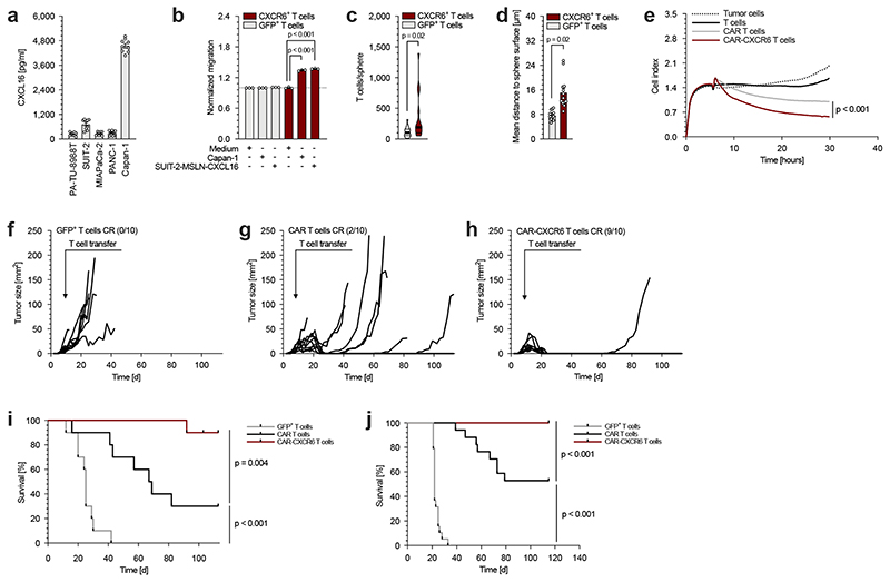
The efficacy of adoptive cell therapy for solid tumours is hampered by the poor accumulation of the transferred T cells in tumour tissue. Here, we show that forced expression of C-X-C chemokine receptor type 6 (whose ligand is highly expressed by human and murine pancreatic cancer cells and tumour-infiltrating immune cells) in antigen-specific T cells enhanced the recognition and lysis of pancreatic cancer cells and the efficacy of adoptive cell therapy for pancreatic cancer. In mice with subcutaneous pancreatic tumours treated with T cells with either a transgenic T-cell receptor or a murine chimeric antigen receptor targeting the tumour-associated antigen epithelial cell adhesion molecule, and in mice with orthotopic pancreatic tumours or patient-derived xenografts treated with T cells expressing a chimeric antigen receptor targeting mesothelin, the T cells exhibited enhanced intratumoral accumulation, exerted sustained anti-tumoral activity and prolonged animal survival only when co-expressing C-X-C chemokine receptor type 6. Arming tumour-specific T cells with tumour-specific chemokine receptors may represent a promising strategy for the realization of adoptive cell therapy for solid tumours.


Conflict of interest statement

Competing interests

Parts of this work have been performed for the doctoral theses of SL, VB, SS, KD and JL at the Ludwig-Maximilians-Universität München. MR, SG, SE and SK are inventors on a patent application related to this work filed by the Ludwig-Maximilians-Universität München. SE and SK received research support from TCR2 Inc and Arcus Biosciences for work on T cell therapies unrelated to the present manuscript. The remaining authors declare no competing interests.

Figures

Fig. 1
Fig. 1. CXCL16 is expressed in murine pancreatic tumours and affects CXCR6-engineered T cells.
(a) C-X-C chemokine expression profile of Panc02-OVA tumours quantified by qPCR (n = 6). (b) ELISA evaluating murine CXCL16 protein concentrations in different organs of Panc02-OVA tumour-bearing mice (n = 12). (c) Expression level of murine CXCL16 by Panc02-OVA tumour cells after stimulation with 20 ng/ml IFN-γ, 20 ng/ml TNF-α or a combination of both, quantified using ELISA. (d) CXCL16 levels of CXCL16-knockout Panc02-OVA (n = 10) and CRISPR control Panc02-OVA tumours (n = 15) determined using CXCL16 ELISA. (e) CD11c- and CD11c+ cells were isolated from Panc02-OVA tumour tissue by FACS sorting, and qPCR was used to analyse CXCL16 expression levels of both populations (n = 10 mice). (f) Trans-well migration of GFP- and CXCR6-transduced T cells towards descending concentrations of recombinant murine CXCL16. After 3 h the number of migrated T cells was quantified by flow cytometry. (g) Target cell lysis of CXCL16-overexpressing Panc02-OVA by CXCR6- and GFP-transduced OT-1 T cells following migration through a permeable membrane (suppl. fig.2h). After a migration period of 3 h, migrated T cells and tumour cells were co-cultured for further 1.5 h. (h) ELISA revealing time-dependent activation levels of CXCR6- and GFP-transduced OT-1 T cells upon co-culture with Panc02-OVA tumour cells. E:T ratio 5:1. (i) Panc02-OVA tumour cells were co-incubated with GFP- and CXCR6-transduced OT-1 T cells, and lysis of tumour cells was measured after 6.5 h. (j) Adherence of GFP- or CXCR6-transduced T cells to a CXCL16-coated (9 pmol) or control BSA-coated (9 pmol) surface. As an additional control, T cells were pre-incubated with soluble recombinant CXCL16 (2 μg/ml). (k) Membrane expression of CXCR6 upon stimulation with 200 ng recombinant CXCL16 or CCL1 (arrow) indicating intracellular trafficking and receptor recycling. In vitro experiments (c, d, f, g, h, I, j) show mean values ± SEM of at least two biological replicates and are representative of three independent experiments (n = 3). p-values are based on two-sided unpaired t-test. Data shown in k are representative for two independent experiments (n = 2). Ex vivo experiments shown are representative of n = 2 (a, d) or n = 3 (b, e). Data shown in e are comprised of two independent experiments (n = 2). Analyses of differences between groups were performed using unpaired Mann-Whitney test.
Fig. 2
Fig. 2. CXCR6-transduced T cells induce tumour regression.
(a) Tumour growth curves of Panc02-OVA-bearing mice with adoptive transfer of 107 GFP- or CXCR6-transduced OT-1 T cells (n= 5 mice per group). T cells were transferred when tumours were palpable (day 5). 2 out of 5 mice treated with CXCR6-transduced T cells showed complete response (CR). (b) C57BL/6 mice inoculated with CXCL16-knockout Panc02-OVA (clone 55) or (c) CRISPR control Panc02-OVA (clone 50) were treated with a single i.v. injection of 107 GFP- or CXCR6-transduced OT-1 T cells (n = 5-12 mice per group). (d) Tumour growth of subcutaneous E.G7-OVA-CXCL16 tumours following treatment with a single injection of 107 mCherry- or CXCR6-transduced OT-1 T cells (n = 4-5 mice per group). (e) Tumour growth of subcutaneous Panc02-OVA-pCAM tumour with adoptive transfer of 107 T cells transduced with either anti-EpCAM-CAR or anti-EpCAM-CAR-CXCR6 (n = 5 mice per group). (f) Tumour growth of subcutaneous Panc02-OVA-EpCAM tumours with adoptive transfer of 107 T cells transduced with anti-EpCAM-CAR, anti-EpCAM-CAR-CXCR3, anti-EpCAM-CAR-CXCR6 or anti-EpCAM-CAR-CCR4 (n = 10-14 mice per group). Experiments shown are representative of two (b, c, d, e, f) or three independent (a) experiments. Analyses of differences between groups were performed using two-way ANOVA with correction for multiple testing by the Bonferroni method.
Fig. 3
Fig. 3. CXCR6-transduced T cells are recruited into tumour tissue.
(a) Flow cytometry analysis evaluating the number of OT-1 T cells in Panc02-OVA bearing mice after treatment with CD45.1+ GFP- and CD90.1+ CXCR6-transduced OT-1 T cells in a ratio of 1:1 (n = 5 mice). (b, c) Ex vivo quantification of tumour-infiltrating CXCR6- or GFP-transduced OT-1 T cells in Panc02-OVA tumours by two-photon microscopy (n = 5 mice per group). (d) Flow cytometry analysis quantifying homing of mCherry- and CXCR6-transduced T cells into Panc02 tumours (n = 3 mice per group). (e, f) Before flow cytometry analysis, tumour infiltration and T cell velocity was investigated using DSFC and intravital imaging (n = 4 mice per group). (g) Representative coronal and axial granzyme B PET image taken from Panc02 (left shoulder; white circle) and Panc02-OVA (right shoulder, green circle) tumour-bearing mice treated with either CXCR6-transduced or GFP-transduced (mock) OT-1 T cells. (h) In order to assess granzyme B levels, tracer accumulation in tumour in relation to heart (background radioactivity) was measured (n = 4 mice per group). Experiments shown are representative of two (d, e, f) or three independent (a, b, c) experiments with n = 3-5 mice per group. Data shown in h are comprised of two independent experiments with n = 4 mice per group. Analyses of differences between groups were performed using unpaired Mann-Whitney test or two-way ANOVA with correction for multiple testing by the Bonferroni method.
Fig. 4
Fig. 4. CXCL16 expressed by human pancreatic cancer cells enhances cytotoxic activity of engineered T cells.
(a) Secretion of CXCL16 by human pancreatic cancer cells was measured by ELISA. (b) Migration capability of GFP- and CXCR6-transduced human T cells toward Capan-1 and MSLN-CXCL16-overexpressing SUIT-2 supernatants. The number of migrated cells was normalized to the medium control condition. (c, d) Number of sphere-penetrating GFP- and CXCR6-transduced human T cells and infiltration depth into spheres formed by HEK overexpressing human CXCL16. (e) In a combined migration-cytotoxicity assay anti-MSLN-CAR and anti-MSLN-CAR-CXCR6-transduced human T cells are compared with regard of specific migratory and cytotoxic efficiency. T cells migrated towards MSLN-CXCL16-overexpressing SUIT-2 tumour cells (Suppl. fig. 6e) followed by CAR-mediated cytotoxicity presented by real-time target cell lysis. (f - i) In a subcutaneous xenograft model, MSLN-CXCL16-overexpressing SUIT-2 tumour bearing mice were treated with GFP-transduced (f), anti-MSLN-CAR-transduced (g) or anti-MSLN-CAR-CXCR6 co-transduced T cells (h). Tumour growth and survival was measured over 110 days (n = 10 mice per group). One mouse of the anti-MSLN-CAR-CXCR6 treated group was sacrificed on day 103 post tumour injection due to (unclear) neck swelling, presumably unrelated to subcutaneous tumour injection and was therefore censored at the timepoint. (i). (j) For orthotopic treatment experiments, SUIT-2-MSLN-CXCL16 tumour cells were implanted into the pancreas. Mice were treated with a single i.v. injection of either non-transduced human T cells, anti-MSLN-CAR- or anti-MSLN-CAR-CXCR6-transdcued T cells. Tumour growth and survival was monitored over 115 days (n = 17-20 mice per group). In vitro experiments (b, e) show mean values ± SEM of at least two biological replicates and are representative of three independent experiments (n = 3). Data shown in a, c and d are comprised of three independent experiments (n = 3) each with three biological replicates. In vivo experiments (f – j) are summarized from two independent experiments. p-values are based on two-sided unpaired t-test or two-way ANOVA with correction for multiple testing by the Bonferroni method. Comparison of survival rates was performed with the Log-rank (Mantel-Cox) test.
Fig. 5
Fig. 5. CXCL16 is expressed by PDAC specimens and attracts CXCR6 transduced T cells.
(a) Gene expression analysis (mRNA) of pancreatic cancer specimens in comparison to healthy pancreatic tissue (n = 36 PDAC patients and n = 12 healthy controls) using NanoString nCounter® System. (b) TCGA data analysis comparing CXCL16 expression (mRNA) by PDAC and healthy tissue (n = 178 PDAC patients and n = 165 healthy controls). (c) Representative images of PDAC specimens stained for CXCL16. (d) Quantification of CXCL16 expression by tumour cells and tumour-infiltrating immune cells validated by immunohistochemical staining of CXCL16 in PDAC specimens (n = 399 with three biopsies per patient). (e) Single cell RNA (scRNA) sequencing analysis of PDAC and healthy pancreas tissue comparing CXCL16 expression levels. (f) CXCL16 levels in plasma of PDAC patients and healthy specimens quantified by ELISA (n = 10 PDAC patients and n = 11 healthy specimens). (g) Activation level of human T cells (quantified by IFN-γ concentrations) following co-culture with pancreatic cancer PDO (summarized data from independent co-cultures with 3 different PDO specimens: B34, B54 and B61; n = 3). (h) Representative images showing confocal analysis of pancreatic cancer PDO (specimen B61) infiltrated by GFP- or CXCR6-transduced T cells. (i) Quantification of GFP- and CXCR6-transduced T cells penetrating into PDO (summarized from specimen B34 and B48; n = 2) in the absence or presence of an anti-CXCL16 neutralization antibody. (j) For PDX experiments, PDO (MGH1247) were heterotopically implanted in NCG mice and treated with non-transduced, anti-MSLN-CAR or anti-MSLN-CAR-CXCR6-transduced T cells. Tumour growth was monitored for 35 days post ACT (n = 5 mice per group). (k) PDX tumour weight after treatment with either non-transduced, anti-MSLN-CAR or anti-MSLN-CAR-CXCR6-transduced T cells. (l) Quantification of GFP- and CXCR6-transduced T cells after penetration into surgical ovarian cancer (OC) specimens of seven patients. Analyses of differences between groups in a, b and f were performed using unpaired Mann-Whitney test. Data shown in g are comprised of three independent experiments (n = 3). Data shown in i are comprised of two independent experiments with mean values ± SEM of at least 10 organoids per condition (i; n = 2). p-values are based on two-sided unpaired t-test. Data shown in j, k and l resulted from one single experiment (n = 1). Differences in tumour growth were analysed by using two-way ANOVA with correction for multiple testing by the Bonferroni method and differences in tumour weight were analysed by using unpaired Mann-Whitney test. Data shown in l are mean values ± SEM of seven OC specimen co-cultures with the same T cell donor. p-vales in k are based Wilcoxon signed-rank test.

Comment in

References

    1. Rosenberg SA, Restifo NP. Adoptive cell transfer as personalized immunotherapy for human cancer. Science. 2015;348:62–68. doi: 10.1126/science.aaa4967. - DOI - PMC - PubMed
    1. Kobold S, et al. Immunotherapy in tumors. Deutsches Arzteblatt international. 2015;112:809–815. doi: 10.3238/arztebl.2015.0809. - DOI - PMC - PubMed
    1. Sheridan C. First approval in sight for Novartis’ CAR-T therapy after panel vote. Nat Biotechnol. 2017;35:691–693. doi: 10.1038/nbt0817-691. - DOI - PubMed
    1. Ahmed N, et al. Human epidermal growth factor receptor 2 (HER2) - specific chimeric antigen recpetor-modified T cells for the immunotherapy of HER2-positive sarcoma. J Clin Oncol. 2015;33:1688–1696. - PMC - PubMed
    1. Adusumilli PS, et al. Regional delivery of mesothelin-targeted CAR T cell therapy generates potent and long-lasting CD4-dependent tumor immunity. Sci Transl Med. 2014;6:261–151. doi: 10.1126/scitranslmed.3010162. - DOI - PMC - PubMed

Publication types