Review
. 2021 Aug;21(8):481-499.
doi: 10.1038/s41568-021-00363-z. Epub 2021 Jun 3.

Interleukins in cancer: from biology to therapy

Affiliations
Review

Interleukins in cancer: from biology to therapy

Daria Briukhovetska et al. Nat Rev Cancer. 2021 Aug.

Abstract

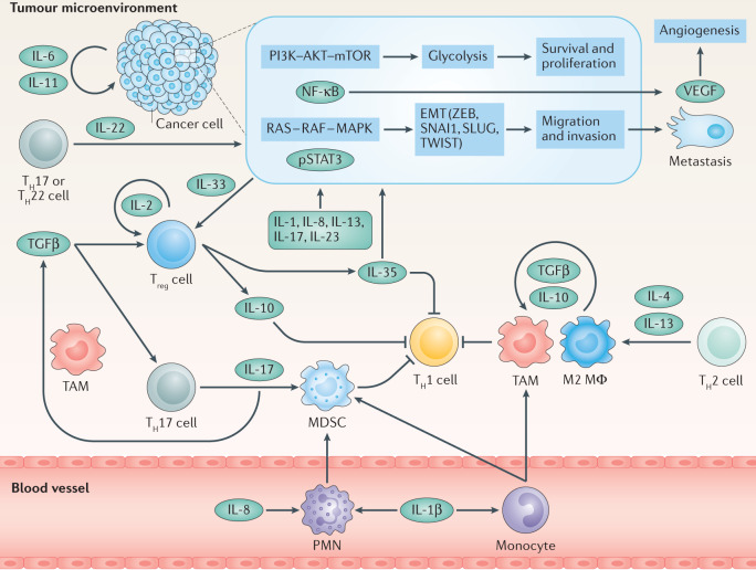
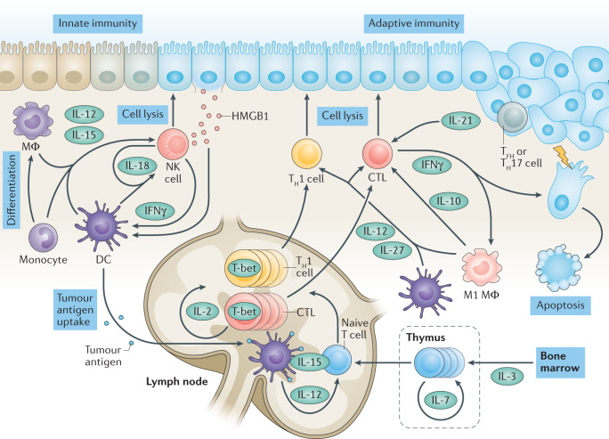
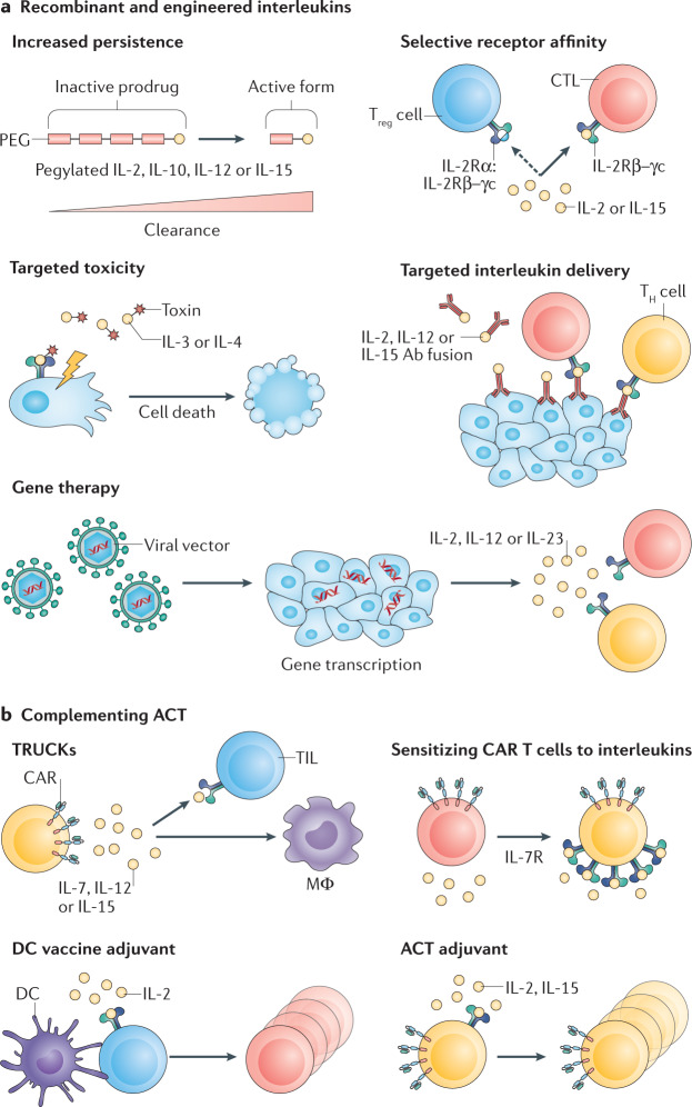
Interleukins and associated cytokines serve as the means of communication for innate and adaptive immune cells as well as non-immune cells and tissues. Thus, interleukins have a critical role in cancer development, progression and control. Interleukins can nurture an environment enabling and favouring cancer growth while simultaneously being essential for a productive tumour-directed immune response. These properties of interleukins can be exploited to improve immunotherapies to promote effectiveness as well as to limit side effects. This Review aims to unravel some of these complex interactions.


Conflict of interest statement

J.D. has received remuneration from Novartis for work unrelated to this Review. P.L. is an unpaid consultant to, or involved in clinical trials for, Amgen, AstraZeneca, Baim Institute, Beren Therapeutics, Esperion Therapeutics, Genentech, Kancera, Kowa Pharmaceuticals, Medimmune, Merck, Norvo Nordisk, Novartis, Pfizer and Sanofi-Regeneron. P.L. is a member of scientific advisory board for Amgen, Caristo, Cartesian, Corvidia Therapeutics, CSL Behring, DalCor Pharmaceuticals, Dewpoint, Kowa Pharmaceuticals, Olatec Therapeutics, Medimmune, Novartis, PlaqueTec and XBiotech, Inc. P.L.’s laboratory has received research funding in the past 2 years from Novartis. P.L. is on the Board of Directors of XBiotech, and has a financial interest in Xbiotech, a company developing therapeutic human antibodies. P.L.’s interests were reviewed and are managed by Brigham and Women’s Hospital and Partners HealthCare in accordance with their conflict-of-interest policies. S.K. and S.E. are inventors named on several patents in the field of immuno-oncology unrelated to the present work. S.K. and S.E. received research support from TCR2 Inc. and Arcus Bioscience for work unrelated to this Review. S.K. and S.E. have licensed intellectual property to TCR2 Inc. S.K. has received honoraria from GlaxoSmithKline and Novartis. D.B. and C.A.D. declare no competing interests.

Figures

Fig. 1
Fig. 1. Interleukins in carcinogenesis.
Persistent inflammation in response to pathogen-associated molecular patterns (PAMPs) and danger-associated molecular patterns triggers activation of nuclear factor-κB (NF-κB), which primes pro-IL-1β production, and nucleotide-binding oligomerization domain (NOD)-like receptor (NLR) family pyrin domain-containing 3 (NLRP3) inflammasome activation, which causes the release of active IL-1β from fibroblasts, epithelial cells and myeloid cells such as dendritic cells (DCs), monocytes and macrophages (MΦ). In turn, IL-33 derived from tumour-initiating cells recruits macrophages, which upon activator protein 1 (AP-1) signalling produce transforming growth factor-β (TGFβ) that suppresses the function of cytotoxic T lymphocytes (CTLs). IL-1β induces production of nitric oxide (NO) and reactive oxygen species (ROS) by epithelial cells, which may cause DNA damage, and promotes the production of IL-6 and IL-11 from epithelial and myeloid cells, and IL-22 from type 3 innate lymphoid cells (ILC3s) and γδ T cells. Under homeostatic conditions, IL-22 facilitates DNA repair caused by bacterial genotoxins, but in transformed cells, IL-6 and IL-11 together with IL-22 rapidly induce phosphorylation (P) of signal transducer and activator of transcription 3 (STAT3). Activation of STAT3 signalling is observed in multiple types of cancer and induces proliferation, survival, stemness, epithelial–mesenchymal transition (EMT) and migration of transformed cells. IL-1β together with TGFβ induces differentiation of T helper 17 (TH17) cells, which upon IL-23 stimulation from DCs secrete IL-17A and IL-17F (IL-17A/F). IL-17, which typically activates NF-κB to mediate wound-healing signalling, and may exacerbate nascent tumour outgrowth.
Fig. 2
Fig. 2. Tumour microenvironment.
Immune evasion and tumour progression rely on cancer cell-intrinsic and cancer cell-extrinsic cytokine signalling. Several cancer types were demonstrated to overexpress certain cytokines, for example IL-6 or IL-11, which may act in an autocrine manner to activate phosphoinositide 3-kinase (PI3K)–AKT–mTOR signalling to upregulate glycolysis and induce metabolic reprogramming, nuclear factor kappa-κB (NF-κB), rat sarcoma (RAS)–rapidly accelerated fibrosarcoma (RAF)–mitogen-activated protein kinase (MAPK) and signal transducer and activator of transcription 3 (STAT3). These pathways in turn can lead to epithelial–mesenchymal transition (EMT), increased proliferation, reduced apoptosis, increased migration and production of cytokines, such as IL-8, metalloproteinases and vascular endothelial growth factor (VEGF), which induces angiogenesis. Other cytokines, such as IL-1β, IL-13, IL-17, IL-22, IL-23 and IL-35 can also induce EMT and, thus, tumour progression. Tumour-secreted IL-8, in turn, induces recruitment of polymorphonuclear leukocytes (PMNs). Together with monocytes, they differentiate into myeloid-derived suppressor cells (MDSCs), which inhibit T helper 1 (TH1) responses. MDSCs, tumour-associated macrophages (TAMs) and M2 macrophages (MΦ), polarized by TH2-type cytokines, contribute to the pool of transforming growth factor-β (TGFβ) to shape an immunosuppressive microenvironment. In turn, TGFβ together with IL-33 promotes differentiation of regulatory T cells (Treg cells), which bear a high-affinity IL-2 receptor (IL-2R) and are a major source of IL-10 that under chronic conditions suppresses antitumour responses. Alternatively, together with IL-6, TGFβ promotes the differentiation of TH17 cells to produce IL-17 and promote further MDSC recruitment and differentiation. pSTAT3, phosphorylated STAT3; ZEB, zinc-finger E-box-binding homeobox.
Fig. 3
Fig. 3. Interleukin-based cancer control.
Natural killer (NK) cells bear a set of receptors that allow the recognition and elimination of transformed cells. Danger-associated molecular patterns, such as high mobility group protein B1 (HMGB1), which are released from malignant cells, are processed by resident antigen-presenting cells, such as dendritic cells (DCs) and macrophages (MΦ). In turn, these cells produce IL-12 and IL-15 to promote the cytotoxic activity of NK cells and cytotoxic T lymphocytes (CTLs) and induce interferon-γ (IFNγ) release. Resident and recruited monocytes upon priming differentiate into macrophage-like cells and contribute to the IL-12 and IFNγ pool. DCs loaded with tumour antigens migrate into the draining lymph nodes, where they present processed antigens together with major histocompatibility complex class II molecules to naive T cells. Naive T cells originate from the lymphoid progenitors in the bone marrow, where they require an IL-3 proliferation signal, and further IL-7-promoted development in the thymus. IL-12 secreted by DCs in the lymph node triggers expression of T-box transcription factor T-bet (also known as TBX21), which defines T helper 1 (TH1) cell polarization. Upon stimulation, TH1 cells and CTLs migrate to the tumour site and produce IL-2, which leads to rapid lymphocyte proliferation (represented by a circular arrow) and amplification of antigen-specific responses. CTLs and TH1 cells use perforin and granzymes to kill tumour cells and release IFNγ, which can directly induce apoptosis of tumour cells and primes M1 macrophage polarization. DCs together with M1 macrophages produce IL-12 necessary to sustain TH1 cell polarization and amplify IFNγ production. Cytotoxic effects may be also enhanced by IL-10 secreted by M1 macrophages, IL-27 from macrophages and DCs and IL-21 from TH17 cells and T follicular helper (TFH) cells.
Fig. 4
Fig. 4. Mechanisms of interleukin therapy.
a | Recombinant and engineered cytokines. Increased persistence: prolonging the half-life and controlling toxic effects by the progressive release of the active drug from conjugated polymers or Fc tags. Targeted toxicity: interleukin–toxin fusion proteins target the toxin to cells bearing the interleukin receptor, leading to cell death. Gene therapy: to avoid systemic toxic effects, the interleukin is expressed directly at the tumour site. Selective receptor affinity: interleukins can be engineered to alter their receptor affinity, thereby increasing efficacy or reducing side effects. Targeted interleukin delivery: by coupling of interleukins to tumour-targeting antibodies (Abs), they reach higher concentrations at the tumour site while decreasing side effects associated with high systemic concentrations. b | Complementing adoptive cell therapy (ACT). T cells redirected for antigen-unrestricted cytokine-initiated killing (TRUCKs): expression of interleukins by ACT cells leads to an accumulation of the interleukin at the tumour site, thereby avoiding systemic toxicity and mounting non-ACT immune responses by the activation of endogenous T cells (tumour-infiltrating T cells (TILs)) for targeted cancer cell killing, as well as macrophages, which can also mount an innate immune response against cancer cells that do not express the antigen. Sensitizing chimeric antigen receptor (CAR) T cells to interleukins: expression of interleukin receptors (for example, IL-7R) on ACT cells increases the likelihood of signalling at low interleukin concentrations. Dendritic cell (DC) vaccine adjuvant: DC vaccination can be accompanied by therapy with interleukins (for example, IL-2), enhancing the activation of DC-primed T cells. ACT adjuvant: increasing persistence, survival, proliferation and activation of ACT by interleukin therapy (for example, IL-2 or IL-15).

References

    1. Dunn GP, Old LJ, Schreiber RD. The three Es of cancer immunoediting. Annu. Rev. Immunol. 2004;22:329–360. doi: 10.1146/annurev.immunol.22.012703.104803. - DOI - PubMed
    1. Dunn GP, Bruce AT, Ikeda H, Old LJ, Schreiber RD. Cancer immunoediting: from immunosurveillance to tumor escape. Nat. Immunol. 2002;3:991–998. doi: 10.1038/ni1102-991. - DOI - PubMed
    1. Hanahan D, Weinberg RA. Hallmarks of cancer: the next generation. Cell. 2011;144:646–674. doi: 10.1016/j.cell.2011.02.013. - DOI - PubMed
    1. Bruni D, Angell HK, Galon J. The immune contexture and Immunoscore in cancer prognosis and therapeutic efficacy. Nat. Rev. Cancer. 2020 doi: 10.1038/s41568-020-0285-7. - DOI - PubMed
    1. Pages F, et al. International validation of the consensus Immunoscore for the classification of colon cancer: a prognostic and accuracy study. Lancet. 2018;391:2128–2139. doi: 10.1016/S0140-6736(18)30789-X. - DOI - PubMed

Publication types