Review
. 2021 Jul;17(7):426-439.
doi: 10.1038/s41584-021-00621-2. Epub 2021 Jun 3.

The role of HIF proteins in maintaining the metabolic health of the intervertebral disc

Affiliations
Review

The role of HIF proteins in maintaining the metabolic health of the intervertebral disc

Elizabeth S Silagi et al. Nat Rev Rheumatol. 2021 Jul.

Abstract

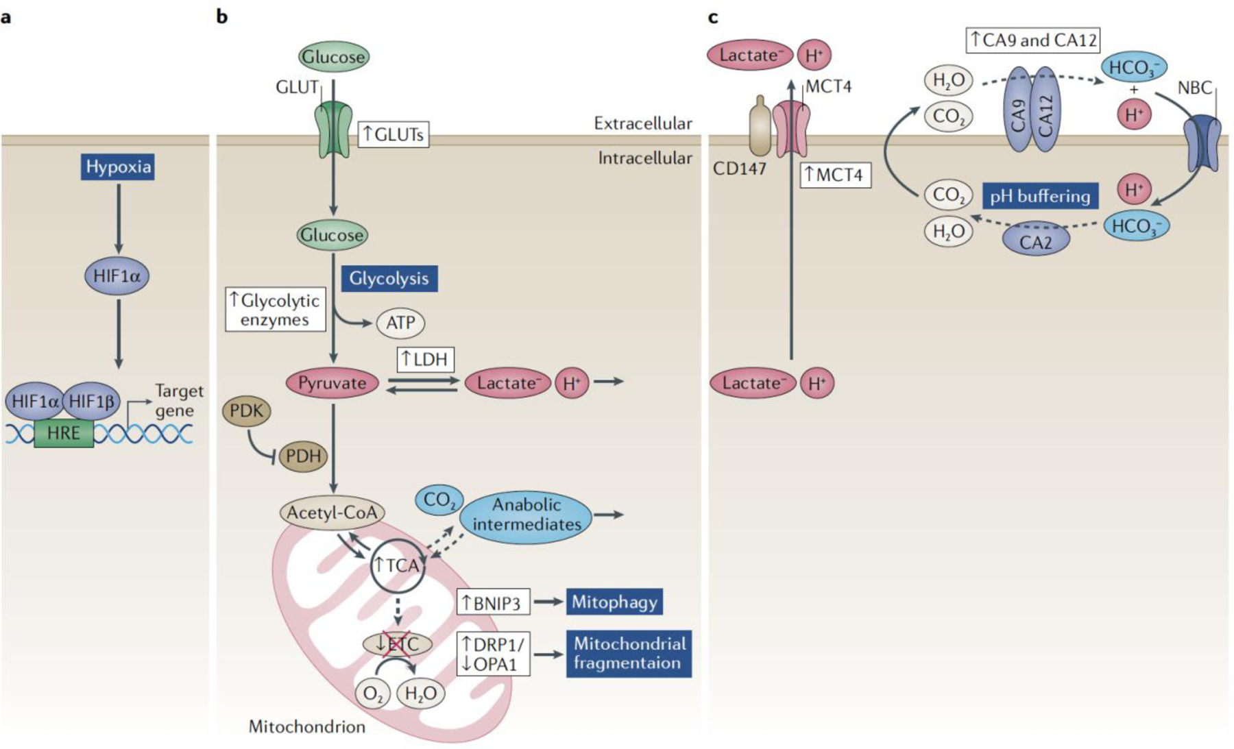
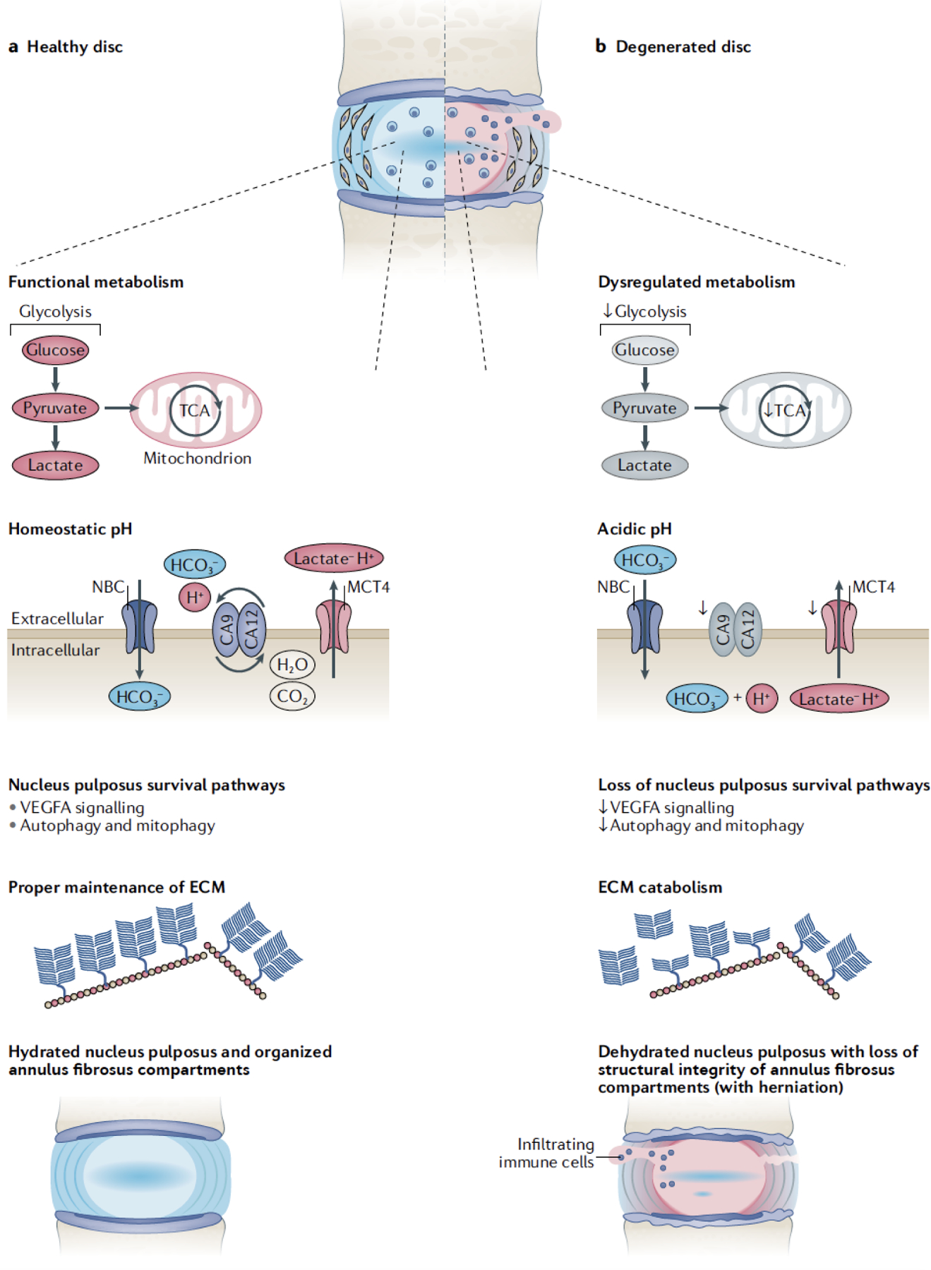
The physiologically hypoxic intervertebral disc and cartilage rely on the hypoxia-inducible factor (HIF) family of transcription factors to mediate cellular responses to changes in oxygen tension. During homeostatic development, oxygen-dependent prolyl hydroxylases, circadian clock proteins and metabolic intermediates control the activities of HIF1 and HIF2 in these tissues. Mechanistically, HIF1 is the master regulator of glycolytic metabolism and cytosolic lactate levels. In addition, HIF1 regulates mitochondrial metabolism by promoting flux through the tricarboxylic acid cycle, inhibiting downsteam oxidative phosphorylation and controlling mitochondrial health through modulation of the mitophagic pathway. Accumulation of metabolic intermediates from HIF-dependent processes contribute to intracellular pH regulation in the disc and cartilage. Namely, to prevent changes in intracellular pH that could lead to cell death, HIF1 orchestrates a bicarbonate buffering system in the disc, controlled by carbonic anhydrase 9 (CA9) and CA12, sodium bicarbonate cotransporters and an intracellular H+/lactate efflux mechanism. In contrast to HIF1, the role of HIF2 remains elusive; in disorders of the disc and cartilage, its function has been linked to both anabolic and catabolic pathways. The current knowledge of hypoxic cell metabolism and regulation of HIF1 activity provides a strong basis for the development of future therapies designed to repair the degenerative disc.


Conflict of interest statement

Competing Interests: None to disclose for all authors.

Figures

Figure 1.
Figure 1.. Regulation of HIF-1α in hypoxic NP cells.
A) Schematic of the intervertebral disc tissue compartments and vasculature. The absence of vasculature in disc compartments makes the NP tissue physiologically hypoxic resulting in robust HIF-1α expression. B) Oxygen dependent mechanisms of HIF-α regulation. In the presence of sufficient O2, PHD2 hydroxylates proline residues in the ODD of HIF-1α targeting it for VHL-mediated polyubiquitination and 26S proteasomal degradation. PHD2 function can be blocked by two mechanisms: 1) Lactate accumulation generates metabolic intermediates, including pyruvate and succinate, which compete with the PHD2 substrate, 2-OG, and inhibit PHD activity. 2) Class I and II HDACs directly inhibit HIF-PHD2 axis. Unlike PHD2, PHD3 serves as a cofactor for transcriptional activation of C-TAD dependent target genes. In NP cells, HIF-1 function is refractory to FIH mediated inhibition. C). Oxygen-independent mechanisms of HIF-α regulation. HIF-1α can be targeted for 26S degradation by HSP70 possibly through displacement of HSP90. In NP cells, HIF-1α is a circadian clock-regulated gene. BMAL1 and RORα synergize to upregulate N-TAD and C-TAD dependent target genes, without evidence of direct binding to HIF-α. HDAC6 is shown to recruit HSP90 as a cofactor to upregulate HIF target gene expression, whereas CCN2 was reported to block HIF-1α cofactor binding and diminish its activity.
Figure 2.
Figure 2.. HIF-1α -dependent metabolic and pH regulatory pathways in NP cells.
In the hypoxic NP cell, HIF-1α transcriptionally regulates many genes involved with glycolysis and pH regulation; HIF targets are shown in violet boxes, arrows denote up- or down- regulation. HIF-1α promotes glycolytic flux and lactate generation by controlling glucose import through GLUT1 and upregulating glycolytic enzymes. MCT4 facilitates the export of H+/lactate, in order to maintain intracellular pH and the perpetuation of pyruvate reduction. HIF-1α also modulates pyruvate entry into the mitochondrial TCA cycle through PDH-PDK1 axis, an area ripe for future investigations in disc cells. Although TCA cycle function is preserved in the NP, mitochondrial ETC is inhibited by hypoxia; arrows denote up- or down-regulation of the pathways. In order to maintain healthy mitochondrial activity, hypoxia and HIF-1α modulates autophagic and mitophagic pathways; HIF-targets shown in green boxes; arrows denote up- or down-regulation. Overall, to tightly control the intracellular pH in glycolytic NP cells, HIF-1α orchestrates a HCO3- buffering system, governed by CA9/12 and NBCs, and fueled by recycled and TCA-cycle derived CO2.
Figure 3.
Figure 3.. Pathological link between loss of HIF-1α function and intervertebral disc degeneration.
A) Schematic of a healthy intervertebral disc. Healthy NP cells are characterized by functional glycolytic and TCA cycle flux. They possess multiple pathways to buffer intracellular H+ production and maintain homeostatic pHi, including H+/lactate extrusion by MCT4 and HCO3- buffering by the CA9/CA12/NBC axis. Functional NP tissue compartments are maintained by hypoxia and HIF-dependent survival pathways- i.e. VEGFA signaling, autophagy, and mitophagy. Healthy NP tissue possess a chondroitin-sulfate proteoglycan-rich ECM which are responsible for the disc’s biomechanical function. B) Degenerated intervertebral discs. This phenotype recapitulates the fate of discs lacking HIF function and activity. Loss of HIF-1α signaling diminishes target gene expression required for cell metabolism and intracellular pH buffering. Dysregulation of the critical NP cell survival pathways and acidosis results in NP cell death and increased matrix breakdown. Compromised ECM and diminished biomechanical function makes the tissue susceptible to herniations, immune cell activation and pain.

Similar articles

Cited by

References

    1. Kaelin WG The von Hippel-Lindau tumour suppressor protein: O2 sensing and cancer. Nature Reviews Cancer vol. 8 865–873 (2008). - PubMed
    1. Shen C & Kaelin WG The VHL/HIF axis in clear cell renal carcinoma. Seminars in Cancer Biology vol. 23 18–25 (2013). - PMC - PubMed
    1. Schödel J & Ratcliffe PJ Mechanisms of hypoxia signalling: new implications for nephrology. Nature Reviews Nephrology vol. 15 641–659 (2019). - PubMed
    1. Semenza GL Hypoxia-inducible factors in physiology and medicine. Cell 148, 399–408 (2012). - PMC - PubMed
    1. Schito L & Semenza GL Hypoxia-Inducible Factors: Master Regulators of Cancer Progression. Trends in Cancer vol. 2 758–770 (2016). - PubMed

Publication types

MeSH terms

Substances