. 2020 Apr;42(2):6-10.
doi: 10.1042/bio04202007. Epub 2020 Apr 8.

Clock-in, clock-out: circadian timekeeping between tissues

Affiliations

Clock-in, clock-out: circadian timekeeping between tissues

Jacob G Smith et al. Biochem (Lond). 2020 Apr.

Abstract

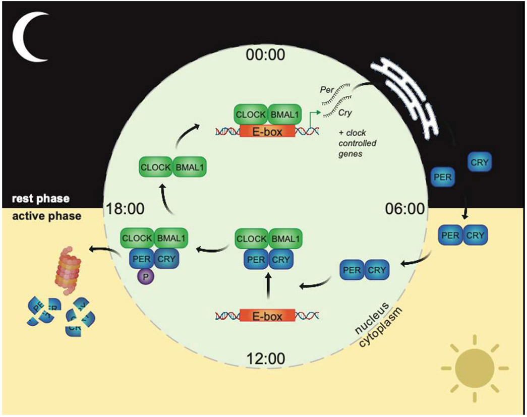
Life evolved in the presence of alternating periods of light and dark that accompany the daily rotation of the Earth on its axis. This offered an advantage for organisms able to regulate their physiology to anticipate these daily cycles. In each light-sensitive organism studied, spanning single-celled bacteria to complex mammals, there exist timekeeping mechanisms able to control physiology over the course of 24 hours. Endowed with internal timekeeping, organisms can put their previously stored energy to the most efficient use, selectively ramping up biological processes at specific times of day or night according to when they'll be needed. Humans have evolved to be more active during the day (diurnal), likely due to the increased opportunities for foraging or hunting in our evolutionary past, and this daily activity is accompanied by an upregulation of genes involved in metabolism to increase the energy available for such behaviours. Remarkably, this happens without conscious thought, due to a complex organism-wide signalling apparatus known as the circadian clock network, that conveys time information between cells and tissues.


Figures

Figure 1.
Figure 1.. The molecular clockwork.
The transcription factors CLOCK and BMAL1 bind DNA E-boxes as a pair, inducing expression of Per and Cry mRNAs. After export to the endoplasmic reticulum, Per/Cry mRNAs are translated into PER/CRY proteins, which build up in the cytoplasm before passing back into the nucleus and repressing CLOCK/BMAL1 transcriptional activity by several mechanisms, including direct binding and dissociation of CLOCK/BMAL from DNA. Phosphorylation (P) of PER2 by specific kinases triggers degradation of the PER/CRY complex by the ubiquitin-proteasome pathway. CLOCK/BMAL1 are subsequently derepressed, and the cycle starts anew. Additional mechanisms, including auxiliary feedback loops, chromatin topology, protein modifications and mRNA processing, work in concert with the core loop to drive 24h hour rhythms at the genome, proteome and signalling level. Figure created using Biorender.com
Figure 2.
Figure 2.. Mammalian circadian network organisation.
Light is the major zeitgeber for the central clock in the SCN, readjusting timing daily to be aligned with the light/dark cycle. The SCN communicates with clocks in the rest of the body to synchronise their timing. Clocks in peripheral tissues can also influence each other’s timing, through release of soluble factors in the bloodstream. For peripheral tissues, external factors such as food intake or exercise are important zeitgebers. Signalling from the SCN to peripheral tissues is considered dominant, though peripheral clocks can influence central clock function in certain contexts. Figure created using Biorender.com
Figure 3.
Figure 3.. Peripheral clock signalling output.
Local clocks in peripheral tissues control the rhythmic release of soluble signalling factors into the bloodstream. For many peripheral tissues, the effect of clock-controlled output on timing in other tissues is not well defined. Figure created using Biorender.com
Figure 4.
Figure 4.. Peripheral clock signalling to brain.
Recent research has revealed that local peripheral clocks can feedback to the brain and alter timing of circadian behavioural cycles, through as-of-yet poorly defined mechanisms. Figure created using Biorender.com

References

    1. Koronowski KB, Kinouchi K, Welz PS, Smith JG, Zinna VM, Shi J, Samad M, Chen S, Magnan CN, Kinchen JM, Li W3, Baldi P, Benitah SA, Sassone-Corsi P. Defining the Independence of the Liver Circadian Clock. Cell. 2019. May 30;177(6):1448–1462.e14. doi: 10.1016/j.cell.2019.04.025. - DOI - PMC - PubMed
    1. Crosby P, Hamnett R, Putker M, Hoyle NP, Reed M, Karam CJ, Maywood ES, Stangherlin A, Chesham JE, Hayter EA, Rosenbrier-Ribeiro L, Newham P, Clevers H, Bechtold DA, O'Neill JS. Insulin/IGF-1 Drives PERIOD Synthesis to Entrain Circadian Rhythms with Feeding Time. Cell. 2019. May 2;177(4):896–909.e20. doi: 10.1016/j.cell.2019.02.017. Epub 2019 Apr 25. - DOI - PMC - PubMed
    1. Reinke H, Asher G. Crosstalk between metabolism and circadian clocks (Review) Nat Rev Mol Cell Biol. 2019. April;20(4):227–241. doi: 10.1038/s41580-018-0096-9. - DOI - PubMed
    1. Dyar KA, Lutter D, Artati A, Ceglia NJ, Liu Y, Armenta D, Jastroch M, Schneider S, de Mateo S, Cervantes M, Abbondante S, Tognini P, Orozco-Solis R, Kinouchi K, Wang C, Swerdloff R, Nadeef S, Masri S, Magistretti P, Orlando V, Borrelli E, Uhlenhaut NH, Baldi P, Adamski J, Tschöp MH, Eckel-Mahan K, Sassone-Corsi P. Atlas of Circadian Metabolism Reveals System-wide Coordination and Communication between Clocks. Cell. 2018. September 6;174(6):1571–1585.e11. doi: 10.1016/j.cell.2018.08.042. - DOI - PMC - PubMed
    1. Perrin L, Loizides-Mangold U, Skarupelova S, Pulimeno P, Chanon S, Robert M, Bouzakri K, Modoux C, Roux-Lombard P, Vidal H, Lefai E, Dibner C. Human skeletal myotubes display a cell-autonomous circadian clock implicated in basal myokine secretion. Mol Metab. 2015. August 6;4(11):834–45. doi: 10.1016/j.molmet.2015.07.009. eCollection 2015 November. - DOI - PMC - PubMed

LinkOut - more resources