

From Microscopy to Nanoscopy: Defining an Arabidopsis thaliana Meiotic Atlas at the Nanometer Scale
- PMID: 34084178
- PMCID: PMC8167036
- DOI: 10.3389/fpls.2021.672914
From Microscopy to Nanoscopy: Defining an Arabidopsis thaliana Meiotic Atlas at the Nanometer Scale
Abstract
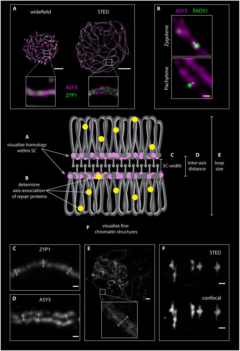
Visualization of meiotic chromosomes and the proteins involved in meiotic recombination have become essential to study meiosis in many systems including the model plant Arabidopsis thaliana. Recent advances in super-resolution technologies changed how microscopic images are acquired and analyzed. New technologies enable observation of cells and nuclei at a nanometer scale and hold great promise to the field since they allow observing complex meiotic molecular processes with unprecedented detail. Here, we provide an overview of classical and advanced sample preparation and microscopy techniques with an updated Arabidopsis meiotic atlas based on super-resolution microscopy. We review different techniques, focusing on stimulated emission depletion (STED) nanoscopy, to offer researchers guidance for selecting the optimal protocol and equipment to address their scientific question.
Keywords: Arabidopsis; cytology; immunofluorescence; meiosis; super-resolution microscopy.
Copyright © 2021 Sims, Schlögelhofer and Kurzbauer.
Conflict of interest statement
The authors declare that the research was conducted in the absence of any commercial or financial relationships that could be construed as a potential conflict of interest.
Figures


Similar articles
-
Molecular cell biology of male meiotic chromosomes and isolation of male meiocytes in Arabidopsis thaliana.Methods Mol Biol. 2014;1110:217-30. doi: 10.1007/978-1-4614-9408-9_10. Methods Mol Biol. 2014. PMID: 24395259
-
AIE Nanoparticles with High Stimulated Emission Depletion Efficiency and Photobleaching Resistance for Long-Term Super-Resolution Bioimaging.Adv Mater. 2017 Nov;29(43). doi: 10.1002/adma.201703643. Epub 2017 Oct 4. Adv Mater. 2017. PMID: 28977700
-
Recent advances in STED and RESOLFT super-resolution imaging techniques.Spectrochim Acta A Mol Biomol Spectrosc. 2020 Apr 15;231:117715. doi: 10.1016/j.saa.2019.117715. Epub 2019 Nov 4. Spectrochim Acta A Mol Biomol Spectrosc. 2020. PMID: 31748155 Review.
-
Meiotic chromosome synapsis and recombination in Arabidopsis thaliana: new ways of integrating cytological and molecular approaches.Chromosome Res. 2014 Jun;22(2):179-90. doi: 10.1007/s10577-014-9426-8. Chromosome Res. 2014. PMID: 24941912 Review.
-
Immunolocalization protocols for visualizing meiotic proteins in Arabidopsis thaliana: method 3.Methods Mol Biol. 2013;990:109-18. doi: 10.1007/978-1-62703-333-6_11. Methods Mol Biol. 2013. PMID: 23559207
Cited by
-
Epigenetic regulation during meiosis and crossover.Physiol Mol Biol Plants. 2023 Dec;29(12):1945-1958. doi: 10.1007/s12298-023-01390-w. Epub 2023 Nov 20. Physiol Mol Biol Plants. 2023. PMID: 38222277 Free PMC article. Review.
-
Meiotic double-strand break repair DNA synthesis tracts in Arabidopsis thaliana.PLoS Genet. 2024 Jul 16;20(7):e1011197. doi: 10.1371/journal.pgen.1011197. eCollection 2024 Jul. PLoS Genet. 2024. PMID: 39012914 Free PMC article.
-
Caught in the Act: Live-Cell Imaging of Plant Meiosis.Front Plant Sci. 2021 Dec 21;12:718346. doi: 10.3389/fpls.2021.718346. eCollection 2021. Front Plant Sci. 2021. PMID: 34992616 Free PMC article. Review.
-
Fast and Precise: How to Measure Meiotic Crossovers in Arabidopsis.Mol Cells. 2022 May 31;45(5):273-283. doi: 10.14348/molcells.2022.2054. Mol Cells. 2022. PMID: 35444069 Free PMC article. Review.
-
Conservation and divergence of meiotic DNA double strand break forming mechanisms in Arabidopsis thaliana.Nucleic Acids Res. 2021 Sep 27;49(17):9821-9835. doi: 10.1093/nar/gkab715. Nucleic Acids Res. 2021. PMID: 34458909 Free PMC article.
References
Publication types
LinkOut - more resources
Full Text Sources