Review
. 2021 Jun;8(2):021315.
doi: 10.1063/5.0044713.

Photodynamic viral inactivation: Recent advances and potential applications

Affiliations
Review

Photodynamic viral inactivation: Recent advances and potential applications

Jace A Willis et al. Appl Phys Rev. 2021 Jun.

Abstract

Antibiotic-resistant bacteria, which are growing at a frightening rate worldwide, has put the world on a long-standing alert. The COVID-19 health crisis reinforced the pressing need to address a fast-developing pandemic. To mitigate these health emergencies and prevent economic collapse, cheap, practical, and easily applicable infection control techniques are essential worldwide. Application of light in the form of photodynamic action on microorganisms and viruses has been growing and is now successfully applied in several areas. The efficacy of this approach has been demonstrated in the fight against viruses, prompting additional efforts to advance the technique, including safety use protocols. In particular, its application to suppress respiratory tract infections and to provide decontamination of fluids, such as blood plasma and others, can become an inexpensive alternative strategy in the fight against viral and bacterial infections. Diverse early treatment methods based on photodynamic action enable an accelerated response to emerging threats prior to the availability of preventative drugs. In this review, we evaluate a vast number of photodynamic demonstrations and first-principle proofs carried out on viral control, revealing its potential and encouraging its rapid development toward safe clinical practice. This review highlights the main research trends and, as a futuristic exercise, anticipates potential situations where photodynamic treatment can provide a readily available solution.


Figures

FIG. 1.
FIG. 1.
Left, Jablonski diagram depicting energy transfer through an excited photosensitizer. Right, subsequent molecular interactions resulting from excited photosensitizer interaction with molecular oxygen.
FIG. 2.
FIG. 2.
Visualization of typical interaction radii compared to virion and cell sizes. From left to right, ROS travels up to about 20 nm in organic environments, singlet oxygen survives much longer and can travel about 75 nm, HIV virions average around 100 nm in diameter, SARS-CoV-2 is approximately 125 nm in diameter, human erythrocytes (red blood cells) average 7.81 μm in diameter, and average human cells vary greatly but average roughly 100 μm.
FIG. 3.
FIG. 3.
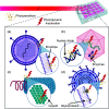
Schematic illustration of photodynamic inactivation of various enveloped and non-enveloped viruses. (a) Demonstrates inactivation of envelope proteins in an enveloped virus, (b) of viral nucleic acids and enzymes within the capsid of a non-enveloped virus, (c) of the capsid itself in a helical virus, and (d) of glycoproteins on the viral envelope surface of a complex structure virus.
FIG. 4.
FIG. 4.
Dose threshold distribution model curves from a theoretical death fraction curve [f(D)] and its derivative [g(D)] as a dose distribution curve, explaining the parameters Dth and FWHM.
FIG. 5.
FIG. 5.
Diagram of percentage of deaths of 99%, 95%, and 90% for a porphyrin considering the relationship between photosensitizer concentration and light dose.
FIG. 6.
FIG. 6.
Endotracheal tube: (A) PVC-based, (B) functionalized with curcumin. Source: Zangirolami et al.
FIG. 7.
FIG. 7.
Representative diagram of Endotracheal tube (ETT) positioning. Source: Zangirolami et al.

References

    1. Tischer B. K. and Osterrieder N., “ Herpesviruses—A zoonotic threat?,” Vet. Microbiol. 140, 266–270 (2010).10.1016/j.vetmic.2009.06.020 - DOI - PMC - PubMed
    1. Irwin K. K., Renzette N., Kowalik T. F., and Jensen J. D., “ Antiviral drug resistance as an adaptive process,” Virus Evol. 2, vew014 (2016).10.1093/ve/vew014 - DOI - PMC - PubMed
    1. Kimberlin D. W. and Whitley R. J., “ Antiviral resistance: Mechanisms, clinical significance, and future implications,” J. Antimicrob. Chemother. 37, 403–421 (1996).10.1093/jac/37.3.403 - DOI - PubMed
    1. Moller K. I., Kongshoj B., Philipsen P. A., Thomsen V. O., and Wulf C., “ How Finsen's light cured lupus vulgaris,” Photodermatology 21, 118–124 (2005).10.1111/j.1600-0781.2005.00159.x - DOI - PubMed
    1. Hideg E., Jansen M. A. K., and Strid A., “ UV-B exposure, ROS, and stress: Inseparable companions or loosely linked associates?,” Trends Plant Sci. 18, 107–115 (2013).10.1016/j.tplants.2012.09.003 - DOI - PubMed

LinkOut - more resources