. 2019 Dec 9;11(4):1122-1131.
doi: 10.1039/c9sc04189c.

Chemical modification of the adeno-associated virus capsid to improve gene delivery

Affiliations

Chemical modification of the adeno-associated virus capsid to improve gene delivery

Mathieu Mével et al. Chem Sci. .

Abstract

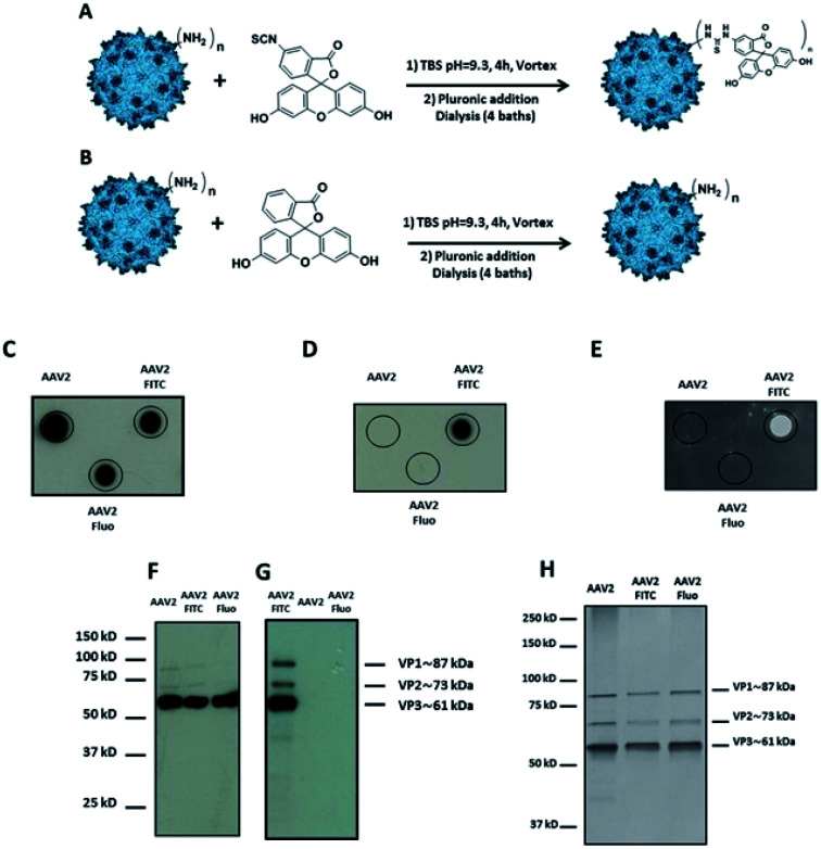
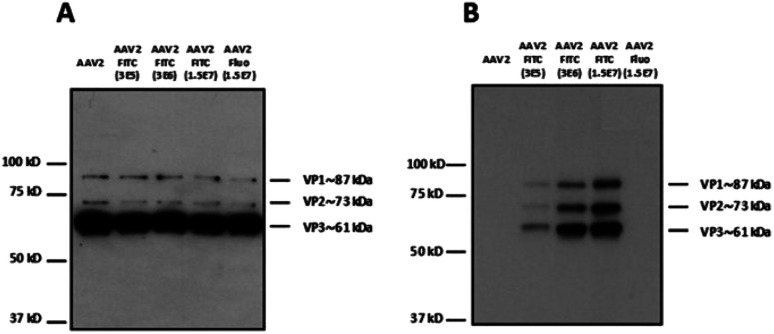
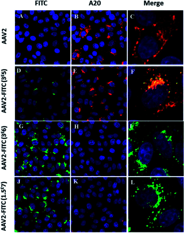
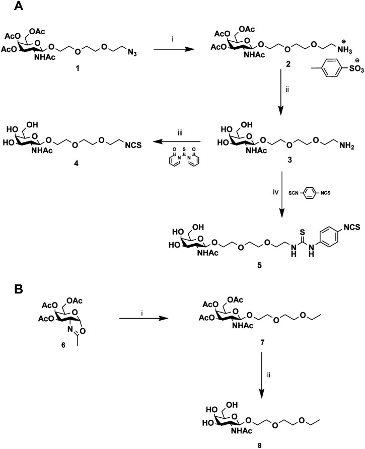
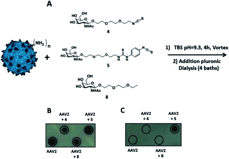
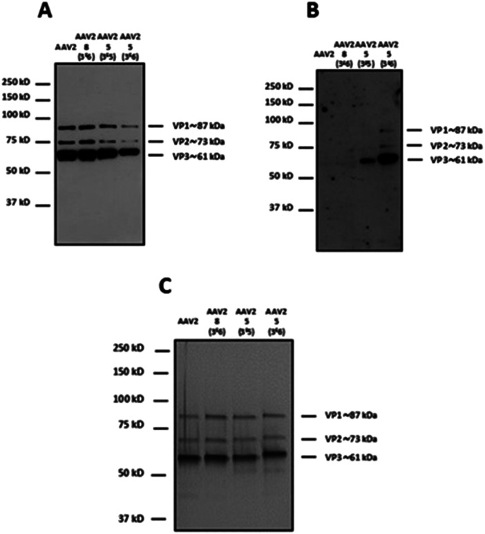
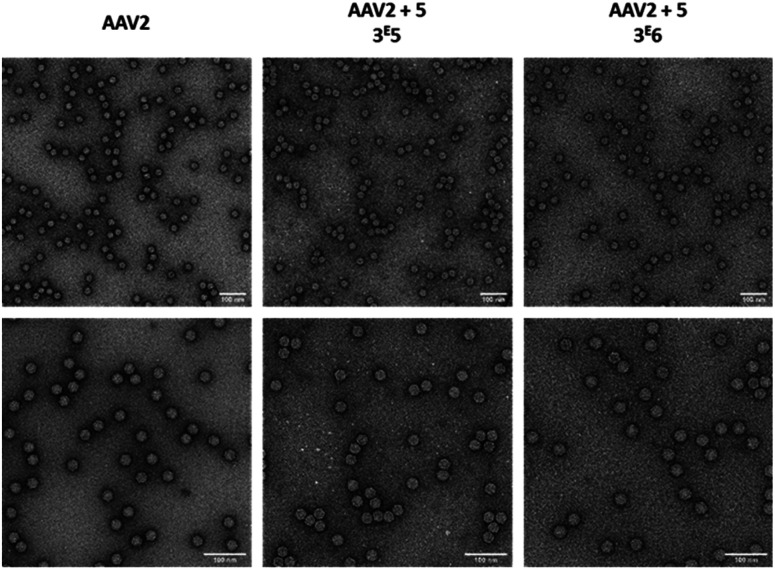
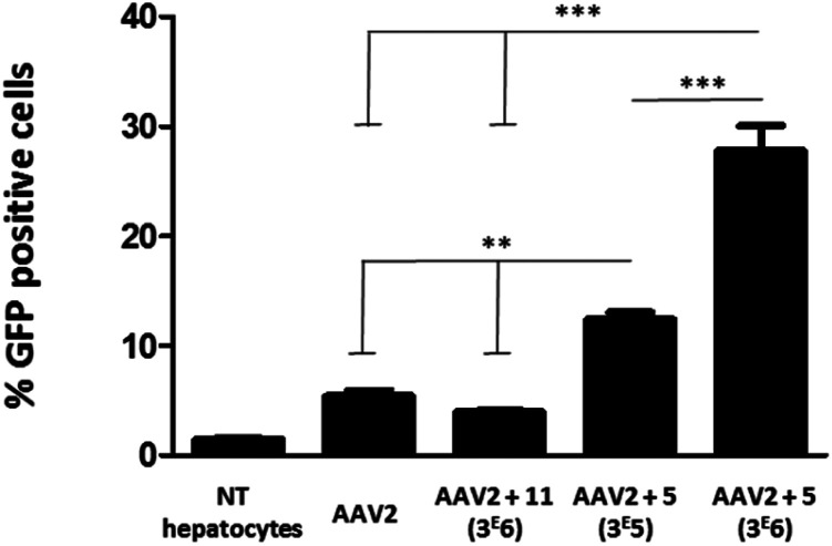
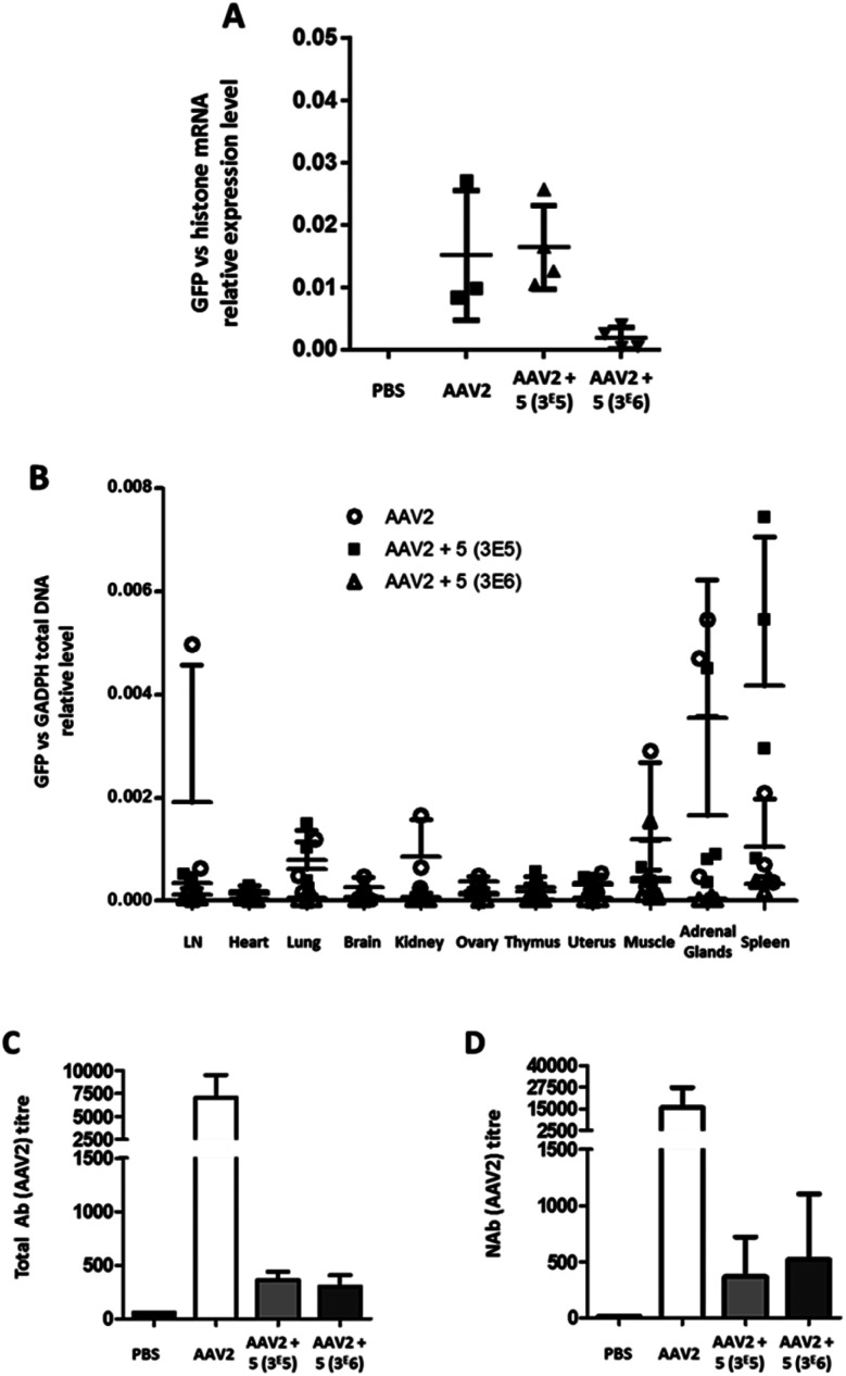
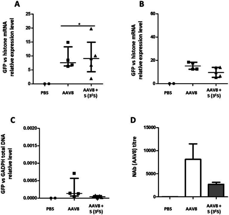
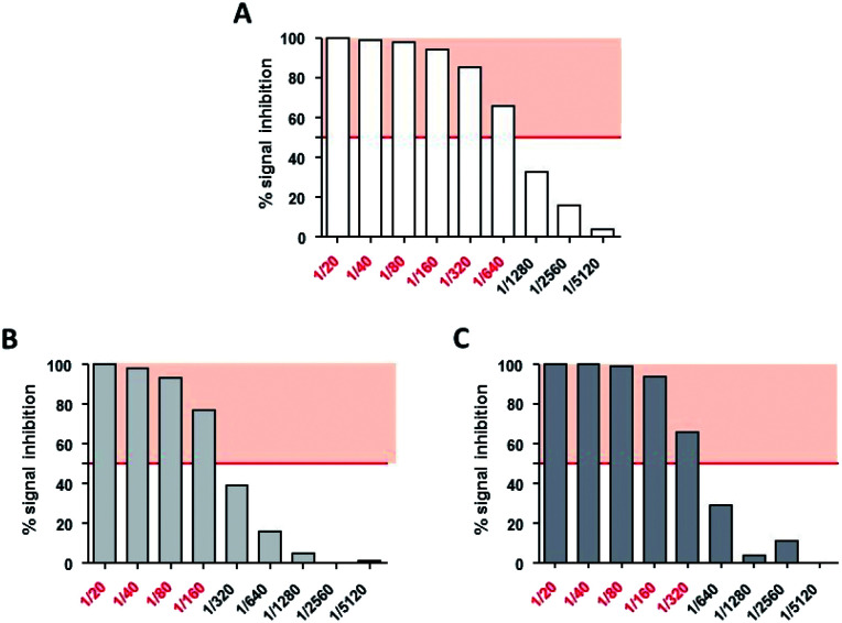
Gene delivery vectors based on adeno-associated virus (AAV) are highly promising due to several desirable features of this parent virus, including a lack of pathogenicity, efficient infection of dividing and non-dividing cells and sustained maintenance of the viral genome. However, the conclusion from clinical data using these vectors is that there is a need to develop new AAVs with a higher transduction efficiency and specificity for relevant target tissues. To overcome these limitations, we chemically modified the surface of the capsid of AAV vectors. These modifications were achieved by chemical coupling of a ligand by the formation of a thiourea functionality between the amino group of the capsid proteins and the reactive isothiocyanate motif incorporated into the ligand. This strategy does not require genetic engineering of the capsid sequence. The proof of concept was first evidenced using a fluorophore (FITC). Next, we coupled the N-acetylgalactosamine ligand onto the surface of the AAV capsid for asialoglycoprotein receptor-mediated hepatocyte-targeted delivery. Chemically-modified capsids also showed reduced interactions with neutralizing antibodies. Taken together, our findings reveal the possibility of creating a specific engineered platform for targeting AAVs via chemical coupling.


Conflict of interest statement

M. M., D. D., are E. A. are inventors on a patent including the technology described in this manuscript. JPC and GGA are employees of Vivet Therapeutics.

Figures

Fig. 1
Fig. 1. Covalent coupling of FITC onto the capsid of AAV2 via primary amino groups. (A) 1012 vg of AAV2-GFP vectors were added to a solution of FITC (3 × 10⁵ eq.) in TBS buffer (pH 9.3) and incubated for 4 h at RT. (B) The procedure was carried out with fluorescein (3 × 10⁵ eq.) as a control. (C–E) 109 vg of each condition was analyzed by dot blot using the A20 antibody that recognizes the assembled capsid (C) or using an anti-FITC antibody (D) or by direct fluorescence emission (E). (F and G) 5 × 10⁸ vg of the same samples were analyzed by western blot using a polyclonal antibody to detect denatured AAV capsid proteins (F) or using an anti-FITC antibody (G). (H) 1010 vg of each condition was analyzed by silver nitrate staining.
Fig. 2
Fig. 2. Modulation of the number of FITC molecules on the capsid of AAV. (A) 5 × 10⁸ vg of the samples were analyzed by western blot using a polyclonal antibody to detect denatured AAV capsid proteins or (B) an anti-FITC antibody to detect VP capsid proteins chemically modified with FITC molecules.
Fig. 3
Fig. 3. Confocal imaging of modulated AAV2-FITC. (A–C) HeLa cells transduced with AAV2 and incubated with A20 primary antibody and Al647 secondary antibody. Red fluorescence from A20 immunolabeling (B) was detected. (D–F) HeLa cells transduced with AAV2-3 × 10⁵ FITC and incubated with A20 primary antibody and Al647 secondary antibody. Green FITC fluorescence (D) and red fluorescence from A20 immunolabeling (E) were detected. Colocalisation of FITC and A20-Al647 (F). (G–I) HeLa Cells were transduced with AAV2-3 × 10⁶ FITC and incubated with A20 primary antibody and Al647 secondary antibody. Green FITC fluorescence from AAV2-3 × 10⁶ FITC was detected (G) whereas the detection of red fluorescence from A20 immunolabeling was very low (H). (J–L) HeLa cells were transduced with AAV2-1.5 × 10⁷ FITC and incubated with A20 primary antibody and Al647 secondary antibody. Green FITC fluorescence from AAV2-1.5 × 10⁷ FITC was detected (J) whereas the detection of red fluorescence from A20 immunolabeling was very low (L). Cell nuclei were counterstained with DAPI (blue); scale bars: 25 μm for FITC and A20 images; 5 μm for merged images. All the controls are detailed in the ESI.
Scheme 1
Scheme 1. Synthesis of GalNAc derivatives with Alk-NCS, Aryl-NCS coupling functionality and without the coupling functionality. (A) (i) MeOH, APTS, H2, Pd–C (100%), (ii) MeOH/H2O, IRN78 (77%), (iii) DMF, 1,1′-thiocarbonyldi-2(1H)-pyridone (88%), (iv) DMF, p-phenylene diisothiocyanate (85%). (B) (i) DCM, 2-(2-ethoxyethoxy)ethanol, molecular sieves (73%), (ii) MeOH/H2O, IRN78 (77%).
Fig. 4
Fig. 4. Identification of the reactive function for the covalent coupling of GalNAc ligands on the capsid of AAV2 via primary amino groups. (A) 1012 vg of AAV2-GFP vectors were added to a solution of compound 4 or compound 5 (3 × 10⁵ eq.) in TBS buffer (pH 9.3) and incubated for 4 h at RT. The same experimental procedure was followed for compound 8 (3 × 10⁵ eq.) in TBS at pH 9.3 as a control. (B and C) AAV2 control and samples of AAV2 vectors incubated with GalNAc ligands in TBS buffer (AAV2 + 4, AAV2 + 5 and AAV2 + 8) were analyzed by dot blot. 1010 vg of each condition was analyzed using the A20 antibody that recognizes the intact capsid (B) or using soybean-FITC lectin that recognizes the N-acetylgalactosamine sugar (C).
Fig. 5
Fig. 5. Modulation of the number of GalNAc molecules on the capsid of AAV vectors. 1012 vg of AAV2-GFP vectors were added to a solution of 5 (3 × 10⁵ and 3 × 10⁶ eq.) in TBS buffer (pH 9.3) and incubated for 4 h at RT. The same experimental procedure was followed but substituting 5 by 8 (3 × 10⁶ eq.) as a control. 5 × 10⁸ vg of the samples was analyzed by western blot using a polyclonal antibody against the capsid proteins to detect VP1, VP2 and VP3 proteins (A) or using FITC-soybean lectin (B). (C) 1010 vg of each condition was analyzed by silver nitrate staining. The capsid protein molecular weight is indicated at the right of the images according to a protein ladder.
Fig. 6
Fig. 6. Electron microscopy images of GalNAc-AAV2 particles. Unmodified AAV2 vectors (left) and AAV2 vectors modified with 3 × 10⁵ (middle) or 3 × 10⁶ equivalent (right) of 5 were negatively stained with uranyl acetate and examined by transmission electron microscopy at a ×50 000 (upper panel) or ×80 000 (lower panel) nominal magnification. Scale bars: 100 nm.
Fig. 7
Fig. 7. Transduction of primary mouse hepatocytes with native AAV2 and AAV2 vectors chemically modified with GalNAc and mannose ligands. Primary mouse hepatocytes (1.7 × 10⁵ cells per well) were incubated in P24 plates and transduced with the native AAV2 control, AAV2 + 11 (3 × 10⁶), AAV2 + 5 (3 × 10⁵) and AAV2 + 5 (3 × 10⁶) at a MOI of 105. All AAV vectors encoded for GFP. The percentage of GFP positive cells was measured by FACS analysis 72 h after the transduction. Non-transduced cells (NT hepatocytes) were used as a control for the fluorescence background. Three replicates of each condition were analyzed by ANOVA test (***p < 0.001, **p < 0.01). Data are represented as mean ± SD.
Fig. 8
Fig. 8. Liver transduction profile of GalNAc-AAV2 and development of anti-AAV2 antibodies. Groups of mice were injected with unmodified AAV2 eGFP or GalNAc-AAV2 carrying a GFP transgene at molar ratios of 3 × 10⁵ or 3 × 10⁶. A control group received PBS only. Twenty-one days after administration, the mice were sacrificed and organs were extracted. (A) The GFP mRNA expression levels in liver samples were normalized against endogenous histone mRNA expression. In addition, a biodistribution analysis was performed (B). Total (C) and neutralizing (D) antibody levels in serum were determined. Medians and range (A and B) or means and standard deviation (C and D) are shown. N = 2–6 animals per group.
Fig. 9
Fig. 9. Liver transduction profile of GalNAc-AAV8 and development of anti-AAV8 antibodies. Groups of mice were injected with unmodified AAV8 eGFP or GalNAc-AAV8 carrying a GFP transgene at a molar ratio of 3 × 10⁵. A control group received PBS only. Ten (A) and 21 days (B) after administration mice were sacrificed and organs extracted. (A and B) The GFP mRNA expression levels in liver samples were normalized against endogenous histone mRNA expression. GFP total DNA normalized to GAPDH in the spleen (C). Neutralizing antibody levels in serum were determined (D). Medians and range (A–C) or means and standard deviation (D) are shown. N = 2–6 animals per group.
Fig. 10
Fig. 10. Neutralizing assays of AAV2 and GalNAc-AAV2 on HeLa cells. HeLa cells were incubated with AAV2 LacZ (A), GalNac-AAV2 LacZ (3 × 10⁵) (B) or GalNac-AAV2 LacZ (3 × 10⁶) (C) in the presence of various dilutions of AAV2-neutralizing serum (1/20 to 1/5120). After measurement of beta-galactosidase activity, neutralizing titers were expressed as the highest dilution of serum that inhibited more than 50% (red police) of AAV2, GalNac-AAV2 LacZ (3 × 10⁵) or GalNac-AAV2 (3 × 10⁶) signals without incubation with neutralizing serum (IC50).

Similar articles

Cited by

References

    1. Zincarelli C. Soltys S. Rengo G. Rabinowitz J. E. Mol. Ther. 2008;16:1073–1080. doi: 10.1038/mt.2008.76. - DOI - PubMed
    1. Mingozzi F. High K. A. Nat. Rev. Genet. 2011;12:341–355. doi: 10.1038/nrg2988. - DOI - PubMed
    1. Buning H. EMBO Mol. Med. 2013;5:1–3. doi: 10.1002/emmm.201202291. - DOI - PMC - PubMed
    1. Gaudet D. Methot J. Dery S. Brisson D. Essiembre C. Tremblay G. Tremblay K. de Wal J. Twisk J. van den Bulk N. Sier-Ferreira V. van Deventer S. Gene Ther. 2013;20:361–369. doi: 10.1038/gt.2012.43. - DOI - PMC - PubMed
    1. Russell S. Bennett J. Wellman J. A. Chung D. C. Yu Z. F. Tillman A. Wittes J. Pappas J. Elci O. McCague S. Cross D. Marshall K. A. Walshire J. Kehoe T. L. Reichert H. Davis M. Raffini L. George L. A. Hudson F. P. Dingfield L. Zhu X. Haller J. A. Sohn E. H. Mahajan V. B. Pfeifer W. Weckmann M. Johnson C. Gewaily D. Drack A. Stone E. Wachtel K. Simonelli F. Leroy B. P. Wright J. F. High K. A. Maguire A. M. Lancet. 2017;390:849–860. doi: 10.1016/S0140-6736(17)31868-8. - DOI - PMC - PubMed