Review
. 2021 Jun 4;12(1):67.
doi: 10.1186/s13244-021-01001-w.

Diagnostic methods and therapeutic options of uveal melanoma with emphasis on MR imaging-Part II: treatment indications and complications

Affiliations
Review

Diagnostic methods and therapeutic options of uveal melanoma with emphasis on MR imaging-Part II: treatment indications and complications

Pietro Valerio Foti et al. Insights Imaging. .

Abstract

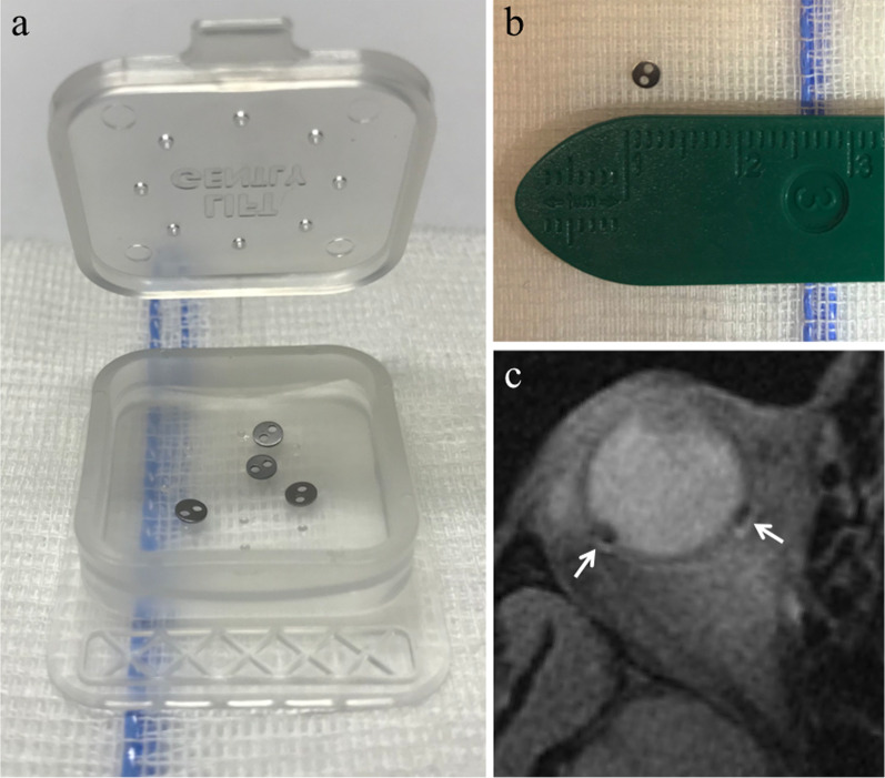
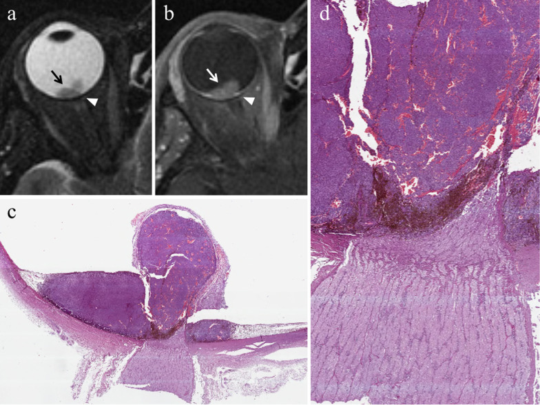
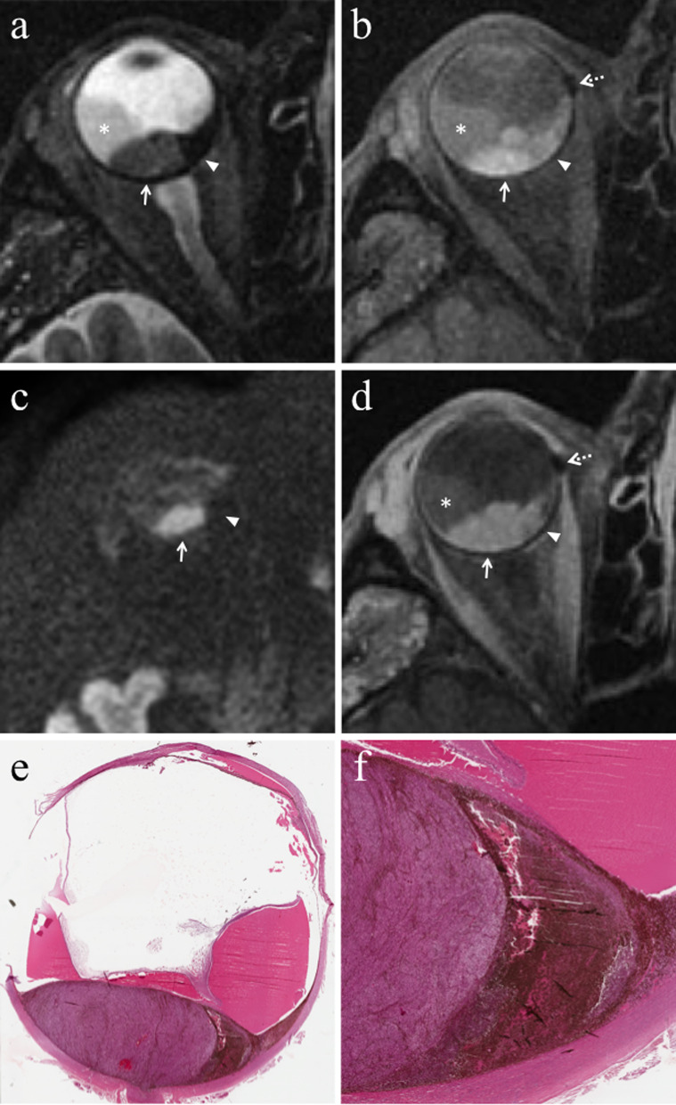
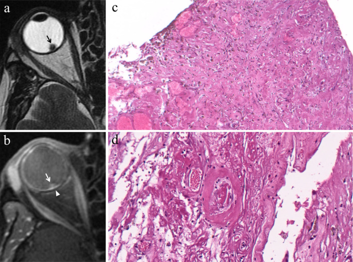
Therapy of uveal melanoma aims to preserve the eye and its function and to avoid metastatic dissemination. The treatment choice is difficult and must keep into account several factors; the therapeutic strategy of uveal melanoma should therefore be personalized, sometimes requiring to combine different treatment techniques. Nowadays globe-sparing radiotherapy techniques are often preferred to enucleation. Plaque brachytherapy, the most commonly used eye-preserving therapy, is suitable for small- and medium-sized uveal melanomas. Proton beam radiotherapy is indicated for tumours with noticeable size, challenging shape and location, but is more expensive and less available than brachytherapy. Enucleation is currently restricted to advanced tumours, uveal melanomas with orbital or optic nerve involvement, blind and painful eyes because of treatment-related complications (neovascular glaucoma, chronic inflammatory processes). The effect of proton beam therapy on neoplastic tissue is related to direct cytotoxic action of the radiations, impairment of neoplastic vascular supply and immunologic response. Complications after radiotherapy are frequent and numerous and mainly related to tumour thickness, radiation dose and distance between the tumour and optic nerve. The purpose of this pictorial review is to provide the radiologists with awareness about diagnostic methods and therapeutic options of uveal melanoma. In the present second section, we discuss the therapeutic management of uveal melanoma, describing the main ocular-conserving radiotherapic techniques. We subsequently present an overview of the effects of radiations on neoplastic tissue. Lastly, we review ocular complications following radiotherapy that should be evaluated by radiologists during follow-up MRI examinations.

Keywords: Brachytherapy; Eye; Magnetic resonance imaging; Melanoma; Proton therapy.


Conflict of interest statement

The authors declare no competing interests in the manuscript.

Figures

Fig. 1
Fig. 1
Tantalum clips for proton-beam radiotherapy. a, b Tantalum clips (2.5 mm in diameter). c Fat-suppressed T1-weighted image shows the artefact produced by the clips (white arrows); the artefact is negligible since tantalum is a nonmagnetic metal
Fig. 2.
Fig. 2.
3D computer model of the patient’s eye/tumor for proton-beam radiotherapy. Eyeplan treatment planning system: (a) eye side, (b) beam axis and (c) optical-axis-centered fundus isodose views
Fig. 3
Fig. 3
A 55-year-old woman with a choroidal melanoma of the right eye infiltrating the optic disc. Axial (a) T2-weighted turbo spin-echo STIR and (b) contrast-enhanced fat-suppressed T1-weighted images demonstrate an intraocular lesion along the posterior aspect of globe (white arrows), infiltrating the optic disc (white arrowheads). c On low magnification, histological examination shows a strong overlap with MR imaging: a poorly pigmented mass located in the posterior segment of the eye, at the level of the optic disc (H&E, original magnification 25×). d Higher magnification confirms the MR findings, demonstrating an early infiltration of the emergence of the optic nerve by the melanoma (H&E, original magnification 50×)
Fig. 4
Fig. 4
A 29-year-old man with a choroidal melanoma of the right eye treated with proton-beam radiotherapy. The patient underwent secondary enucleation about three years after radiotherapy. Axial a T2-weighted turbo spin-echo STIR and b contrast-enhanced fat-suppressed T1-weighted images display an intraocular lesion along the posterior aspect of globe (white arrows). On T2-weighted image, a central well-marginated hypointense area is detectable within the mass (white arrowhead); it represents radiotherapy-related necrosis and its low signal intensity is due to the dispersion of melanin pigment. On contrast-enhanced T1-weighted image, the above-mentioned area appears relatively hypointense (white arrowhead) compared to the surrounding enhancing viable neoplastic tissue. c Histological examination showing an “abrupt transition” between a radiotherapy-related necrotic area with dispersion of melanin pigment (on the right) and the vital tumor tissue (on the left) (H&E, original magnification 100×)
Fig. 5
Fig. 5
A 60-year-old man with choroidal melanoma of the right eye treated with proton-beam radiotherapy. The patient underwent secondary enucleation about three years after radiotherapy because of local recurrence. Axial a T2-weighted turbo spin-echo STIR, b fat-suppressed T1-weighted, (c) DW (b = 1000 s/mm2) and (d) contrast-enhanced fat-suppressed T1-weighted images show an intraocular mass along the posterior aspect of globe, at the level of the optic disc. The central and lateral portions of the lesion (white arrows) exhibit intermediate signal intensity on T2-weighted image and high signal intensity on T1-weighted image and represent viable tumor. The medial portion of the mass demonstrates low signal intensity on T2-weighted image and moderately high signal intensity on T1-weighted image; it represents radiation-induced necrosis with dispersion of melanin pigment, responsible for the low T2 signal. Note the well-defined border between the two distinct portions of the lesion, particularly evident on T2-weighted image. On (d) axial contrast-enhanced fat-suppressed T1-weighted image, the viable tumor demonstrates mild enhancement (white arrow) compared to relatively lower signal intensity of the medial necrotic part (white arrowhead). On c DW image, the viable neoplastic tissue displays high signal intensity (white arrow), a finding consistent with restricted diffusion due to high cellularity, whereas the necrotic part (white arrowhead) is hypointense, lacking of restricted diffusion. Laterally to the lesion a retinal detachment is detectable (white asterisks), with intermediate signal intensity on T2- and T1-weighted images, without enhancement after contrast agent administration. Along the medial outer edge of the sclera, a small metal artefact due to tantalum clip is appreciable (white dotted arrows in b and d). e Histological examination: low magnification showing a poorly pigmented melanoma, protruding into the posterior ocular segment and containing a necrotic component (on the right) (H&E, original magnification 25×); f Higher magnification demonstrating the "abrupt transition" between vital tumor tissue (on the left) and necrosis with abundant dispersed melanin (on the right) (H&E, original magnification 50×)
Fig. 6
Fig. 6
A 58-year-old man with residual fibrotic scar after proton-beam radiotherapy for choroidal melanoma of the right eye. The patient underwent secondary enucleation two years after radiotherapy because of neovascular glaucoma. a Axial T2-weighted turbo spin-echo STIR image demonstrates an intraocular small rounded hypointense mass along the posterior aspect of globe (black arrow). On b contrast-enhanced fat-suppressed T1-weighted image, the mass exhibits mild enhancement (white arrow). Note the pronounced enhancement of the choroid below the irradiated lesion due to radiation-related neoangiogenesis (white arrowhead). c Histological examination showing a well-circumscribed sclero-hemorrhagic nodule composed of ectatic vessels, fibrosis with scattered reactive fibroblasts and abundant component of melanophages; no neoplastic cells are visible (H&E, original magnification 200×). d On higher magnification, within the choroid below the abovementioned nodule, ectatic and thick-walled vessels are evident (H&E, original magnification 400×)
Fig. 7
Fig. 7
An 82-year-old man with a choroidal melanoma of the left eye treated with plaque brachytherapy. The patient underwent secondary enucleation seven years after radiotherapy because of painful eye and local recurrence. Axial a T2-weighted turbo spin-echo STIR and b fat-suppressed T1-weighted images. Along the medial aspect of the left globe an intraocular mass (arrows) exhibits inhomogeneous signal intensity with irregularly edged hypointense areas on T2-weighted image (white arrowhead in a), due to radiation-induced fibrotic alterations. The anterior chamber and the vitreous body of the left eye demonstrate high signal intensity on T1-weighted image (black asterisk in b), a finding consistent with extensive vitreous hemorrhage. Note the difference with the physiological water-like signal intensity of the contralateral eye. c Histological low magnification showing the heterogeneous appearance of the tumor, composed of poorly pigmented spindle cells, intermingled with dense and eosinophilic intratumoral fibrotic areas (H&E, original magnification 25×). d On higher magnification, the fibrotic nature of the above-described eosinophilic areas is well documented: neoplastic spindle cells are mixed to multiple deposits of dense collagen fibres, representing the main effect of radiotherapy (H&E, original magnification 100×)
Fig. 8
Fig. 8
A 72-year-old woman with radiation-induced panuveitis and diffuse choroidal detachment after proton-beam radiotherapy for uveal melanoma of the left eye. The patient underwent secondary enucleation four years after radiotherapic treatment because of radiation-related inflammatory complications. Axial (a) T2-weighted turbo spin-echo STIR and b contrast-enhanced fat-suppressed T1-weighted images. The choroid is diffusely thickened and detached and displays pronounced enhancement after contrast agent administration (white arrows in b). On T2-weighted image, an exudative collection into the suprachoroidal space is detectable along the lateral aspect of the left eye (white arrowhead). Note the diffuse edematous thickening of the periocular tissues that appear hyperintense on T2-weighted image (white asterisk) and demonstrate marked enhancement on contrast-enhanced T1-weighted image (black asterisk). On c axial DW image (b = 1000 s/mm2), the periocular tissues and detached choroid exhibit restricted diffusion with high signal intensity (white dotted arrow). d Medium magnification showing the histological equivalent of radiologically identified panuveitis: the suprachoroidal compartment is entirely replaced by an acute/suppurative inflammatory process, rich in neutrophils (H&E, original magnification 50×). e Histopathology confirms the presence of a radiotherapy-related choroidal detachment (H&E, original magnification 50×)
Fig. 9
Fig. 9
A 37-year-old man with radiation-induced endophthalmitis after proton-beam radiotherapy for choroidal melanoma of the right eye. The patient underwent secondary enucleation three years after radiotherapy because of drug-resistant neovascular glaucoma and painful eye, associated with local recurrence. a Coronal T2-weighted FLAIR (from brain MRI scan) and b axial fat-suppressed T1-weighted images show increased signal intensity of the vitreous body of the right eye (white asterisks) due to vitreous inflammation with protein leakage; note the difference with the physiological water-like signal intensity of the contralateral eye. Along the superior aspect of the right globe, a dome-shaped intraocular mass is detectable, consistent with local recurrence of the choroidal melanoma (white arrow in a). On (c) coronal contrast-enhanced fat-suppressed T1-weighted image, the mass demonstrates enhancement (white arrow)
Fig. 10
Fig. 10
A 58-year-old man with radiation-induced chronic conjunctivitis after proton-beam radiotherapy for choroidal melanoma of the right eye. The same patient as in Fig. 6. a Contrast-enhanced fat-suppressed T1-weighted image reveals thickening and marked enhancement of the conjunctiva of the right eye (white arrow). The right lacrimal gland is also enlarged with noticeable enhancement (white arrowhead). Note the residual irradiated tumor along the posterior aspect of the globe (dotted white arrow). b Sub-conjunctival inflammatory infiltrate, mainly composed of lymphocytes, ulcerating the overlying stratified squamous epithelium (H&E, original magnification 150×)
Fig. 11
Fig. 11
A 65-year-old man with radiation-induced cataract after proton-beam radiotherapy for choroidal melanoma of the left eye. a Axial and b coronal fat-supressed T1-weighted images show a subcapsular peripheral hyperintense rim of the left lens (white arrows), consistent with radiation-induced cataract. Note the slight hyperintensity of the vitreous body (white asterisk in a) due to intraocular inflammation. A small metal artefact (white arrowhead in a), due tantalum clip, is detectable along the medial aspect of the sclera
Fig. 12
Fig. 12
A 75-year-old woman with radiation-induced vitreous hemorrhage after proton-beam radiotherapy for choroidal melanoma of the left eye. The patient underwent secondary enucleation one year after radiotherapy because of painful eye. Axial (a) T2-weighted FLAIR (from brain MRI scan), b fat-suppressed T1-weighted, c T2-weighted turbo spin-echo and (d) DW (b = 1000 s/mm2) images. The anterior chamber and the vitreous body of the left eye demonstrate inhomogeneous high signal intensity on T2-weighted FLAIR and T1-weighted images (white asterisks). On T2-weighted image, an intraocular fluid–fluid level, with relative hypointensity of the declivous portion (white asterisk), is recognizable within the left globe. Note the difference with the physiological water-like signal intensity of the contralateral eye. On DW image, the left vitreous body exhibits restricted diffusion with high signal intensity (black asterisk). The findings are consistent with extensive vitreous hemorrhage. A dome-shaped intraocular lesion (white arrows in a and c) is recognizable along the lateral aspect of the left globe
Fig. 13
Fig. 13
A 58-year-old man with neovascular glaucoma after proton-beam radiotherapy for choroidal melanoma of the right eye. The same patient as in Fig. 6. a Axial and (b) sagittal contrast-enhanced fat-supressed T1-weighted images display marked enhancement of the ciliary body and the anterior portion of the choroid (white arrows), a finding consistent with anterior segment neovascularization leading to neovascular glaucoma. Note the residual irradiated tumor along the posterior aspect of the globe (white arrowhead in b). Small hypointense artefacts produced by the tantalum clips are appreciable along the posterior edge of the sclera (white dotted arrows in a). c Histological examination showing a florid angiogenic process surrounding a ciliary body (H&E, original magnification 150×). d At higher magnification, angiogenesis consisting of numerous congested end ectatic vessels is well documented (H&E, original magnification 400×)
Fig. 14
Fig. 14
A 37-year-old man with radiation-induced optic neuropathy after proton-beam radiotherapy for choroidal melanoma of the right eye. The same patient as in Fig. 9. a, b Axial T2-weighted turbo spin-echo STIR images show an intraocular lesion (white arrowhead in b) along the posterosuperior aspect of the globe. The right optic nerve appears thinned and slightly hyperintense (white arrows) compared to the contralateral one (white dotted arrows). c Histological examination showing the optic nerve tissue with diffuse degenerative changes: a diffuse fibrosis, extensively dissociating the nerve fibres is evident (H&E, original magnification 50×). d Histological detail showing the presence of fibrosis, microcalcifications and microcystic changes, affecting the optic nerve and consisting with a radiotherapy-related degenerative process (H&E, original magnification 150×)
Fig. 15
Fig. 15
A 75-year-old woman with inflammatory swelling of the lacrimal gland after proton-beam radiotherapy for choroidal melanoma of the left eye. The same patient as in Fig. 12. Axial a T2-weighted turbo spin-echo STIR and b contrast-enhanced fat-suppressed T1-weighted images demonstrate an enlarged left lacrimal gland with marked enhancement (white arrows). A dome-shaped intraocular lesion (white arrowheads) is recognizable along the lateral aspect (temporal area) of left globe, adjacent to the lacrimal gland. Note the inhomogeneous signal intensity of the left vitreous body on T2- and T1-weighted sequences (asterisks), a finding consistent with vitreous hemorrhage
Fig. 16
Fig. 16
An 82-year-old man with radiation-induced periocular fibrotic adhesions after brachytherapy for choroidal melanoma of the right eye. The same patient as in Fig. 7. Sagittal T1-weighted image shows soft tissue thickening along the superior outer edge of the sclera (white arrows), a finding consistent with fibrotic adhesions

References

    1. Damato BE. Treatment selection for uveal melanoma. Dev Ophthalmol. 2012;49:16–26. doi: 10.1159/000328251. - DOI - PubMed
    1. Tarlan B, Kıratlı H. Uveal melanoma: current trends in diagnosis and management. Turk J Ophthalmol. 2016;46(3):123–137. doi: 10.4274/tjo.37431. - DOI - PMC - PubMed
    1. Reichstein D, Karan K. Plaque brachytherapy for posterior uveal melanoma in 2018: improved techniques and expanded indications. Curr Opin Ophthalmol. 2018;29(3):191–198. doi: 10.1097/ICU.0000000000000468. - DOI - PubMed
    1. Groenewald C, Konstantinidis L, Damato B. Effects of radiotherapy on uveal melanomas and adjacent tissues. Eye. 2013;27(2):163–171. doi: 10.1038/eye.2012.249. - DOI - PMC - PubMed
    1. Qiang Z, Cairns JD. Laser photocoagulation treatment of choroidal melanoma. Aust N Z J Ophthalmol. 1993;21(2):87–92. doi: 10.1111/j.1442-9071.1993.tb00759.x. - DOI - PubMed

LinkOut - more resources