

Analyzing Centrioles and Cilia by Expansion Microscopy
- PMID: 34085228
- PMCID: PMC8344367
- DOI: 10.1007/978-1-0716-1538-6_18
Analyzing Centrioles and Cilia by Expansion Microscopy
Abstract
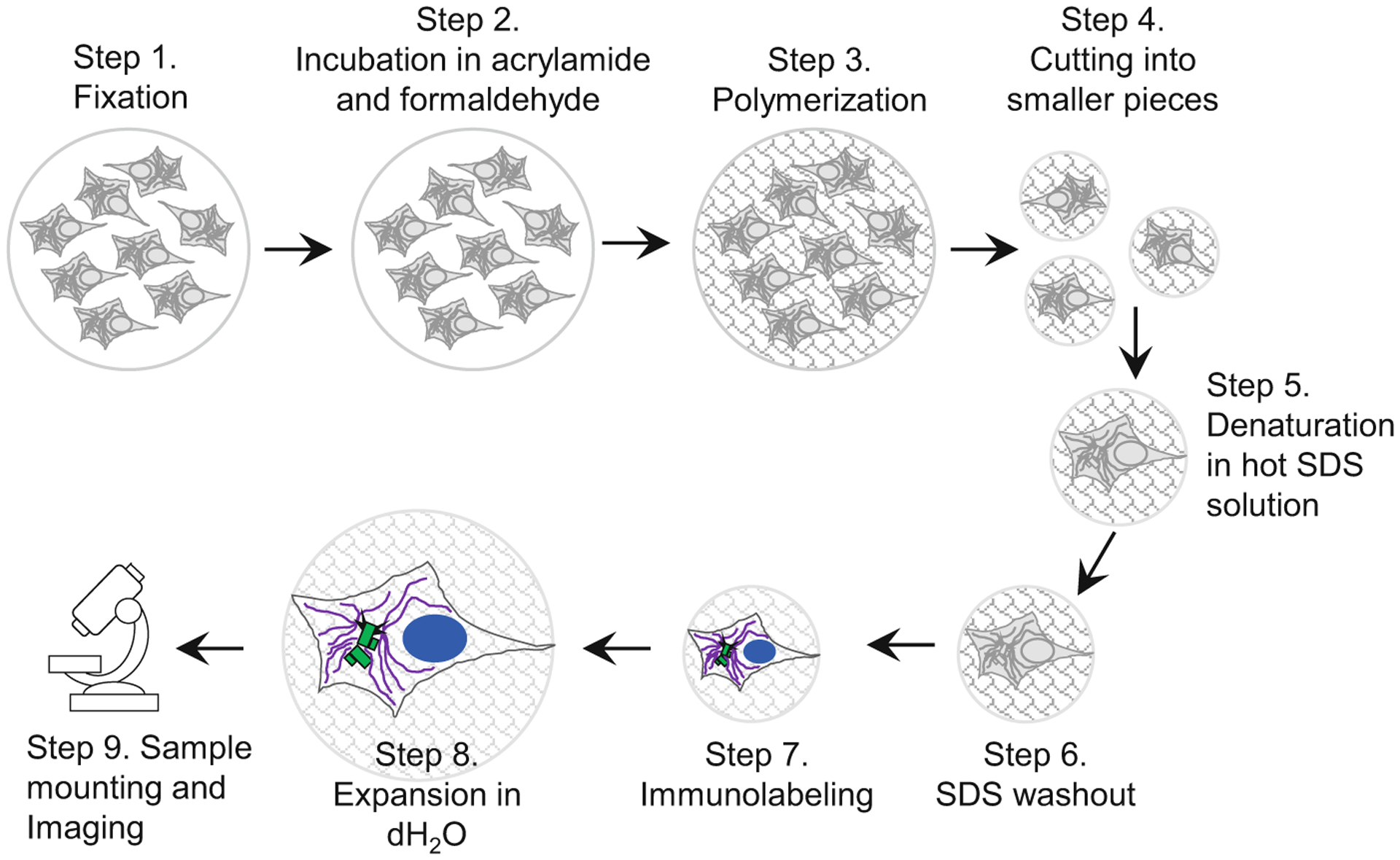
Expansion microscopy is an imaging method based on isotropic physical expansion of biological samples, which improves optical resolution and allows imaging of subresolutional cellular components by conventional microscopes. Centrioles are small microtubule-based cylindrical structures that build centrosomes and cilia, two organelles essential for vertebrates. Due to a centriole's small size, electron microscopy has traditionally been used to study centriole length and ultrastructural features. Recently, expansion microscopy has been successfully used as an affordable and accessible alternative to electron microscopy in the analysis of centriole and cilia length and structural features. Here, we describe an expansion microscopy approach for the analysis of centrioles and cilia in large populations of mammalian adherent and nonadherent cells and multiciliated cultures.
Keywords: Centriole; Centriole length; Centrosome; Cilia; Expansion microscopy.
Figures

References
-
- Schermelleh L et al. (2019) Super-resolution microscopy demystified. Nat Cell Biol 21(1):72–84 - PubMed
Publication types
MeSH terms
Grants and funding
LinkOut - more resources
Full Text Sources
