Candidalysin triggers epithelial cellular stresses that induce necrotic death
- PMID: 34085369
- PMCID: PMC8460601
- DOI: 10.1111/cmi.13371
Candidalysin triggers epithelial cellular stresses that induce necrotic death
Abstract
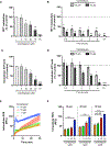
Candida albicans is a common opportunistic fungal pathogen that causes a wide range of infections from superficial mucosal to hematogenously disseminated candidiasis. The hyphal form plays an important role in the pathogenic process by invading epithelial cells and causing tissue damage. Notably, the secretion of the hyphal toxin candidalysin is essential for both epithelial cell damage and activation of mucosal immune responses. However, the mechanism of candidalysin-induced cell death remains unclear. Here, we examined the induction of cell death by candidalysin in oral epithelial cells. Fluorescent imaging using healthy/apoptotic/necrotic cell markers revealed that candidalysin causes a rapid and marked increase in the population of necrotic rather than apoptotic cells in a concentration dependent manner. Activation of a necrosis-like pathway was confirmed since C. albicans and candidalysin failed to activate caspase-8 and -3, or the cleavage of poly (ADP-ribose) polymerase. Furthermore, oral epithelial cells treated with candidalysin showed rapid production of reactive oxygen species, disruption of mitochondria activity and mitochondrial membrane potential, ATP depletion and cytochrome c release. Collectively, these data demonstrate that oral epithelial cells respond to the secreted fungal toxin candidalysin by triggering numerous cellular stress responses that induce necrotic death. TAKE AWAYS: Candidalysin secreted from Candida albicans causes epithelial cell stress. Candidalysin induces calcium influx and oxidative stress in host cells. Candidalysin induces mitochondrial dysfunction, ATP depletion and epithelial necrosis. The toxicity of candidalysin is mediated from the epithelial cell surface.
© 2021 The Authors. Cellular Microbiology published by John Wiley & Sons Ltd.
Conflict of interest statement
Conflict of interest
The authors declare no conflicts of interest
Figures


Similar articles
-
Candidalysin biology and activation of host cells.mBio. 2025 Jun 11;16(6):e0060324. doi: 10.1128/mbio.00603-24. Epub 2025 Apr 28. mBio. 2025. PMID: 40293285 Free PMC article. Review.
-
Candida albicans-Induced Epithelial Damage Mediates Translocation through Intestinal Barriers.mBio. 2018 Jun 5;9(3):e00915-18. doi: 10.1128/mBio.00915-18. mBio. 2018. PMID: 29871918 Free PMC article.
-
Variations in candidalysin amino acid sequence influence toxicity and host responses.mBio. 2024 Aug 14;15(8):e0335123. doi: 10.1128/mbio.03351-23. Epub 2024 Jul 2. mBio. 2024. PMID: 38953356 Free PMC article.
-
Nanobody-mediated neutralization of candidalysin prevents epithelial damage and inflammatory responses that drive vulvovaginal candidiasis pathogenesis.mBio. 2024 Mar 13;15(3):e0340923. doi: 10.1128/mbio.03409-23. Epub 2024 Feb 13. mBio. 2024. PMID: 38349176 Free PMC article.
-
Candidalysin: discovery and function in Candida albicans infections.Curr Opin Microbiol. 2019 Dec;52:100-109. doi: 10.1016/j.mib.2019.06.002. Epub 2019 Jul 6. Curr Opin Microbiol. 2019. PMID: 31288097 Free PMC article. Review.
Cited by
-
Acanthus ilicifolius Methanolic Extract for Oral Candidiasis Treatment through Tongue Epithelial STAT3 and Cell Death Evaluation.Eur J Dent. 2023 Oct;17(4):1201-1206. doi: 10.1055/s-0042-1760298. Epub 2023 Feb 10. Eur J Dent. 2023. PMID: 36764307 Free PMC article.
-
Pathogenesis and virulence of Candida albicans.Virulence. 2022 Dec;13(1):89-121. doi: 10.1080/21505594.2021.2019950. Virulence. 2022. PMID: 34964702 Free PMC article. Review.
-
Candidalysin biology and activation of host cells.mBio. 2025 Jun 11;16(6):e0060324. doi: 10.1128/mbio.00603-24. Epub 2025 Apr 28. mBio. 2025. PMID: 40293285 Free PMC article. Review.
-
Protective effects of gallic acid and SGK1 inhibitor on oxidative stress and cardiac damage in an isolated heart model of ischemia/reperfusion injury in rats.Iran J Basic Med Sci. 2023 Mar;26(3):308-315. doi: 10.22038/IJBMS.2023.68045.14874. Iran J Basic Med Sci. 2023. PMID: 36865044 Free PMC article.
-
Raman Spectroscopic Algorithms for Assessing Virulence in Oral Candidiasis: The Fight-or-Flight Response.Int J Mol Sci. 2024 Oct 24;25(21):11410. doi: 10.3390/ijms252111410. Int J Mol Sci. 2024. PMID: 39518963 Free PMC article.
References
Publication types
MeSH terms
Substances
Grants and funding
LinkOut - more resources
Full Text Sources
Medical

